

Search Count: 39
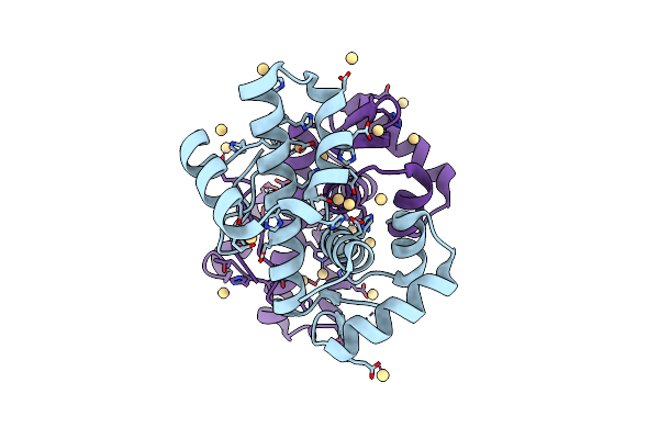 |
Manganese-Dependent Transcriptional Repressor Dr2539 Complexed With Cadmium (Sad 7 Kev)
Organism: Deinococcus radiodurans
Method: X-RAY DIFFRACTION Resolution:2.50 Å Release Date: 2024-05-08 Classification: DNA BINDING PROTEIN Ligands: CD |
Organism: Deinococcus radiodurans
Method: X-RAY DIFFRACTION
Release Date: 2024-05-08
Ligands: CD
 |
Manganese-Dependent Transcriptional Repressor Dr2539 Complexed With Cadmium
Organism: Deinococcus radiodurans
Method: X-RAY DIFFRACTION Resolution:2.00 Å Release Date: 2024-05-08 Classification: DNA BINDING PROTEIN Ligands: CD |
Organism: Deinococcus radiodurans
Method: X-RAY DIFFRACTION
Release Date: 2024-05-08
Ligands: CD
 |
Manganese-Dependent Transcriptional Repressor Dr2539 Complexed With Manganese And Bound To Dr1709 Promotor
Organism: Deinococcus radiodurans
Method: X-RAY DIFFRACTION Resolution:2.20 Å Release Date: 2024-05-08 Classification: DNA BINDING PROTEIN Ligands: MN, GOL |
Organism: Deinococcus radiodurans
Method: X-RAY DIFFRACTION
Release Date: 2024-05-08
Ligands: MN, GOL
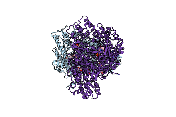 |
Crystal Structure Of Human Glycine Decarboxylase (P-Protein)
Organism: Homo sapiens
Method: X-RAY DIFFRACTION Resolution:2.30 Å Release Date: 2019-11-20 Classification: OXIDOREDUCTASE Ligands: PLP, EDO, BCT |
Organism: Homo sapiens
Method: X-RAY DIFFRACTION
Release Date: 2019-11-20
Ligands: PLP, EDO, BCT
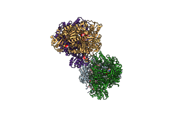 |
Crystal Structure Of Neanderthal Glycine Decarboxylase (P-Protein)
Organism: Homo sapiens neanderthalensis
Method: X-RAY DIFFRACTION Resolution:2.10 Å Release Date: 2019-11-20 Classification: OXIDOREDUCTASE Ligands: PLP, EDO, PEG, GOL |
Organism: Homo sapiens neanderthalensis
Method: X-RAY DIFFRACTION
Release Date: 2019-11-20
Ligands: PLP, EDO, PEG, GOL
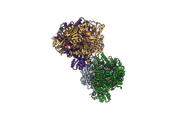 |
Crystal Structure Of Human Glycine Decarboxylase (P-Protein) Bound With Pyridoxyl-Glycine-5'-Monophosphate
Organism: Homo sapiens
Method: X-RAY DIFFRACTION Resolution:2.00 Å Release Date: 2019-11-20 Classification: OXIDOREDUCTASE Ligands: PLG, EDO, GOL, BCT, PEG |
Organism: Homo sapiens
Method: X-RAY DIFFRACTION
Release Date: 2019-11-20
Ligands: PLG, EDO, GOL, BCT, PEG
 |
Crystal Structure Of Neanderthal Adenylosuccinate Lyase (Adsl)
Organism: Homo sapiens neanderthalensis
Method: X-RAY DIFFRACTION Resolution:1.70 Å Release Date: 2018-05-30 Classification: LYASE Ligands: PEG, GOL, CL |
Organism: Homo sapiens neanderthalensis
Method: X-RAY DIFFRACTION
Release Date: 2018-05-30
Ligands: PEG, GOL, CL
 |
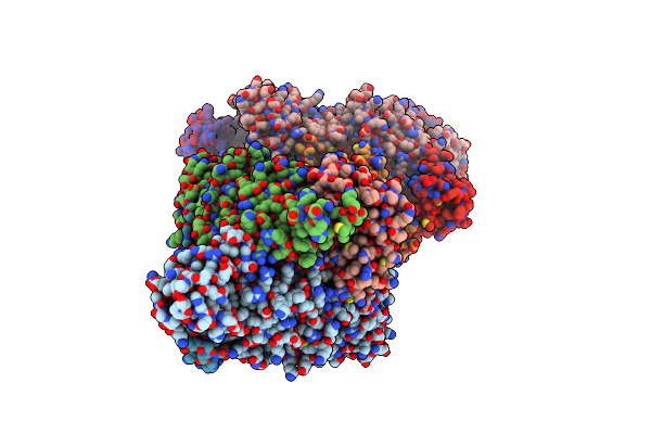
Crystal Structure Of Neanderthal Adenylosuccinate Lyase (Adsl) In Complex With Its Products Amp And Fumarate
Organism: Homo sapiens neanderthalensis
Method: X-RAY DIFFRACTION Resolution:2.30 Å Release Date: 2018-05-30 Classification: LYASE Ligands: CL, AMP, FUM, PEG, 2SA, GOL |
Organism: Homo sapiens neanderthalensis
Method: X-RAY DIFFRACTION
Release Date: 2018-05-30
Ligands: CL, AMP, FUM, PEG, 2SA, GOL
 |
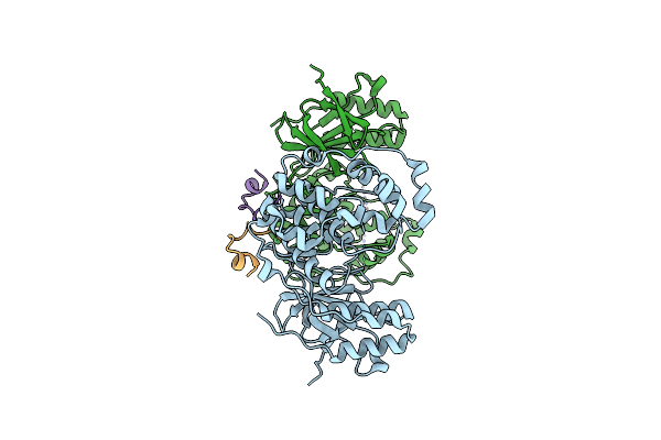
Crystal Structure Of Neanderthal Adenylosuccinate Lyase (Adsl)In Complex With Its Products Aicar And Fumarate
Organism: Homo sapiens neanderthalensis
Method: X-RAY DIFFRACTION Resolution:2.40 Å Release Date: 2018-05-30 Classification: LYASE Ligands: SSS, CL, FUM, AMZ |
Organism: Homo sapiens neanderthalensis
Method: X-RAY DIFFRACTION
Release Date: 2018-05-30
Ligands: SSS, CL, FUM, AMZ
 |
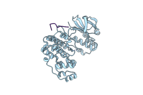
Structure Of Mapk14 With Bound The Kim Domain Of The Toxoplasma Protein Gra24
Organism: Homo sapiens, Toxoplasma gondii
Method: X-RAY DIFFRACTION Resolution:2.80 Å Release Date: 2016-10-26 Classification: TRANSFERASE |
Organism: Homo sapiens, Toxoplasma gondii
Method: X-RAY DIFFRACTION
Release Date: 2016-10-26
 |
Structure Of Dead Kinase Mapk14 With Bound The Kim Domain Of Mkk6
Organism: Homo sapiens
Method: X-RAY DIFFRACTION Resolution:2.40 Å Release Date: 2016-10-26 Classification: TRANSFERASE |
Organism: Homo sapiens
Method: X-RAY DIFFRACTION
Release Date: 2016-10-26
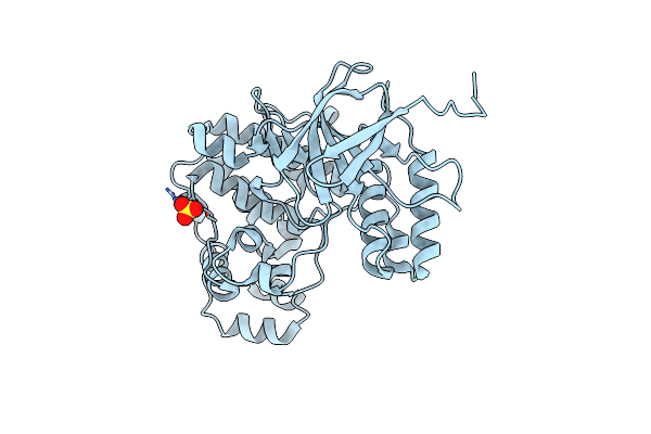 |
Structure Of Inactive Mapk14 With Ordered Activation Loop
Organism: Homo sapiens
Method: X-RAY DIFFRACTION Resolution:2.42 Å Release Date: 2016-01-20 Classification: TRANSFERASE Ligands: SO4 |
Organism: Homo sapiens
Method: X-RAY DIFFRACTION
Release Date: 2016-01-20
Ligands: SO4
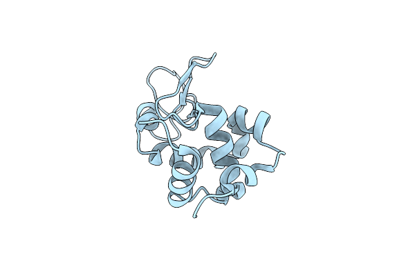 |
Room Temperature Crystal Structure Of Lysozyme Determined By Serial Synchrotron Crystallography (Micro Focused Beam - Crystfel)
Organism: Gallus gallus
Method: X-RAY DIFFRACTION Resolution:1.70 Å Release Date: 2015-05-06 Classification: HYDROLASE Ligands: CL |
Organism: Gallus gallus
Method: X-RAY DIFFRACTION
Release Date: 2015-05-06
Ligands: CL
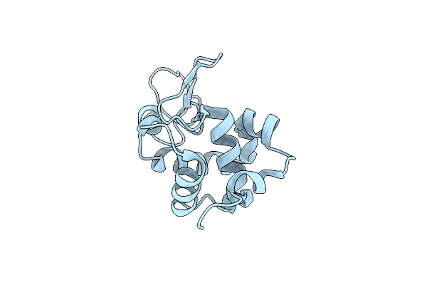 |
Room-Temperature Crystal Structure Of Lysozyme Determined By Serial Synchrotron Crystallography Using A Nano Focused Beam.
Organism: Gallus gallus
Method: X-RAY DIFFRACTION Resolution:1.70 Å Release Date: 2015-05-06 Classification: HYDROLASE Ligands: CL |
Organism: Gallus gallus
Method: X-RAY DIFFRACTION
Release Date: 2015-05-06
Ligands: CL
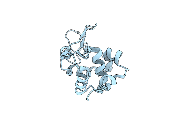 |
Raster-Scanning Protein Crystallography Using Micro And Nano-Focused Synchrotron Beams
Organism: Gallus gallus
Method: X-RAY DIFFRACTION Resolution:1.85 Å Release Date: 2015-05-06 Classification: HYDROLASE Ligands: CL |
Organism: Gallus gallus
Method: X-RAY DIFFRACTION
Release Date: 2015-05-06
Ligands: CL
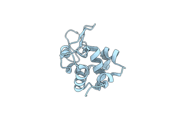 |
Room-Temperature Crystal Structure Of Lysozyme Determined By Serial Synchrotron Crystallography Using A Micro Focused Beam (Conventional Resolution Cut-Off)
Organism: Gallus gallus
Method: X-RAY DIFFRACTION Resolution:1.95 Å Release Date: 2015-05-06 Classification: HYDROLASE Ligands: CL |
Organism: Gallus gallus
Method: X-RAY DIFFRACTION
Release Date: 2015-05-06
Ligands: CL
 |
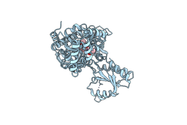
Crystal Structure Of Gh3.12 From Arabidopsis Thaliana In Complex With Ampcpp And Salicylate
Organism: Arabidopsis thaliana
Method: X-RAY DIFFRACTION Resolution:2.81 Å Release Date: 2013-10-02 Classification: LIGASE Ligands: MG, SAL, APC |
Organism: Arabidopsis thaliana
Method: X-RAY DIFFRACTION
Release Date: 2013-10-02
Ligands: MG, SAL, APC
 |
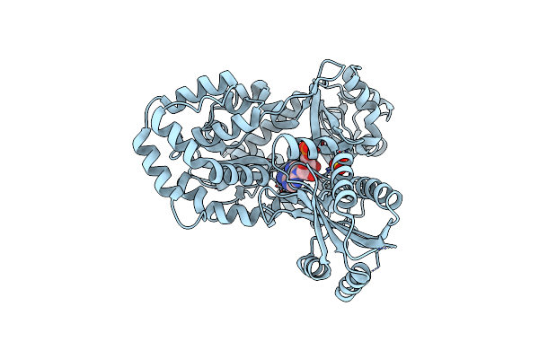
Crystal Structure Of Arabidopsis Thaliana Gh3.11 (Jar1) In Complex With Ja-Ile
Organism: Arabidopsis thaliana
Method: X-RAY DIFFRACTION Resolution:2.01 Å Release Date: 2012-06-20 Classification: LIGASE Ligands: JAI |
Organism: Arabidopsis thaliana
Method: X-RAY DIFFRACTION
Release Date: 2012-06-20
Ligands: JAI
 |
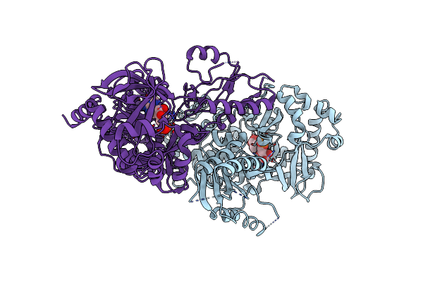
Crystal Structure Of Arabidopsis Gh3.12 (Pbs3) In Complex With Amp
Organism: Arabidopsis thaliana
Method: X-RAY DIFFRACTION Resolution:2.10 Å Release Date: 2012-06-20 Classification: LIGASE Ligands: AMP, SO4 |
Organism: Arabidopsis thaliana
Method: X-RAY DIFFRACTION
Release Date: 2012-06-20
Ligands: AMP, SO4
 |
Crystal Structure Of Seleno-Methionine Derivatized Gh3.12
Organism: Arabidopsis thaliana
Method: X-RAY DIFFRACTION Resolution:2.07 Å Release Date: 2012-06-20 Classification: LIGASE Ligands: SAL, AMP |
Organism: Arabidopsis thaliana
Method: X-RAY DIFFRACTION
Release Date: 2012-06-20
Ligands: SAL, AMP