

Search Count: 16
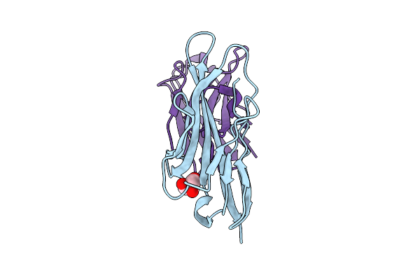 |
Structure Of Single-Domain Antibody Vhh_H4 In Complex With Human Vsig4
Organism: Homo sapiens, Camelidae
Method: X-RAY DIFFRACTION Resolution:1.94 Å Release Date: 2025-10-29 Classification: IMMUNE SYSTEM Ligands: GOL |
Organism: Homo sapiens, Camelidae
Method: X-RAY DIFFRACTION
Release Date: 2025-10-29
Ligands: GOL
 |
Structure Of Single-Domain Antibody Vhh_H5 In Complex With Human Vsig4
Organism: Homo sapiens, Camelidae
Method: X-RAY DIFFRACTION Resolution:1.97 Å Release Date: 2025-10-29 Classification: IMMUNE SYSTEM |
Organism: Homo sapiens, Camelidae
Method: X-RAY DIFFRACTION
Release Date: 2025-10-29
 |
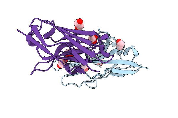
Structure Of The Single-Domain Antibody Vhh_H1 In Complex With Human Vsig4
Organism: Homo sapiens, Camelidae
Method: X-RAY DIFFRACTION Resolution:1.05 Å Release Date: 2025-10-29 Classification: IMMUNE SYSTEM Ligands: SO4 |
Organism: Homo sapiens, Camelidae
Method: X-RAY DIFFRACTION
Release Date: 2025-10-29
Ligands: SO4
 |
Structure Of The Single-Domain Antibody Vhh_H2 In Complex With Human Vsig4
Organism: Homo sapiens
Method: X-RAY DIFFRACTION Resolution:1.45 Å Release Date: 2025-10-29 Classification: IMMUNE SYSTEM Ligands: EDO, IPA |
Organism: Homo sapiens
Method: X-RAY DIFFRACTION
Release Date: 2025-10-29
Ligands: EDO, IPA
 |
Structure Of The Single-Domain Antibody Vhh_H3 In Complex With Human Vsig4
Organism: Homo sapiens
Method: X-RAY DIFFRACTION Resolution:1.90 Å Release Date: 2025-10-29 Classification: IMMUNE SYSTEM Ligands: EDO, PEG |
Organism: Homo sapiens
Method: X-RAY DIFFRACTION
Release Date: 2025-10-29
Ligands: EDO, PEG
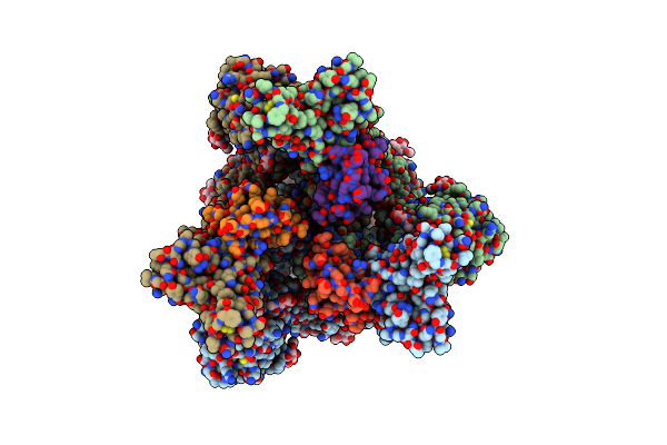 |
Sars-Cov-2 S-Trimer (3 Rbds Up) Bound To Trisb92, Fitted Into Cryo-Em Map
Organism: Severe acute respiratory syndrome coronavirus 2, Homo sapiens
Method: ELECTRON MICROSCOPY Release Date: 2023-04-19 Classification: VIRAL PROTEIN Ligands: NAG |
Organism: Severe acute respiratory syndrome coronavirus 2, Homo sapiens
Method: ELECTRON MICROSCOPY
Release Date: 2023-04-19
Ligands: NAG
 |
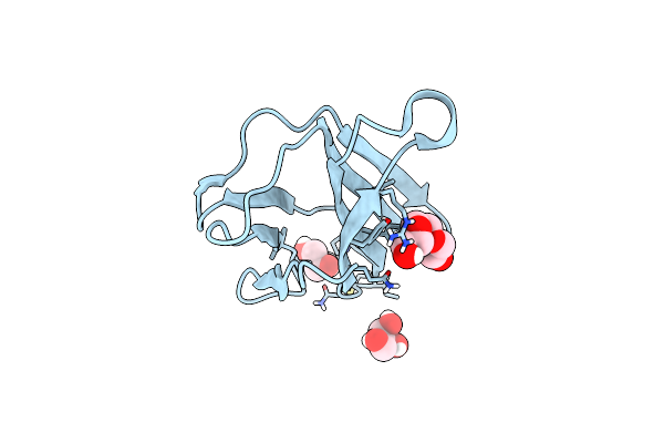
Structure Of Superoxide Dismutase 1 (Sod1) In Complex With Nanobody 2 (Nb2).
Organism: Homo sapiens, Camelus dromedarius
Method: X-RAY DIFFRACTION Resolution:2.19 Å Release Date: 2022-09-28 Classification: METAL BINDING PROTEIN Ligands: CU, ZN |
Organism: Homo sapiens, Camelus dromedarius
Method: X-RAY DIFFRACTION
Release Date: 2022-09-28
Ligands: CU, ZN
 |
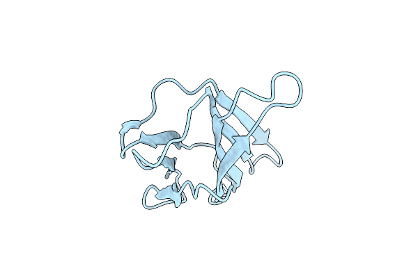
Crystal Structure Of Beta-2-Microglobulin D76E Mutant
Organism: Homo sapiens
Method: X-RAY DIFFRACTION Resolution:1.20 Å Release Date: 2022-08-03 Classification: IMMUNE SYSTEM Ligands: GOL, PGE |
Organism: Homo sapiens
Method: X-RAY DIFFRACTION
Release Date: 2022-08-03
Ligands: GOL, PGE
 |
Crystal Structure Of Beta-2-Microglobulin D76A Mutant
Organism: Homo sapiens
Method: X-RAY DIFFRACTION Resolution:1.20 Å Release Date: 2022-08-03 Classification: IMMUNE SYSTEM |
Organism: Homo sapiens
Method: X-RAY DIFFRACTION
Release Date: 2022-08-03
 |
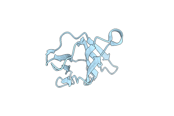
Crystal Structure Of Beta-2-Microglobulin D76S Mutant
Organism: Homo sapiens
Method: X-RAY DIFFRACTION Resolution:1.15 Å Release Date: 2022-08-03 Classification: IMMUNE SYSTEM Ligands: GOL |
Organism: Homo sapiens
Method: X-RAY DIFFRACTION
Release Date: 2022-08-03
Ligands: GOL
 |
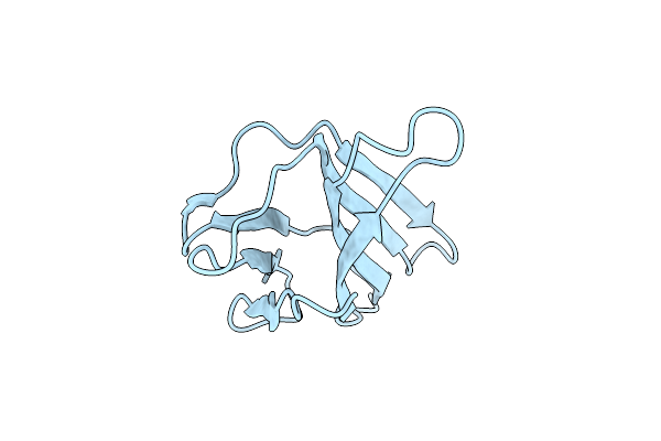
Crystal Structure Of Beta-2-Microglobulin D76G Mutant
Organism: Homo sapiens
Method: X-RAY DIFFRACTION Resolution:1.20 Å Release Date: 2022-08-03 Classification: IMMUNE SYSTEM |
Organism: Homo sapiens
Method: X-RAY DIFFRACTION
Release Date: 2022-08-03
 |
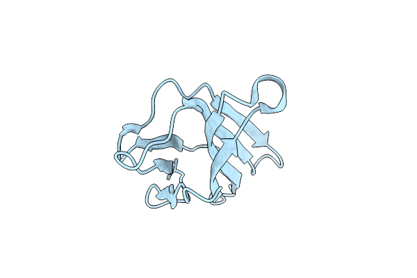
Crystal Structure Of Beta-2-Microglobulin D76Q Mutant
Organism: Homo sapiens
Method: X-RAY DIFFRACTION Resolution:1.51 Å Release Date: 2022-08-03 Classification: IMMUNE SYSTEM |
Organism: Homo sapiens
Method: X-RAY DIFFRACTION
Release Date: 2022-08-03
 |
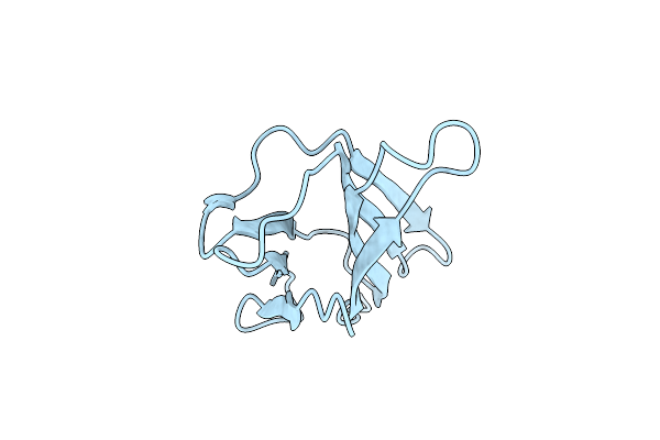
Crystal Structure Of Beta-2-Microglobulin D76Y Mutant
Organism: Homo sapiens
Method: X-RAY DIFFRACTION Resolution:1.25 Å Release Date: 2022-08-03 Classification: IMMUNE SYSTEM |
Organism: Homo sapiens
Method: X-RAY DIFFRACTION
Release Date: 2022-08-03
 |
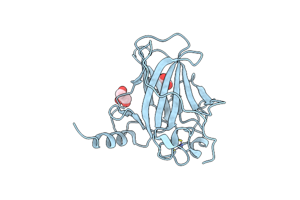
Crystal Structure Of Beta-2-Microglobulin D76K Mutant
Organism: Homo sapiens
Method: X-RAY DIFFRACTION Resolution:1.24 Å Release Date: 2022-08-03 Classification: IMMUNE SYSTEM |
Organism: Homo sapiens
Method: X-RAY DIFFRACTION
Release Date: 2022-08-03
 |
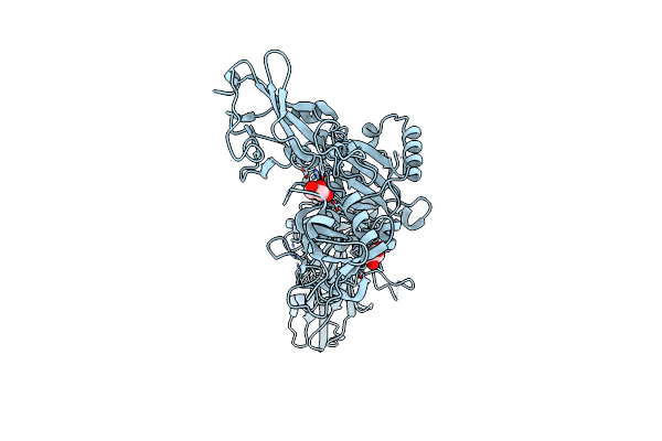
P53 Charged Core
Organism: Homo sapiens
Method: X-RAY DIFFRACTION Resolution:1.67 Å Release Date: 2020-03-11 Classification: CELL CYCLE Ligands: GOL, ZN |
Organism: Homo sapiens
Method: X-RAY DIFFRACTION
Release Date: 2020-03-11
Ligands: GOL, ZN
 |
X-Ray Crystal Structure Of Aggregation-Resistant Protective Antigen Of Bacillus Anthracis (Mutant S559L T576E)
Organism: Bacillus anthracis
Method: X-RAY DIFFRACTION Resolution:1.94 Å Release Date: 2016-01-27 Classification: TOXIN Ligands: CA, GOL |
Organism: Bacillus anthracis
Method: X-RAY DIFFRACTION
Release Date: 2016-01-27
Ligands: CA, GOL