

Search Count: 62
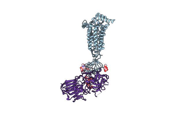 |
Cryo-Em Structure Of Sv2B-Hc-A1 Complex
Organism: Homo sapiens, Clostridium botulinum
Method: ELECTRON MICROSCOPY Release Date: 2025-03-12 Classification: MEMBRANE PROTEIN Ligands: NAG |
Organism: Homo sapiens, Clostridium botulinum
Method: ELECTRON MICROSCOPY
Release Date: 2025-03-12
Ligands: NAG
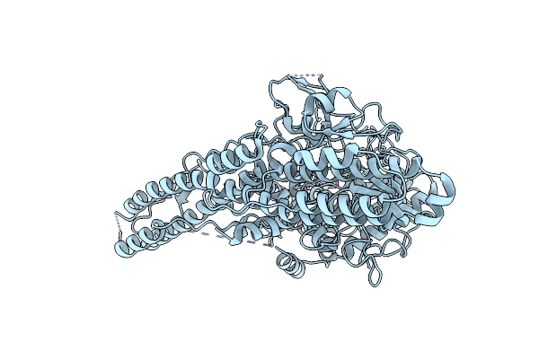 |
Cryo-Em Structure Of Botulinum Neurotoxin A Lc-Hn Domain
Organism: Clostridium botulinum
Method: ELECTRON MICROSCOPY Resolution:3.70 Å Release Date: 2025-03-12 Classification: TOXIN |
Organism: Clostridium botulinum
Method: ELECTRON MICROSCOPY
Release Date: 2025-03-12
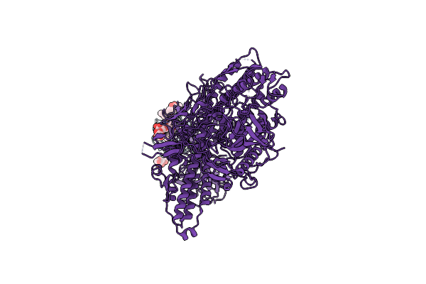 |
Focused Refinement Of Sv2B Luminal Domain And Bont/A1 Complex
Organism: Homo sapiens, Clostridium botulinum
Method: ELECTRON MICROSCOPY Resolution:3.49 Å Release Date: 2025-03-12 Classification: MEMBRANE PROTEIN Ligands: NAG |
Organism: Homo sapiens, Clostridium botulinum
Method: ELECTRON MICROSCOPY
Release Date: 2025-03-12
Ligands: NAG
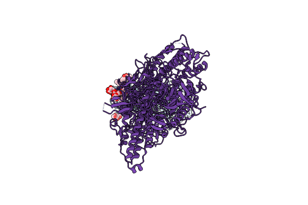 |
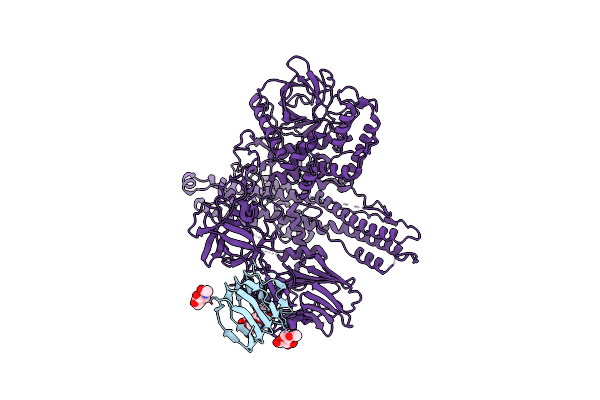
Cryo-Em Structure Of Sv2B-Bont/A1 Complex
Organism: Clostridium botulinum, Homo sapiens
Method: ELECTRON MICROSCOPY Resolution:3.98 Å Release Date: 2025-03-12 Classification: MEMBRANE PROTEIN Ligands: NAG |
Organism: Clostridium botulinum, Homo sapiens
Method: ELECTRON MICROSCOPY
Release Date: 2025-03-12
Ligands: NAG
 |
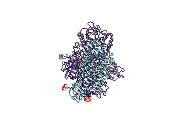
Focused Refinement Of Sv2B-Ld-Bont/A1 At Ph 5.5
Organism: Homo sapiens, Clostridium botulinum
Method: ELECTRON MICROSCOPY Release Date: 2025-03-12 Classification: TOXIN Ligands: NAG |
Organism: Homo sapiens, Clostridium botulinum
Method: ELECTRON MICROSCOPY
Release Date: 2025-03-12
Ligands: NAG
 |
Low-Resolution (5.4 Angstrom) Cryo-Em Structure Of Sv2B-Bont/A1 At Ph 5.5
Organism: Homo sapiens, Clostridium botulinum
Method: ELECTRON MICROSCOPY Release Date: 2025-03-12 Classification: MEMBRANE PROTEIN Ligands: NAG |
Organism: Homo sapiens, Clostridium botulinum
Method: ELECTRON MICROSCOPY
Release Date: 2025-03-12
Ligands: NAG
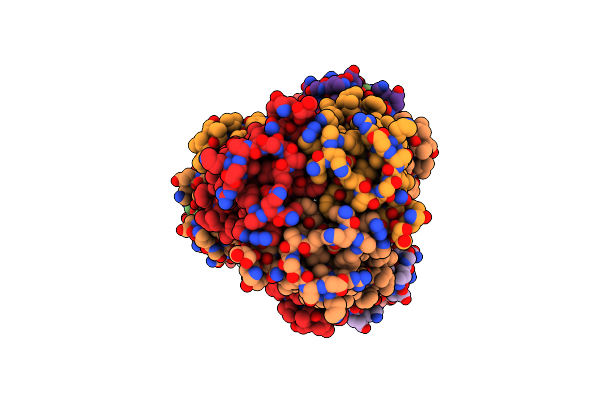 |
Cryo-Em Structure Of Styrene Oxide Isomerase Bound To Benzylamine Inhibitor
Organism: Pseudomonas sp. vlb120, Vicugna pacos
Method: ELECTRON MICROSCOPY Release Date: 2024-04-03 Classification: MEMBRANE PROTEIN Ligands: ABN, HEM |
Organism: Pseudomonas sp. vlb120, Vicugna pacos
Method: ELECTRON MICROSCOPY
Release Date: 2024-04-03
Ligands: ABN, HEM
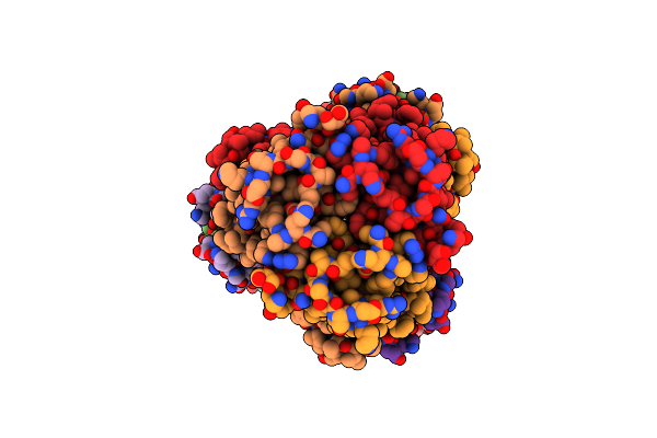 |
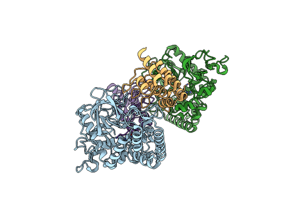
Cryo-Em Structure Of Styrene Oxide Isomerase
Organism: Pseudomonas sp. vlb120, Vicugna pacos
Method: ELECTRON MICROSCOPY Release Date: 2024-04-03 Classification: MEMBRANE PROTEIN Ligands: HEM |
Organism: Pseudomonas sp. vlb120, Vicugna pacos
Method: ELECTRON MICROSCOPY
Release Date: 2024-04-03
Ligands: HEM
 |
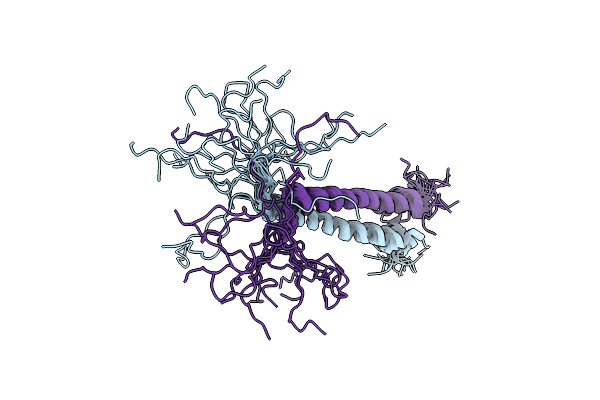
Crystal Structure Of The Lc/A1-Darpin18 Complex
Organism: Clostridium botulinum, Synthetic construct
Method: X-RAY DIFFRACTION Resolution:2.70 Å Release Date: 2023-11-29 Classification: TOXIN Ligands: ZN |
Organism: Clostridium botulinum, Synthetic construct
Method: X-RAY DIFFRACTION
Release Date: 2023-11-29
Ligands: ZN
 |
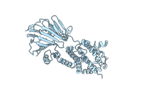
Nmr Assignments And Structure For The Dimeric Kinesin Neck Domain
Organism: Rattus norvegicus
Method: SOLUTION NMR Release Date: 2023-11-01 Classification: MOTOR PROTEIN |
Organism: Rattus norvegicus
Method: SOLUTION NMR
Release Date: 2023-11-01
 |
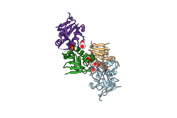
Active Yidc Insertase Crystal Structure With The First Transmembrane Domain Resolved
Organism: Thermotoga maritima (strain atcc 43589 / msb8 / dsm 3109 / jcm 10099)
Method: X-RAY DIFFRACTION Resolution:3.40 Å Release Date: 2021-09-22 Classification: MEMBRANE PROTEIN |
 |
Crystal Structure Of Dna-Binding Domain Of Ompr Of Two-Component System Of Acinetobacter Baumannii
Organism: Acinetobacter baumannii (strain sdf)
Method: X-RAY DIFFRACTION Resolution:2.20 Å Release Date: 2021-08-11 Classification: DNA BINDING PROTEIN Ligands: SO4, PEG, GOL |
Organism: Acinetobacter baumannii (strain sdf)
Method: X-RAY DIFFRACTION
Release Date: 2021-08-11
Ligands: SO4, PEG, GOL
 |
Crystal Structure Of The Catalytic Domain Of Botulinum Neurotoxin Subtype A3
Organism: Clostridium botulinum
Method: X-RAY DIFFRACTION Resolution:2.01 Å Release Date: 2021-04-28 Classification: TOXIN Ligands: ZN |
Organism: Clostridium botulinum
Method: X-RAY DIFFRACTION
Release Date: 2021-04-28
Ligands: ZN
 |
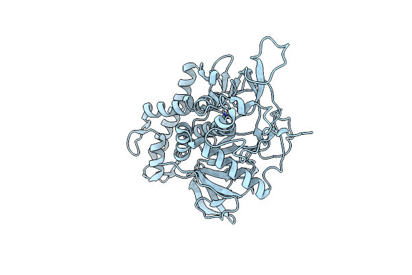
Heterotetrameric Katanin P60:P80 Complex
Organism: Mus musculus
Method: X-RAY DIFFRACTION Resolution:2.00 Å Release Date: 2019-08-07 Classification: HYDROLASE Ligands: PEG, EDO |
Organism: Mus musculus
Method: X-RAY DIFFRACTION
Release Date: 2019-08-07
Ligands: PEG, EDO
 |
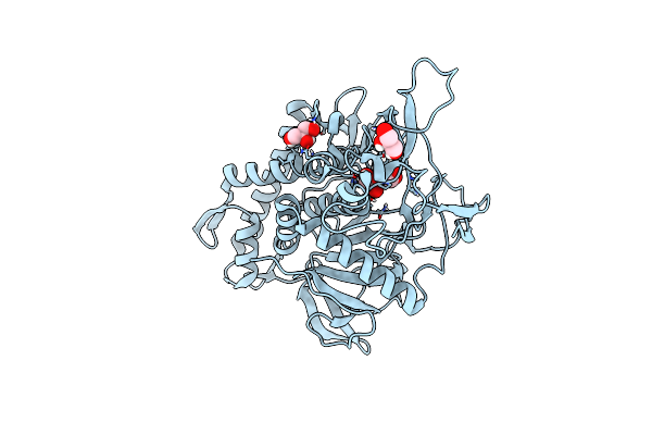
Crystal Structure Of The Light Chain Of Botulinum Neurotoxin X (Residues 2-427)
Organism: Clostridium botulinum
Method: X-RAY DIFFRACTION Resolution:1.31 Å Release Date: 2019-07-31 Classification: TOXIN Ligands: ZN |
Organism: Clostridium botulinum
Method: X-RAY DIFFRACTION
Release Date: 2019-07-31
Ligands: ZN
 |
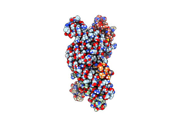
Crystal Structure Of The Light Chain Of Botulinum Neurotoxin X (Residues 2-442)
Organism: Clostridium botulinum
Method: X-RAY DIFFRACTION Resolution:1.45 Å Release Date: 2019-07-31 Classification: TOXIN Ligands: ZN, GOL |
Organism: Clostridium botulinum
Method: X-RAY DIFFRACTION
Release Date: 2019-07-31
Ligands: ZN, GOL
 |
P60P80-Camsap Complex
Organism: Mus musculus
Method: X-RAY DIFFRACTION Resolution:1.70 Å Release Date: 2018-07-11 Classification: HYDROLASE Ligands: EDO, PEG |
Organism: Mus musculus
Method: X-RAY DIFFRACTION
Release Date: 2018-07-11
Ligands: EDO, PEG
 |
Helicobacter Pylori Adhesin Hopq Type I Bound To The N-Terminal Domain Of Human Ceacam1
Organism: Homo sapiens, Helicobacter pylori (strain g27)
Method: X-RAY DIFFRACTION Resolution:2.80 Å Release Date: 2018-06-27 Classification: CELL ADHESION Ligands: BR |
Organism: Homo sapiens, Helicobacter pylori (strain g27)
Method: X-RAY DIFFRACTION
Release Date: 2018-06-27
Ligands: BR
 |
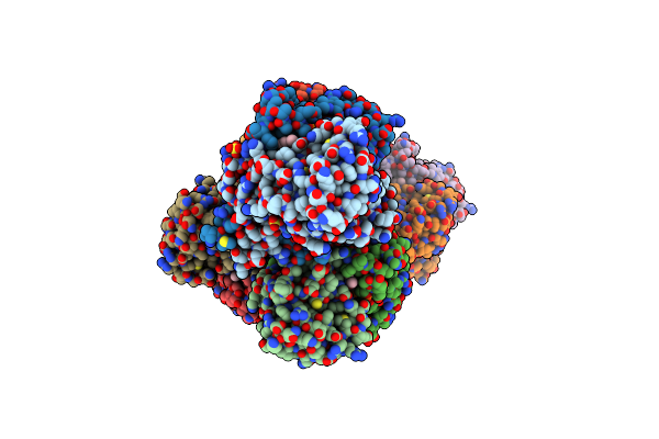
Helicobacter Pylori Adhesin Hopq Type Ii Bound To The N-Terminal Domain Of Human Ceacam1
Organism: Homo sapiens, Helicobacter pylori
Method: X-RAY DIFFRACTION Resolution:2.59 Å Release Date: 2018-06-27 Classification: CELL ADHESION Ligands: SO4 |
Organism: Homo sapiens, Helicobacter pylori
Method: X-RAY DIFFRACTION
Release Date: 2018-06-27
Ligands: SO4
 |
X-Ray Structure Of A Bacterial Adenylyl Cyclase Soluble Domain
Organism: Mycobacterium intracellulare 1956
Method: X-RAY DIFFRACTION Resolution:3.40 Å Release Date: 2017-11-01 Classification: MEMBRANE PROTEIN Ligands: ONM, MN, SO4 |
Organism: Mycobacterium intracellulare 1956
Method: X-RAY DIFFRACTION
Release Date: 2017-11-01
Ligands: ONM, MN, SO4