

Search Count: 29
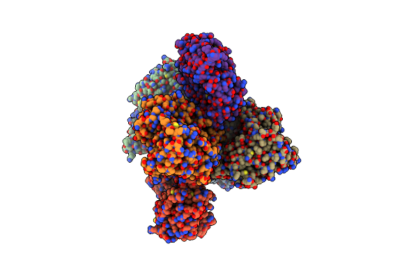 |
Crystal Structure Of E. Coli Bepa
Organism: Escherichia coli (strain k12)
Method: X-RAY DIFFRACTION Resolution:2.60 Å Release Date: 2018-12-26 Classification: HYDROLASE Ligands: ZN, TRS |
Organism: Escherichia coli (strain k12)
Method: X-RAY DIFFRACTION
Release Date: 2018-12-26
Ligands: ZN, TRS
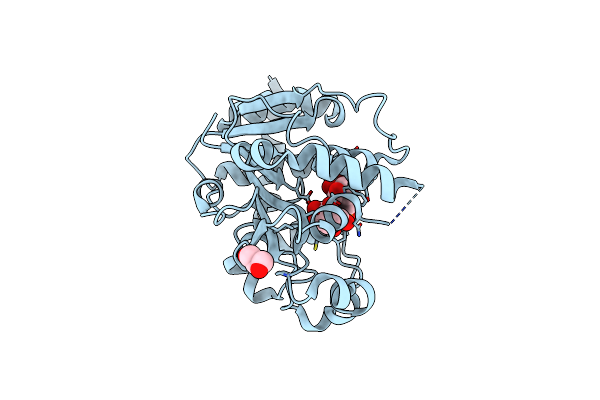 |
Crystal Structure Of Catalytic Domain Of Uggt (Udp-Glucose-Bound Form) From Thermomyces Dupontii
Organism: Thermomyces dupontii
Method: X-RAY DIFFRACTION Resolution:1.40 Å Release Date: 2017-09-27 Classification: TRANSFERASE Ligands: UPG, CA, GOL |
Organism: Thermomyces dupontii
Method: X-RAY DIFFRACTION
Release Date: 2017-09-27
Ligands: UPG, CA, GOL
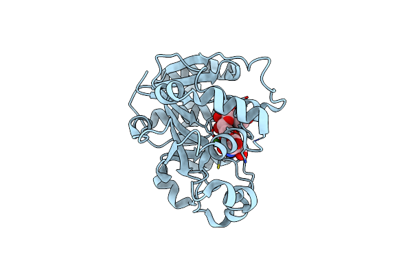 |
Crystal Structure Of Catalytic Domain Of Uggt (Udp-Bound Form) From Thermomyces Dupontii
Organism: Thermomyces dupontii
Method: X-RAY DIFFRACTION Resolution:1.35 Å Release Date: 2017-09-27 Classification: TRANSFERASE Ligands: UDP, TRS, CA |
Organism: Thermomyces dupontii
Method: X-RAY DIFFRACTION
Release Date: 2017-09-27
Ligands: UDP, TRS, CA
 |
Crystal Structure Of Folding Sensor Region Of Uggt From Thermomyces Dupontii
Organism: Thermomyces dupontii
Method: X-RAY DIFFRACTION Resolution:3.10 Å Release Date: 2017-09-27 Classification: TRANSFERASE |
Organism: Thermomyces dupontii
Method: X-RAY DIFFRACTION
Release Date: 2017-09-27
 |
Pyp Mutant - E46Q
Organism: Halorhodospira halophila
Method: NEUTRON DIFFRACTION Resolution:1.49 Å Release Date: 2017-08-30 Classification: SIGNALING PROTEIN Ligands: HC4, DOD |
Organism: Halorhodospira halophila
Method: NEUTRON DIFFRACTION
Release Date: 2017-08-30
Ligands: HC4, DOD
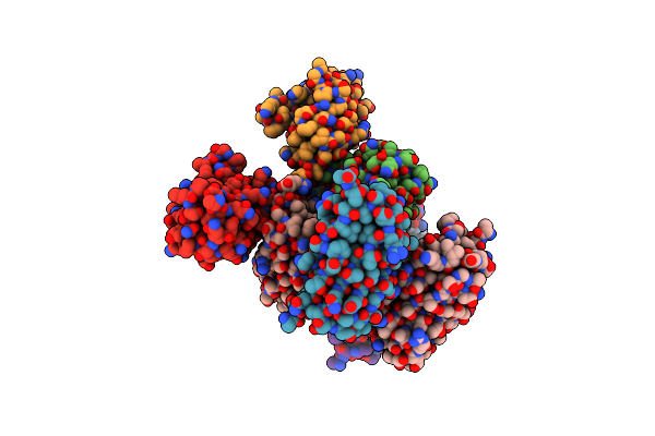 |
Structure Of Monomeric Mutant Of Rei Immunoglobulin Light Chain Variable Domain Crystallized At Ph 6
Organism: Homo sapiens
Method: X-RAY DIFFRACTION Resolution:2.88 Å Release Date: 2017-08-02 Classification: IMMUNE SYSTEM |
Organism: Homo sapiens
Method: X-RAY DIFFRACTION
Release Date: 2017-08-02
 |
Structure Of Monomeric Mutant Of Rei Immunoglobulin Light Chain Variable Domain Crystallized At Ph 8
Organism: Homo sapiens
Method: X-RAY DIFFRACTION Resolution:2.90 Å Release Date: 2017-08-02 Classification: IMMUNE SYSTEM |
Organism: Homo sapiens
Method: X-RAY DIFFRACTION
Release Date: 2017-08-02
 |
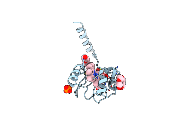
Domain-Swapped Dimer Of Pseudomonas Aeruginosa Cytochrome C551
Organism: Pseudomonas aeruginosa pao1
Method: X-RAY DIFFRACTION Resolution:1.50 Å Release Date: 2015-04-22 Classification: ELECTRON TRANSPORT Ligands: HEC |
Organism: Pseudomonas aeruginosa pao1
Method: X-RAY DIFFRACTION
Release Date: 2015-04-22
Ligands: HEC
 |
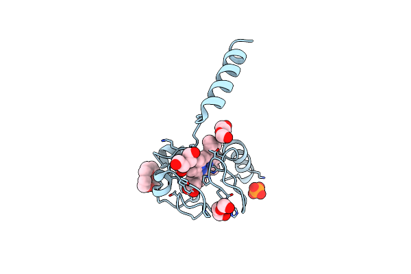
Dimeric Horse Cytochrome C Formed By Refolding From Molten Globule State
Organism: Equus caballus
Method: X-RAY DIFFRACTION Resolution:1.80 Å Release Date: 2014-07-16 Classification: ELECTRON TRANSPORT Ligands: HEC, PG4, PO4, PEG |
Organism: Equus caballus
Method: X-RAY DIFFRACTION
Release Date: 2014-07-16
Ligands: HEC, PG4, PO4, PEG
 |
Dimeric Horse Cytochrome C Obtained By Refolding With Desalting Method
Organism: Equus caballus
Method: X-RAY DIFFRACTION Resolution:1.80 Å Release Date: 2013-12-11 Classification: ELECTRON TRANSPORT Ligands: HEC, PO4, PG4, PEG |
Organism: Equus caballus
Method: X-RAY DIFFRACTION
Release Date: 2013-12-11
Ligands: HEC, PO4, PG4, PEG
 |
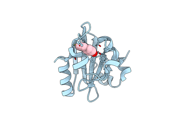
The Pr0 Photocycle Intermediate Of Photoactive Yellow Protein
Organism: Halorhodospira halophila
Method: X-RAY DIFFRACTION Resolution:1.60 Å Release Date: 2012-11-14 Classification: SIGNALING PROTEIN Ligands: HC4 |
Organism: Halorhodospira halophila
Method: X-RAY DIFFRACTION
Release Date: 2012-11-14
Ligands: HC4
 |
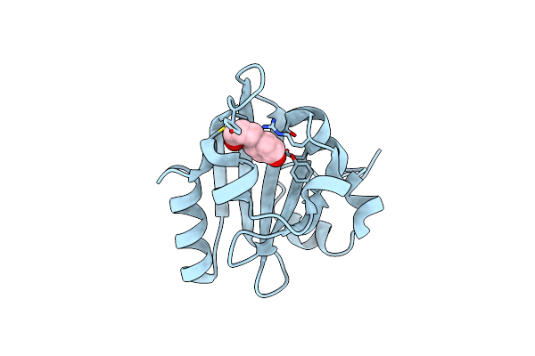
The Pr1 Photocycle Intermediate Of Photoactive Yellow Protein
Organism: Halorhodospira halophila
Method: X-RAY DIFFRACTION Resolution:1.60 Å Release Date: 2012-11-14 Classification: SIGNALING PROTEIN Ligands: HC4 |
Organism: Halorhodospira halophila
Method: X-RAY DIFFRACTION
Release Date: 2012-11-14
Ligands: HC4
 |
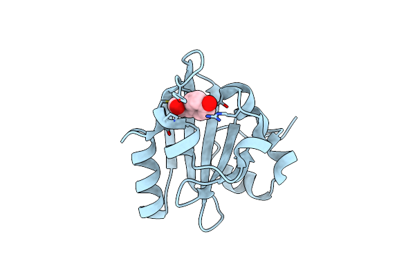
The Pr2 Photocycle Intermediate Of Photoactive Yellow Protein
Organism: Halorhodospira halophila
Method: X-RAY DIFFRACTION Resolution:1.60 Å Release Date: 2012-11-14 Classification: SIGNALING PROTEIN Ligands: HC4 |
Organism: Halorhodospira halophila
Method: X-RAY DIFFRACTION
Release Date: 2012-11-14
Ligands: HC4
 |
The Pb0 Photocycle Intermediate Of Photoactive Yellow Protein
Organism: Halorhodospira halophila
Method: X-RAY DIFFRACTION Resolution:1.60 Å Release Date: 2012-11-14 Classification: SIGNALING PROTEIN Ligands: HC4 |
Organism: Halorhodospira halophila
Method: X-RAY DIFFRACTION
Release Date: 2012-11-14
Ligands: HC4
 |
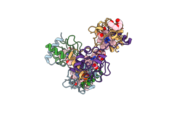
The Crystal Structure Of Arf6-Mklp1 (Mitotic Kinesin-Like Protein 1) Complex
Organism: Mus musculus, Homo sapiens
Method: X-RAY DIFFRACTION Resolution:2.81 Å Release Date: 2012-05-16 Classification: CELL CYCLE/SIGNALING PROTEIN Ligands: GTP, MG, GOL |
Organism: Mus musculus, Homo sapiens
Method: X-RAY DIFFRACTION
Release Date: 2012-05-16
Ligands: GTP, MG, GOL
 |
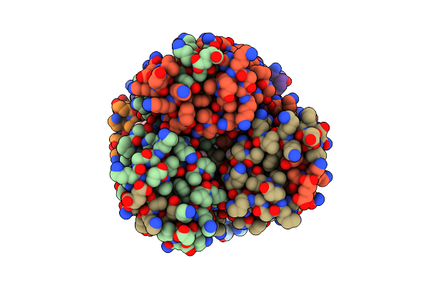
Crystal Structure Of Dimeric Cytochrome C From Horse Heart
Organism: Equus caballus
Method: X-RAY DIFFRACTION Resolution:2.20 Å Release Date: 2010-07-14 Classification: ELECTRON TRANSPORT Ligands: HEC, PG4, PEG, PO4 |
Organism: Equus caballus
Method: X-RAY DIFFRACTION
Release Date: 2010-07-14
Ligands: HEC, PG4, PEG, PO4
 |
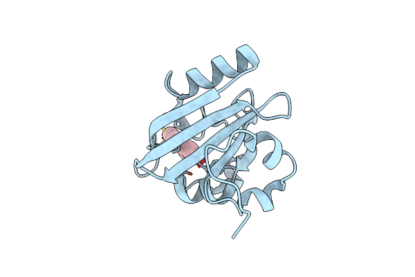
Crystal Structure Of Trimeric Cytochrome C From Horse Heart
Organism: Equus caballus
Method: X-RAY DIFFRACTION Resolution:2.10 Å Release Date: 2010-07-14 Classification: ELECTRON TRANSPORT Ligands: HEC, PG4, PEG, PGE |
Organism: Equus caballus
Method: X-RAY DIFFRACTION
Release Date: 2010-07-14
Ligands: HEC, PG4, PEG, PGE
 |
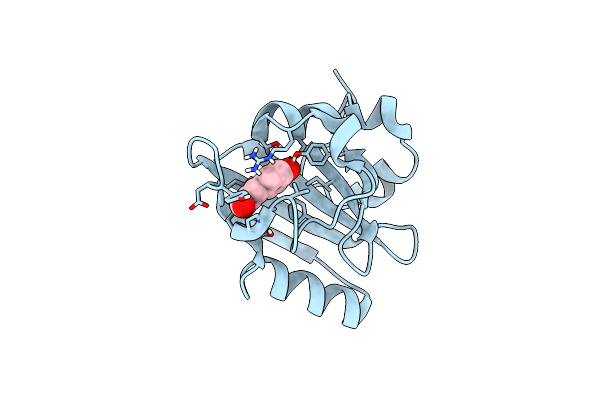
X-Ray Crystal Structure Of Photoactive Yellow Protein, Wild Type, At 295K
Organism: Halorhodospira halophila
Method: X-RAY DIFFRACTION Resolution:1.25 Å Release Date: 2009-03-24 Classification: SIGNALING PROTEIN Ligands: HC4 |
Organism: Halorhodospira halophila
Method: X-RAY DIFFRACTION
Release Date: 2009-03-24
Ligands: HC4
 |
Neutron Crystal Structure Of Photoactive Yellow Protein, Wild Type, At 295K
Organism: Halorhodospira halophila
Method: NEUTRON DIFFRACTION Resolution:1.50 Å Release Date: 2009-03-24 Classification: SIGNALING PROTEIN Ligands: HC4, DOD |
Organism: Halorhodospira halophila
Method: NEUTRON DIFFRACTION
Release Date: 2009-03-24
Ligands: HC4, DOD
 |
Crystal Structure Of Mouse Skd1/Vps4B Apo-Form
Organism: Mus musculus
Method: X-RAY DIFFRACTION Resolution:3.50 Å Release Date: 2008-10-07 Classification: PROTEIN TRANSPORT |
Organism: Mus musculus
Method: X-RAY DIFFRACTION
Release Date: 2008-10-07