

Search Count: 16
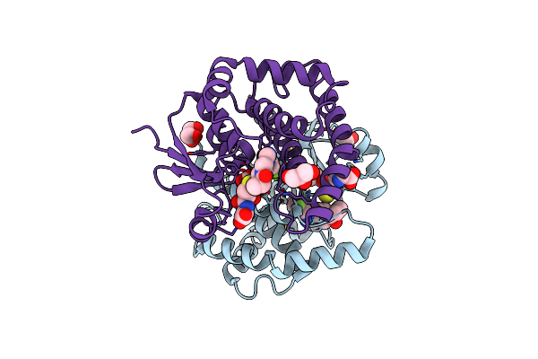 |
Crystal Structure Of Prostaglandin D2 Synthase In Complex With Fragment 1A At 1.84A Resolution.
Organism: Homo sapiens
Method: X-RAY DIFFRACTION Resolution:1.84 Å Release Date: 2021-07-28 Classification: ISOMERASE Ligands: GSH, MG, QPN, GOL |
Organism: Homo sapiens
Method: X-RAY DIFFRACTION
Release Date: 2021-07-28
Ligands: GSH, MG, QPN, GOL
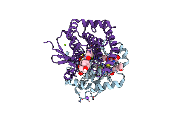 |
H-Pdgs Complexed With A 2-Phenylimidazo[1,2-A]Pyridine-6-Carboxamide Inhibitors
Organism: Homo sapiens
Method: X-RAY DIFFRACTION Resolution:1.88 Å Release Date: 2021-05-26 Classification: Isomerase, Transferase/Inhibitor Ligands: GSH, VH4, MG, NA |
Organism: Homo sapiens
Method: X-RAY DIFFRACTION
Release Date: 2021-05-26
Ligands: GSH, VH4, MG, NA
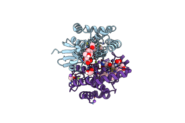 |
H-Pdgs Complexed With A 2-Phenylimidazo[1,2-A]Pyridine-6-Carboxamide Inhibitors
Organism: Homo sapiens
Method: X-RAY DIFFRACTION Resolution:1.13 Å Release Date: 2021-05-26 Classification: Isomerase, Transferase/Inhibitor Ligands: CL, EDO, GSH, VH7, MG, NA |
Organism: Homo sapiens
Method: X-RAY DIFFRACTION
Release Date: 2021-05-26
Ligands: CL, EDO, GSH, VH7, MG, NA
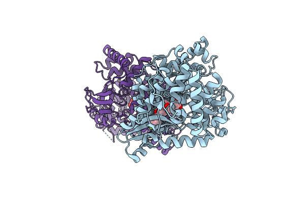 |
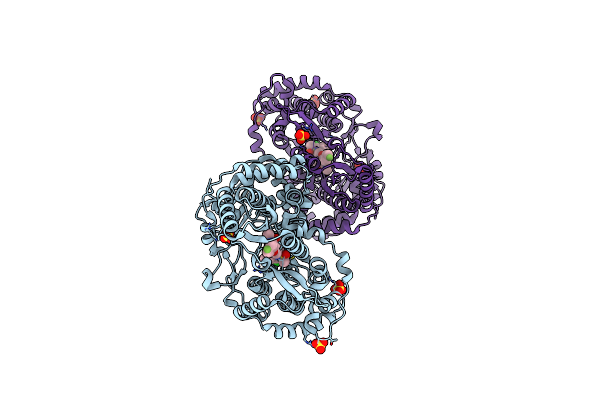
Hcv Ns5B 1B N316 Bound To Compound 18
Organism: Hepatitis c virus subtype 1b
Method: X-RAY DIFFRACTION Resolution:2.30 Å Release Date: 2019-09-04 Classification: VIRAL PROTEIN Ligands: K4J |
Organism: Hepatitis c virus subtype 1b
Method: X-RAY DIFFRACTION
Release Date: 2019-09-04
Ligands: K4J
 |
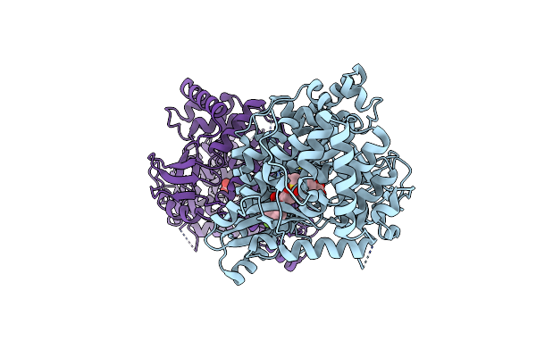
Hcv Ns5B 1A Y316 Bound To Compound 49
Organism: Hepacivirus c
Method: X-RAY DIFFRACTION Resolution:1.95 Å Release Date: 2019-09-04 Classification: VIRAL PROTEIN Ligands: SO4, K4P |
Organism: Hepacivirus c
Method: X-RAY DIFFRACTION
Release Date: 2019-09-04
Ligands: SO4, K4P
 |
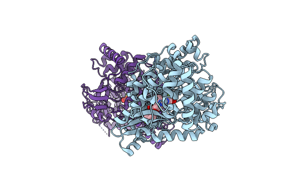
Hcv Ns5B 1B N316 Bound To Compound 18
Organism: Hepatitis c virus genotype 1b (isolate bk)
Method: X-RAY DIFFRACTION Resolution:2.00 Å Release Date: 2019-09-04 Classification: VIRAL PROTEIN Ligands: K4S |
Organism: Hepatitis c virus genotype 1b (isolate bk)
Method: X-RAY DIFFRACTION
Release Date: 2019-09-04
Ligands: K4S
 |
Hcv Ns5B 1B N316 Bound To Compound 31
Organism: Hepatitis c virus subtype 1b
Method: X-RAY DIFFRACTION Resolution:2.14 Å Release Date: 2019-09-04 Classification: VIRAL PROTEIN Ligands: K4M |
Organism: Hepatitis c virus subtype 1b
Method: X-RAY DIFFRACTION
Release Date: 2019-09-04
Ligands: K4M
 |
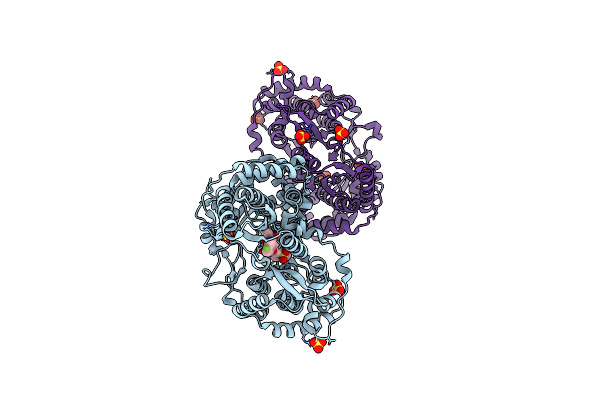
X-Ray Structure Of Receptor Interacting Protein 1 (Rip1)Kinase Domain With A 1-Aminoisoquinoline Inhibitor
Organism: Homo sapiens
Method: X-RAY DIFFRACTION Resolution:2.57 Å Release Date: 2013-11-20 Classification: TRANSFERASE Ligands: MG, Q1A |
Organism: Homo sapiens
Method: X-RAY DIFFRACTION
Release Date: 2013-11-20
Ligands: MG, Q1A
 |
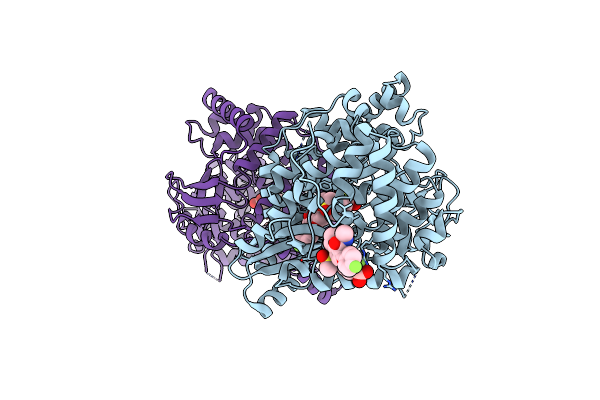
Hcv Ns5B Gt1A C316Y With Gsk5852
Organism: Hepatitis c virus subtype 1a
Method: X-RAY DIFFRACTION Resolution:2.45 Å Release Date: 2013-05-29 Classification: REPLICATION/REPLICATION INHIBITOR Ligands: SO4, 1PV |
Organism: Hepatitis c virus subtype 1a
Method: X-RAY DIFFRACTION
Release Date: 2013-05-29
Ligands: SO4, 1PV
 |
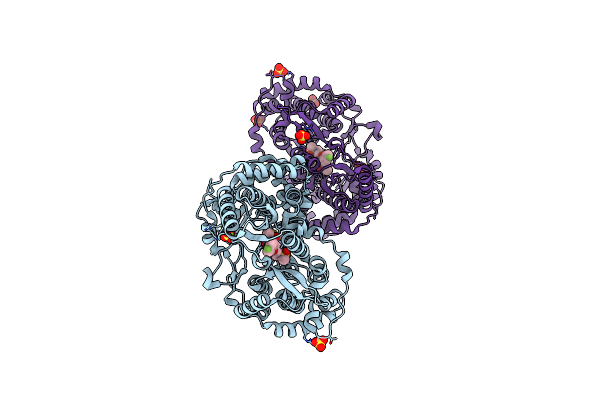
Hcv Ns5B Gt1B N316Y With Gsk5852
Organism: Hepatitis c virus
Method: X-RAY DIFFRACTION Resolution:2.11 Å Release Date: 2013-05-15 Classification: REPLICATION/REPLICATION INHIBITOR Ligands: 1PV |
Organism: Hepatitis c virus
Method: X-RAY DIFFRACTION
Release Date: 2013-05-15
Ligands: 1PV
 |
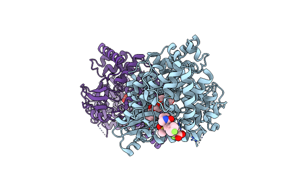
Hcv Ns5B Gt1A With Gsk5852
Organism: Hepatitis c virus subtype 1a
Method: X-RAY DIFFRACTION Resolution:1.70 Å Release Date: 2013-05-15 Classification: REPLICATION/REPLICATION INHIBITOR Ligands: 1PV, SO4 |
Organism: Hepatitis c virus subtype 1a
Method: X-RAY DIFFRACTION
Release Date: 2013-05-15
Ligands: 1PV, SO4
 |
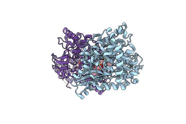
Hcv Ns5B Gt1B N316 With Gsk5852A
Organism: Hepatitis c virus
Method: X-RAY DIFFRACTION Resolution:2.30 Å Release Date: 2013-05-08 Classification: REPLICATION/REPLICATION INHIBITOR Ligands: 1PV |
Organism: Hepatitis c virus
Method: X-RAY DIFFRACTION
Release Date: 2013-05-08
Ligands: 1PV
 |
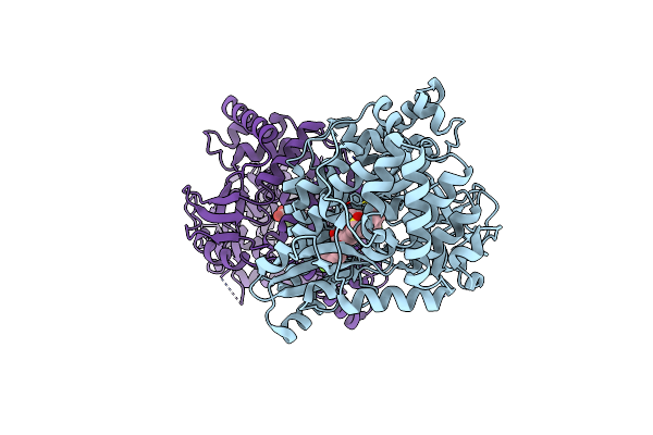
Hcv Ns5B Gt1B N316Y With Cmpd 32
Organism: Hepatitis c virus
Method: X-RAY DIFFRACTION Resolution:1.85 Å Release Date: 2013-05-08 Classification: REPLICATION/REPLICATION INHIBITOR Ligands: 690 |
Organism: Hepatitis c virus
Method: X-RAY DIFFRACTION
Release Date: 2013-05-08
Ligands: 690
 |
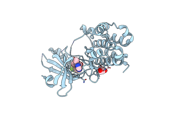
Hcv Ns5B Gt1B N316Y With Cmpd 4
Organism: Hepatitis c virus
Method: X-RAY DIFFRACTION Resolution:2.06 Å Release Date: 2013-05-08 Classification: REPLICATION/REPLICATION INHIBITOR Ligands: 1C0 |
Organism: Hepatitis c virus
Method: X-RAY DIFFRACTION
Release Date: 2013-05-08
Ligands: 1C0
 |
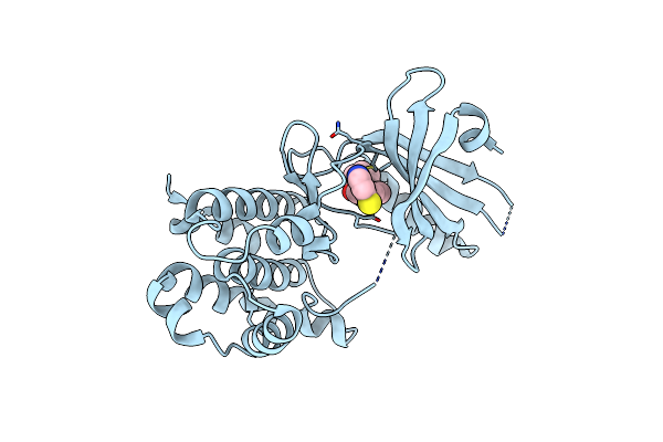
Crystal Structure Of Gsk6414 Bound To Perk (R587-R1092, Delete A660-T867) At 2.28 A Resolution
Organism: Homo sapiens
Method: X-RAY DIFFRACTION Resolution:2.28 Å Release Date: 2012-08-08 Classification: TRANSFERASE/TRANSFERASE INHIBITOR Ligands: GOL, 0WH |
Organism: Homo sapiens
Method: X-RAY DIFFRACTION
Release Date: 2012-08-08
Ligands: GOL, 0WH
 |
Crystal Structure Of Gsk6924 Bound To Perk (R587-R1092, Delete A660-T867) At 2.70 A Resolution
Organism: Homo sapiens
Method: X-RAY DIFFRACTION Resolution:2.70 Å Release Date: 2012-08-08 Classification: TRANSFERASE/TRANSFERASE INHIBITOR Ligands: 924 |
Organism: Homo sapiens
Method: X-RAY DIFFRACTION
Release Date: 2012-08-08
Ligands: 924