

Search Count: 25
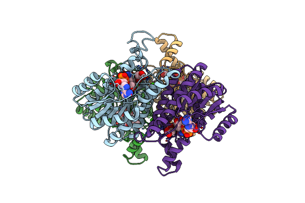 |
Crystal Structure Of D-3-Hydroxybutyrate Dehydrogenase, Prepared In The Presence Of The Substrate D-3-Hydroxybutyrate And Nad(+)
Organism: Alcaligenes faecalis
Method: X-RAY DIFFRACTION Resolution:3.00 Å Release Date: 2012-02-08 Classification: OXIDOREDUCTASE Ligands: CA, CL, NAD, NAI, 3HR, AAE |
Organism: Alcaligenes faecalis
Method: X-RAY DIFFRACTION
Release Date: 2012-02-08
Ligands: CA, CL, NAD, NAI, 3HR, AAE
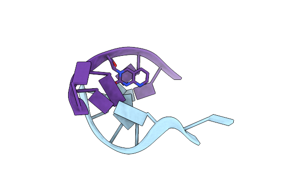 |
Insights Into The Stabilizing Contributions Of A Bicyclic Cytosine Analogue: Crystal Structures Of Dna Duplexes Containing 7,8-Dihydropyrido[2,3-D]Pyrimidin-2-One
|
Method: X-RAY DIFFRACTION
Release Date: 2010-08-11
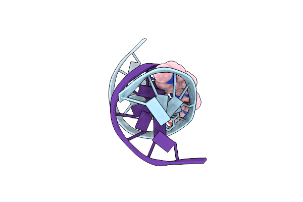 |
Insights Into The Stabilizing Contributions Of A Bicyclic Cytosine Analogue: Crystal Structures Of Dna Duplexes Containing 7,8-Dihydropyrido[2,3-D]Pyrimidin-2-One
Method: X-RAY DIFFRACTION
Resolution:2.90 Å Release Date: 2010-08-11 Classification: DNA Ligands: HT |
Method: X-RAY DIFFRACTION
Release Date: 2010-08-11
Ligands: HT
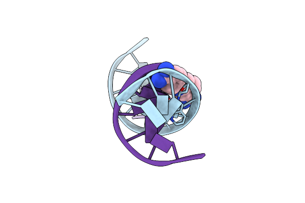 |
Crystal Structure Of A Dna Duplex Containing 7,8-Dihydropyridol[2,3-D]Pyrimidin-2-One
Method: X-RAY DIFFRACTION
Resolution:2.90 Å Release Date: 2010-03-31 Classification: DNA Ligands: DAP |
Method: X-RAY DIFFRACTION
Release Date: 2010-03-31
Ligands: DAP
 |
Crystal Structure Of A Dna Duplex Containing 7,8-Dihydropyridol[2,3-D]Pyrimidin-2-One
Method: X-RAY DIFFRACTION
Resolution:2.90 Å Release Date: 2010-03-31 Classification: DNA Ligands: HT |
Method: X-RAY DIFFRACTION
Release Date: 2010-03-31
Ligands: HT
 |
Crystal Structure Of A Dna Duplex Containing 7,8-Dihydropyridol[2,3-D]Pyrimidin-2-One
|
Method: X-RAY DIFFRACTION
Release Date: 2010-03-31
Ligands: K
 |
Crystal Structure Of A Dna Duplex Containing 7,8-Dihydropyridol[2,3-D]Pyrimidin-2-One
|
Method: X-RAY DIFFRACTION
Release Date: 2010-03-31
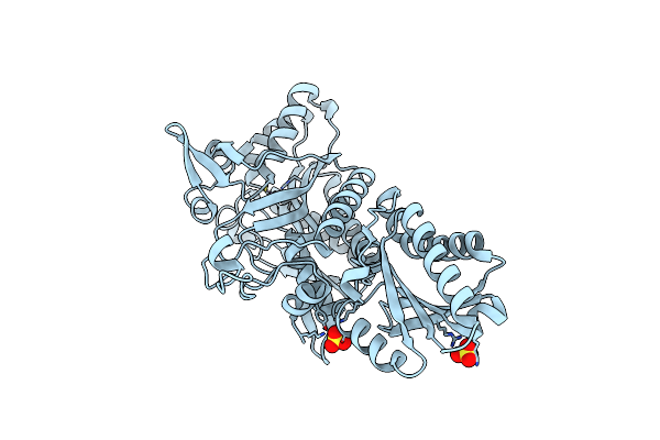 |
Crystal Structure Of Putative Threonyl-Trna Synthetase Thrrs-1 From Aeropyrum Pernix (Selenomethionine Derivative)
Organism: Aeropyrum pernix
Method: X-RAY DIFFRACTION Resolution:2.50 Å Release Date: 2009-10-27 Classification: LIGASE Ligands: SO4, ZN |
Organism: Aeropyrum pernix
Method: X-RAY DIFFRACTION
Release Date: 2009-10-27
Ligands: SO4, ZN
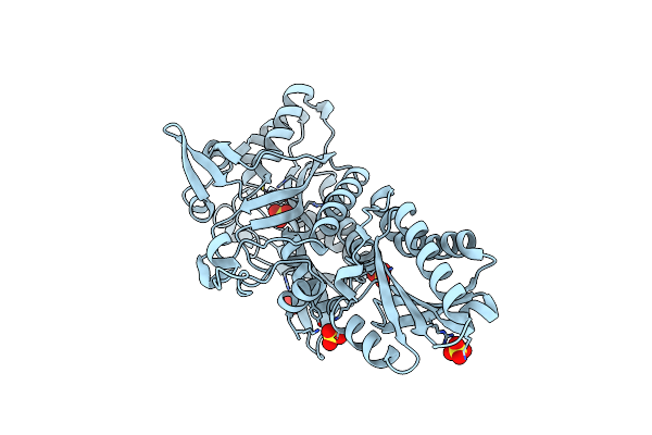 |
Crystal Structure Of Putative Threonyl-Trna Synthetase Thrrs-1 From Aeropyrum Pernix
Organism: Aeropyrum pernix
Method: X-RAY DIFFRACTION Resolution:2.30 Å Release Date: 2009-10-27 Classification: LIGASE Ligands: SO4, ZN |
Organism: Aeropyrum pernix
Method: X-RAY DIFFRACTION
Release Date: 2009-10-27
Ligands: SO4, ZN
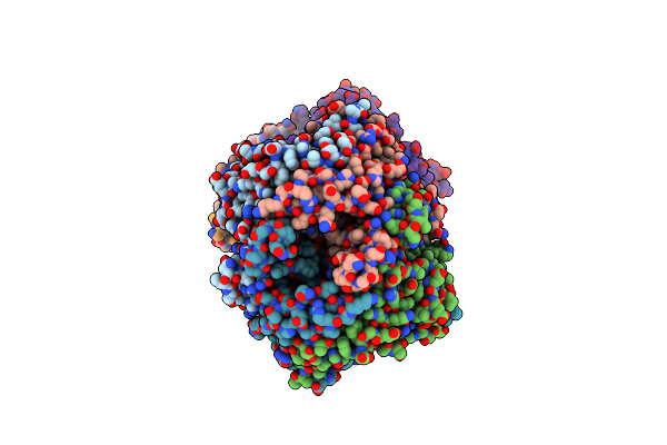 |
Crystal Structure Of Uricase From Arthrobacter Globiformis In Complex With Uric Acid (Substrate)
Organism: Arthrobacter globiformis
Method: X-RAY DIFFRACTION Resolution:1.90 Å Release Date: 2008-05-06 Classification: OXIDOREDUCTASE Ligands: URC |
Organism: Arthrobacter globiformis
Method: X-RAY DIFFRACTION
Release Date: 2008-05-06
Ligands: URC
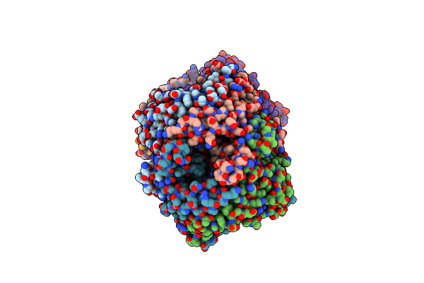 |
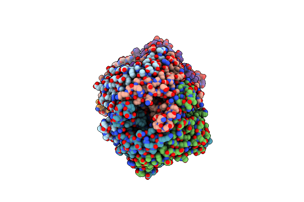
Crystal Structure Of Uricase From Arthrobacter Globiformis In Complex With Allantoate
Organism: Arthrobacter globiformis
Method: X-RAY DIFFRACTION Resolution:1.88 Å Release Date: 2008-05-06 Classification: OXIDOREDUCTASE Ligands: 1AL |
Organism: Arthrobacter globiformis
Method: X-RAY DIFFRACTION
Release Date: 2008-05-06
Ligands: 1AL
 |
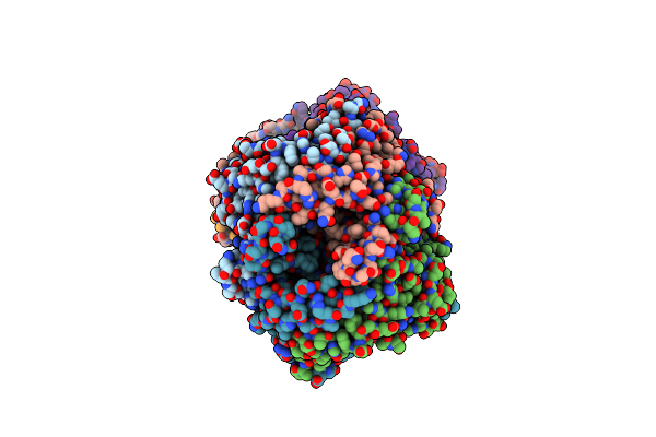
Crystal Structure Of Uricase From Arthrobacter Globiformis In Complex With 8-Azaxanthin (Inhibitor)
Organism: Arthrobacter globiformis
Method: X-RAY DIFFRACTION Resolution:2.24 Å Release Date: 2008-05-06 Classification: OXIDOREDUCTASE Ligands: AZA |
Organism: Arthrobacter globiformis
Method: X-RAY DIFFRACTION
Release Date: 2008-05-06
Ligands: AZA
 |
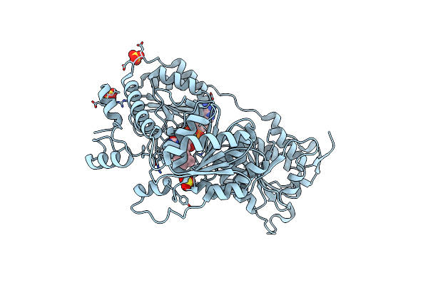
Crystal Structure Of Uricase From Arthrobacter Globiformis
Organism: Arthrobacter globiformis
Method: X-RAY DIFFRACTION Resolution:1.99 Å Release Date: 2008-05-06 Classification: OXIDOREDUCTASE Ligands: NOB, LI |
Organism: Arthrobacter globiformis
Method: X-RAY DIFFRACTION
Release Date: 2008-05-06
Ligands: NOB, LI
 |
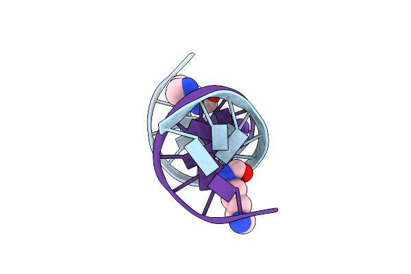
Crystal Structure Of Pyruvate Oxidase From Aerococcus Viridans Containing Fad
Organism: Aerococcus viridans
Method: X-RAY DIFFRACTION Resolution:1.60 Å Release Date: 2007-05-29 Classification: OXIDOREDUCTASE Ligands: SO4, FAD |
Organism: Aerococcus viridans
Method: X-RAY DIFFRACTION
Release Date: 2007-05-29
Ligands: SO4, FAD
 |
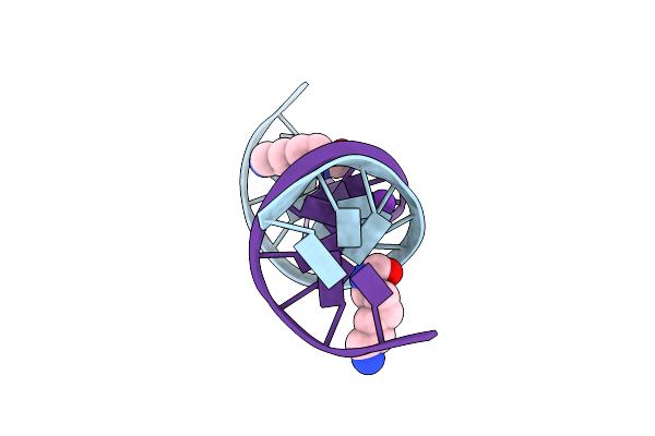
Crystal Structure Of D(Cgcgaatxcgcg) Where X Is 5-(N-Aminohexyl)Carbamoyl-2'-Deoxyuridine
Method: X-RAY DIFFRACTION
Resolution:1.55 Å Release Date: 2007-04-17 Classification: DNA Ligands: MG, CMY, K |
Method: X-RAY DIFFRACTION
Release Date: 2007-04-17
Ligands: MG, CMY, K
 |
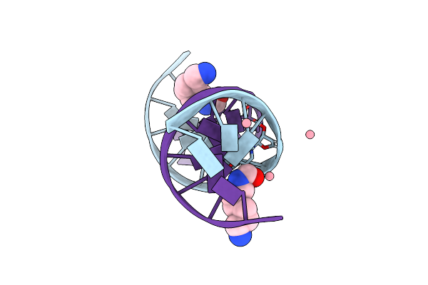
Crystal Structure Of D(Cgcgaatxcgcg) Where X Is 5-(N-Aminohexyl)Carbamoyl-2'-Deoxyuridine
Method: X-RAY DIFFRACTION
Resolution:1.50 Å Release Date: 2007-04-17 Classification: DNA Ligands: CMY, K |
Method: X-RAY DIFFRACTION
Release Date: 2007-04-17
Ligands: CMY, K
 |
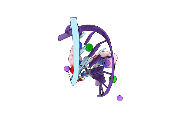
Crystal Structure Of D(Cgcgaatxcgcg) Where X Is 5-(N-Aminohexyl)Carbamoyl-2'-O-Methyluridine
Method: X-RAY DIFFRACTION
Resolution:1.55 Å Release Date: 2007-04-17 Classification: DNA Ligands: CO, MG, CMY |
Method: X-RAY DIFFRACTION
Release Date: 2007-04-17
Ligands: CO, MG, CMY
 |
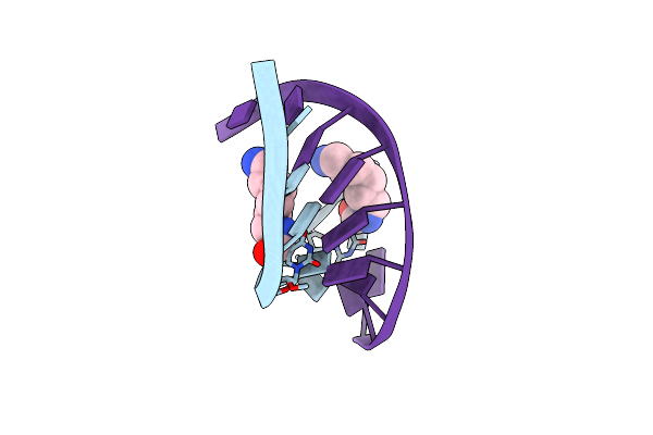
Crystal Structure Of D(Cxctxcttc):R(Gaagaagag) Where X Is 5-(N-Aminohexyl)Carbamoyl-2'-O-Methyluridine
|
Method: X-RAY DIFFRACTION
Release Date: 2007-04-17
 |
Structural Analyses Of Dna:Dna And Rna:Dna Duplexes Containing 5-(N-Aminohexyl)Carbamoyl Modified Uridines
Method: X-RAY DIFFRACTION
Resolution:2.10 Å Release Date: 2007-04-17 Classification: DNA-RNA HYBRID Ligands: CMY |
Method: X-RAY DIFFRACTION
Release Date: 2007-04-17
Ligands: CMY
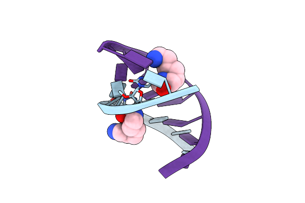 |
Crystal Structure Of D(Cxctxcttc):R(Gaagaagag) Where X Is 5-(N-Aminohexyl)Carbamoyl-2'-O-Methyluridine
Organism: Synthetic construct
Method: X-RAY DIFFRACTION Resolution:2.00 Å Release Date: 2007-04-17 Classification: DNA-RNA HYBRID Ligands: CMY |
Organism: Synthetic construct
Method: X-RAY DIFFRACTION
Release Date: 2007-04-17
Ligands: CMY