

Search Count: 36
 |
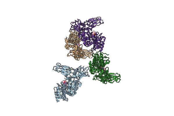
Crystal Structure Of Enterococcus Faecium D63R Penicillin-Binding Protein 5 (Pbp5Fm)
Organism: Enterococcus faecium
Method: X-RAY DIFFRACTION Resolution:3.30 Å Release Date: 2019-04-24 Classification: ANTIBIOTIC Ligands: RB6, SO4 |
Organism: Enterococcus faecium
Method: X-RAY DIFFRACTION
Release Date: 2019-04-24
Ligands: RB6, SO4
 |
Crystal Structure Of Enterococcus Faecium D63R Penicillin-Binding Protein 5 (Pbp5Fm)
Organism: Enterococcus faecium
Method: X-RAY DIFFRACTION Resolution:2.90 Å Release Date: 2019-04-10 Classification: ANTIBIOTIC Ligands: SO4 |
Organism: Enterococcus faecium
Method: X-RAY DIFFRACTION
Release Date: 2019-04-10
Ligands: SO4
 |
Unexpected Tricovalent Binding Mode Of Boronic Acids Within The Active Site Of A Penicillin Binding Protein
Organism: Actinomadura sp.r39
Method: X-RAY DIFFRACTION Resolution:3.10 Å Release Date: 2012-02-29 Classification: HYDROLASE Ligands: B07, SO4, MG |
Organism: Actinomadura sp.r39
Method: X-RAY DIFFRACTION
Release Date: 2012-02-29
Ligands: B07, SO4, MG
 |
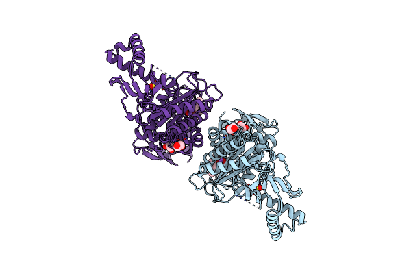
Unexpected Tricovalent Binding Mode Of Boronic Acids Within The Active Site Of A Penicillin Binding Protein
Organism: Actinomadura sp. r39
Method: X-RAY DIFFRACTION Resolution:2.00 Å Release Date: 2012-02-29 Classification: HYDROLASE Ligands: SO4, MG, FP5, ACN, 33D, MES |
Organism: Actinomadura sp. r39
Method: X-RAY DIFFRACTION
Release Date: 2012-02-29
Ligands: SO4, MG, FP5, ACN, 33D, MES
 |
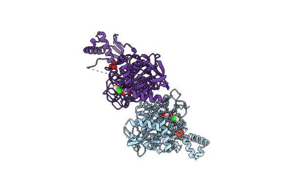
Penicillin-Binding Protein 1B (Pbp-1B) In Complex With An Alkyl Boronate (A01)
Organism: Streptococcus pneumoniae
Method: X-RAY DIFFRACTION Resolution:2.05 Å Release Date: 2011-08-03 Classification: TRANSFERASE Ligands: A01, SO4, CL, NA, EDO |
Organism: Streptococcus pneumoniae
Method: X-RAY DIFFRACTION
Release Date: 2011-08-03
Ligands: A01, SO4, CL, NA, EDO
 |
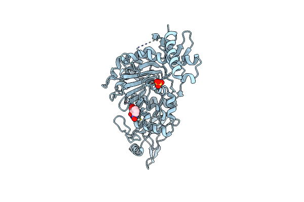
Penicillin-Binding Protein 1B (Pbp-1B) In Complex With An Alkyl Boronate (Za2)
Organism: Streptococcus pneumoniae
Method: X-RAY DIFFRACTION Resolution:1.96 Å Release Date: 2011-08-03 Classification: TRANSFERASE Ligands: ZA2, SO4, CL, NA |
Organism: Streptococcus pneumoniae
Method: X-RAY DIFFRACTION
Release Date: 2011-08-03
Ligands: ZA2, SO4, CL, NA
 |
Penicillin-Binding Protein 1B (Pbp-1B) In Complex With An Alkyl Boronate (Za3)
Organism: Streptococcus pneumoniae
Method: X-RAY DIFFRACTION Resolution:1.78 Å Release Date: 2011-08-03 Classification: TRANSFERASE Ligands: ZA3, SO4, CL, NA |
Organism: Streptococcus pneumoniae
Method: X-RAY DIFFRACTION
Release Date: 2011-08-03
Ligands: ZA3, SO4, CL, NA
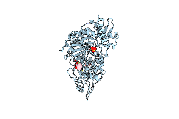 |
Penicillin-Binding Protein 1B (Pbp-1B) In Complex With An Alkyl Boronate (Za4)
Organism: Streptococcus pneumoniae
Method: X-RAY DIFFRACTION Resolution:2.06 Å Release Date: 2011-08-03 Classification: TRANSFERASE Ligands: ZA4, SO4, CL, NA, EDO |
Organism: Streptococcus pneumoniae
Method: X-RAY DIFFRACTION
Release Date: 2011-08-03
Ligands: ZA4, SO4, CL, NA, EDO
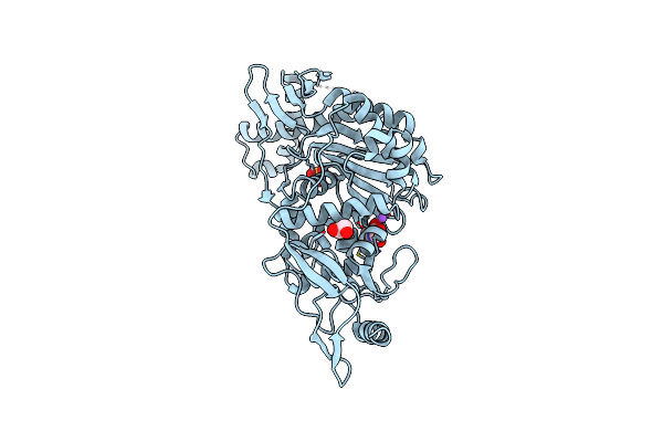 |
Penicillin-Binding Protein 1B (Pbp-1B) In Complex With An Alkyl Boronate (Za5)
Organism: Streptococcus pneumoniae
Method: X-RAY DIFFRACTION Resolution:2.09 Å Release Date: 2011-08-03 Classification: TRANSFERASE Ligands: ZA5, SO4, CL, NA, EDO |
Organism: Streptococcus pneumoniae
Method: X-RAY DIFFRACTION
Release Date: 2011-08-03
Ligands: ZA5, SO4, CL, NA, EDO
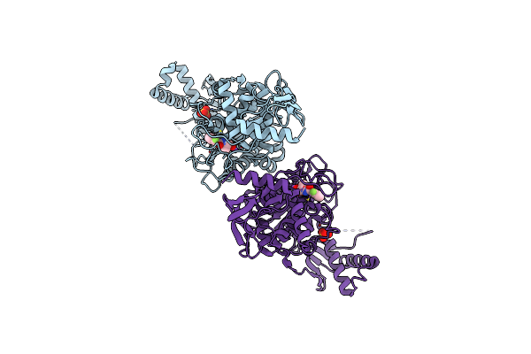 |
Penicillin-Binding Protein 1B (Pbp-1B) In Complex With An Alkyl Boronate (E06)
Organism: Streptococcus pneumoniae
Method: X-RAY DIFFRACTION Resolution:2.07 Å Release Date: 2011-08-03 Classification: TRANSFERASE Ligands: E06, SO4, CL, NA |
Organism: Streptococcus pneumoniae
Method: X-RAY DIFFRACTION
Release Date: 2011-08-03
Ligands: E06, SO4, CL, NA
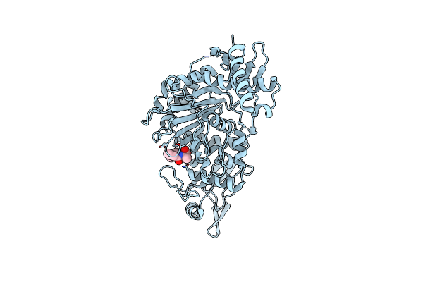 |
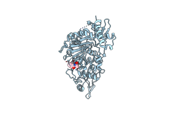
Penicillin-Binding Protein 1B (Pbp-1B) In Complex With An Alkyl Boronate (E08)
Organism: Streptococcus pneumoniae
Method: X-RAY DIFFRACTION Resolution:1.62 Å Release Date: 2011-08-03 Classification: TRANSFERASE Ligands: E08, CL, NA |
Organism: Streptococcus pneumoniae
Method: X-RAY DIFFRACTION
Release Date: 2011-08-03
Ligands: E08, CL, NA
 |
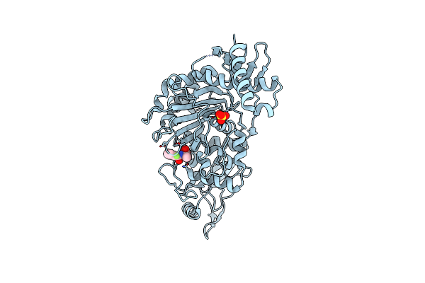
Penicillin-Binding Protein 1B (Pbp-1B) In Complex With An Alkyl Boronate (E07)
Organism: Streptococcus pneumoniae
Method: X-RAY DIFFRACTION Resolution:2.07 Å Release Date: 2011-08-03 Classification: TRANSFERASE Ligands: E07, CL, NA |
Organism: Streptococcus pneumoniae
Method: X-RAY DIFFRACTION
Release Date: 2011-08-03
Ligands: E07, CL, NA
 |
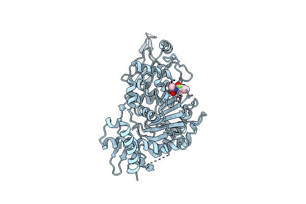
Penicillin-Binding Protein 1B (Pbp-1B) In Complex With An Alkyl Boronate (Eo9)
Organism: Streptococcus pneumoniae
Method: X-RAY DIFFRACTION Resolution:1.88 Å Release Date: 2011-08-03 Classification: TRANSFERASE Ligands: EO9, SO4, CL, NA |
Organism: Streptococcus pneumoniae
Method: X-RAY DIFFRACTION
Release Date: 2011-08-03
Ligands: EO9, SO4, CL, NA
 |
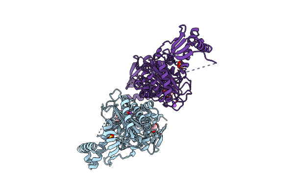
Penicillin-Binding Protein 1B (Pbp-1B) In Complex With An Alkyl Boronate (Z10)
Organism: Streptococcus pneumoniae
Method: X-RAY DIFFRACTION Resolution:1.62 Å Release Date: 2011-08-03 Classification: TRANSFERASE Ligands: Z10, CL, NA |
Organism: Streptococcus pneumoniae
Method: X-RAY DIFFRACTION
Release Date: 2011-08-03
Ligands: Z10, CL, NA
 |
Penicillin-Binding Protein 1B (Pbp-1B) In Complex With An Alkyl Boronate (Z06)
Organism: Streptococcus pneumoniae
Method: X-RAY DIFFRACTION Resolution:2.15 Å Release Date: 2011-08-03 Classification: TRANSFERASE Ligands: Z06, SO4, CL, NA, EDO |
Organism: Streptococcus pneumoniae
Method: X-RAY DIFFRACTION
Release Date: 2011-08-03
Ligands: Z06, SO4, CL, NA, EDO
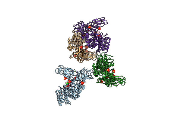 |
Unexpected Tricovalent Binding Mode Of Boronic Acids Within The Active Site Of A Penicillin Binding Protein
Organism: Actinomadura sp
Method: X-RAY DIFFRACTION Resolution:2.70 Å Release Date: 2011-07-27 Classification: HYDROLASE Ligands: BH6, SO4, MG |
Organism: Actinomadura sp
Method: X-RAY DIFFRACTION
Release Date: 2011-07-27
Ligands: BH6, SO4, MG
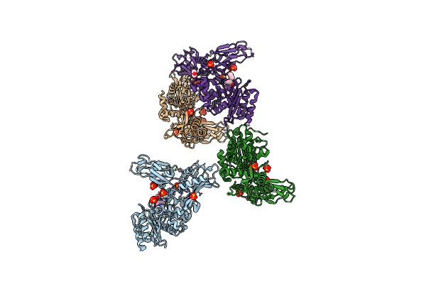 |
Unexpected Tricovalent Binding Mode Of Boronic Acids Within The Active Site Of A Penicillin Binding Protein
Organism: Actinomadura sp. r39
Method: X-RAY DIFFRACTION Resolution:2.60 Å Release Date: 2011-07-27 Classification: HYDROLASE Ligands: FP5, SO4, MG, ACN |
Organism: Actinomadura sp. r39
Method: X-RAY DIFFRACTION
Release Date: 2011-07-27
Ligands: FP5, SO4, MG, ACN
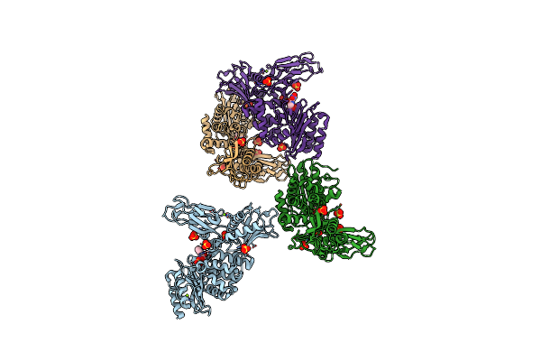 |
Unexpected Tricovalent Binding Mode Of Boronic Acids Within The Active Site Of A Penicillin Binding Protein
Organism: Actinomadura sp. r39
Method: X-RAY DIFFRACTION Resolution:2.50 Å Release Date: 2011-07-27 Classification: HYDROLASE Ligands: ZA3, SO4, MG |
Organism: Actinomadura sp. r39
Method: X-RAY DIFFRACTION
Release Date: 2011-07-27
Ligands: ZA3, SO4, MG
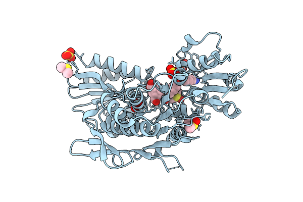 |
Second-Generation Sulfonamide Inhibitors Of Murd: Activity Optimisation With Conformationally Rigid Analogues Of D-Glutamic Acid
Organism: Escherichia coli
Method: X-RAY DIFFRACTION Resolution:1.49 Å Release Date: 2011-05-18 Classification: LIGASE Ligands: 051, DMS, SO4, CL |
Organism: Escherichia coli
Method: X-RAY DIFFRACTION
Release Date: 2011-05-18
Ligands: 051, DMS, SO4, CL
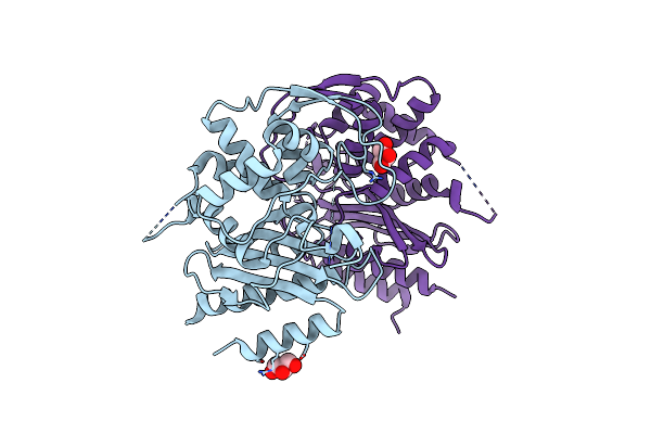 |
Crystal Structure Of Blab Protein From Streptomyces Cacaoi
Organism: Streptomyces cacaoi
Method: X-RAY DIFFRACTION Resolution:1.80 Å Release Date: 2010-10-13 Classification: TRANSCRIPTION Ligands: GOL |
Organism: Streptomyces cacaoi
Method: X-RAY DIFFRACTION
Release Date: 2010-10-13
Ligands: GOL