

Search Count: 23
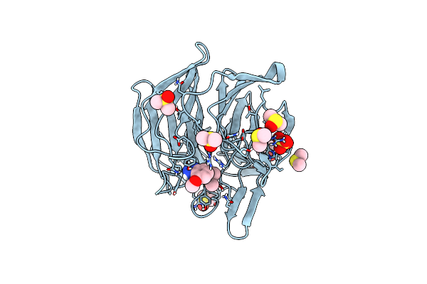 |
Keap1 Kelch Domain Bound To A Small Molecule Fragment
Organism: Mus musculus
Method: X-RAY DIFFRACTION Resolution:1.38 Å Release Date: 2021-04-14 Classification: PEPTIDE BINDING PROTEIN Ligands: DMS, SO4, QHB |
Organism: Mus musculus
Method: X-RAY DIFFRACTION
Release Date: 2021-04-14
Ligands: DMS, SO4, QHB
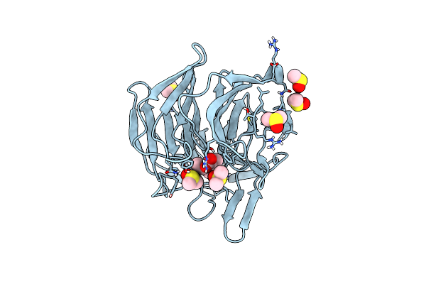 |
Keap1 Kelch Domain Bound To A Small Molecule Fragment
Organism: Mus musculus
Method: X-RAY DIFFRACTION Resolution:1.98 Å Release Date: 2021-04-14 Classification: PEPTIDE BINDING PROTEIN Ligands: DMS, QHN |
Organism: Mus musculus
Method: X-RAY DIFFRACTION
Release Date: 2021-04-14
Ligands: DMS, QHN
 |
Keap1 Kelch Domain Bound To A Small Molecule Inhibitor Of The Keap1-Nrf2 Protein-Protein Interaction
Organism: Mus musculus
Method: X-RAY DIFFRACTION Resolution:1.80 Å Release Date: 2021-04-14 Classification: PEPTIDE BINDING PROTEIN Ligands: DMS, QGZ |
Organism: Mus musculus
Method: X-RAY DIFFRACTION
Release Date: 2021-04-14
Ligands: DMS, QGZ
 |
Keap1 Kelch Domain Bound To A Small Molecule Inhibitor Of The Keap1-Nrf2 Protein-Protein Interaction
Organism: Mus musculus
Method: X-RAY DIFFRACTION Resolution:2.55 Å Release Date: 2021-04-14 Classification: PEPTIDE BINDING PROTEIN Ligands: QHH |
Organism: Mus musculus
Method: X-RAY DIFFRACTION
Release Date: 2021-04-14
Ligands: QHH
 |
Keap1 Kelch Domain Bound To A Small Molecule Inhibitor Of The Keap1-Nrf2 Protein-Protein Interaction
Organism: Mus musculus
Method: X-RAY DIFFRACTION Resolution:1.60 Å Release Date: 2021-04-14 Classification: PEPTIDE BINDING PROTEIN Ligands: SO4, DMS, QHT |
Organism: Mus musculus
Method: X-RAY DIFFRACTION
Release Date: 2021-04-14
Ligands: SO4, DMS, QHT
 |
Keap1 Kelch Domain Bound To A Small Molecule Inhibitor Of The Keap1-Nrf2 Protein-Protein Interaction
Organism: Mus musculus
Method: X-RAY DIFFRACTION Resolution:1.74 Å Release Date: 2021-04-14 Classification: PEPTIDE BINDING PROTEIN Ligands: SO4, DMS, QHK |
Organism: Mus musculus
Method: X-RAY DIFFRACTION
Release Date: 2021-04-14
Ligands: SO4, DMS, QHK
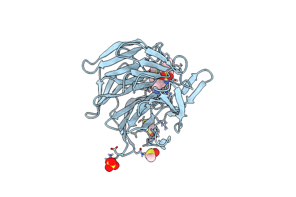 |
Keap1 Kelch Domain Bound To A Small Molecule Inhibitor Of The Keap1-Nrf2 Protein-Protein Interaction
Organism: Mus musculus
Method: X-RAY DIFFRACTION Resolution:2.20 Å Release Date: 2021-04-14 Classification: PEPTIDE BINDING PROTEIN Ligands: DMS, SO4, QJ5 |
Organism: Mus musculus
Method: X-RAY DIFFRACTION
Release Date: 2021-04-14
Ligands: DMS, SO4, QJ5
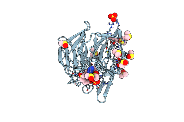 |
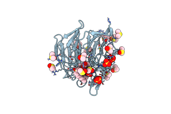
Keap1 Kelch Domain Bound To A Small Molecule Inhibitor Of The Keap1-Nrf2 Protein-Protein Interaction
Organism: Mus musculus
Method: X-RAY DIFFRACTION Resolution:1.28 Å Release Date: 2021-04-14 Classification: PEPTIDE BINDING PROTEIN Ligands: QH5, SO4, DMS |
Organism: Mus musculus
Method: X-RAY DIFFRACTION
Release Date: 2021-04-14
Ligands: QH5, SO4, DMS
 |
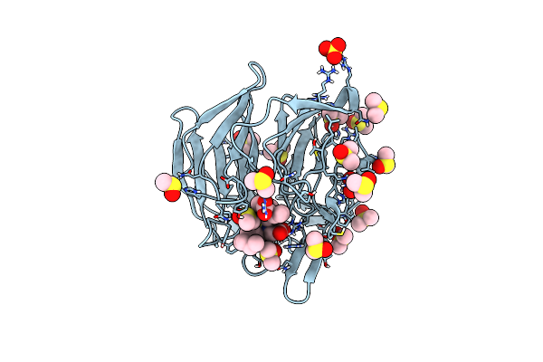
Keap1 Kelch Domain Bound To A Small Molecule Inhibitor Of The Keap1-Nrf2 Protein-Protein Interaction
Organism: Mus musculus
Method: X-RAY DIFFRACTION Resolution:1.21 Å Release Date: 2021-04-14 Classification: PEPTIDE BINDING PROTEIN Ligands: SO4, DMS, QHW |
Organism: Mus musculus
Method: X-RAY DIFFRACTION
Release Date: 2021-04-14
Ligands: SO4, DMS, QHW
 |
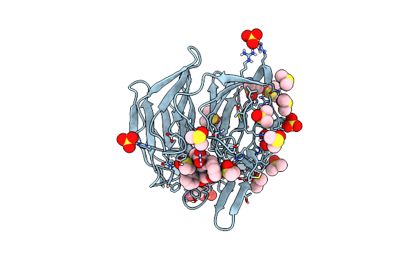
Keap1 Kelch Domain Bound To A Small Molecule Inhibitor Of The Keap1-Nrf2 Protein-Protein Interaction
Organism: Mus musculus
Method: X-RAY DIFFRACTION Resolution:1.29 Å Release Date: 2021-04-14 Classification: PEPTIDE BINDING PROTEIN Ligands: SO4, DMS, QH8 |
Organism: Mus musculus
Method: X-RAY DIFFRACTION
Release Date: 2021-04-14
Ligands: SO4, DMS, QH8
 |
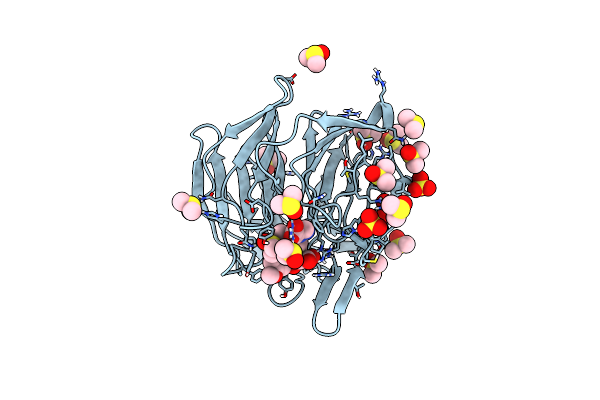
Keap1 Kelch Domain Bound To A Small Molecule Inhibitor Of The Keap1-Nrf2 Protein-Protein Interaction
Organism: Mus musculus
Method: X-RAY DIFFRACTION Resolution:1.38 Å Release Date: 2021-04-14 Classification: PEPTIDE BINDING PROTEIN Ligands: SO4, DMS, QH2 |
Organism: Mus musculus
Method: X-RAY DIFFRACTION
Release Date: 2021-04-14
Ligands: SO4, DMS, QH2
 |
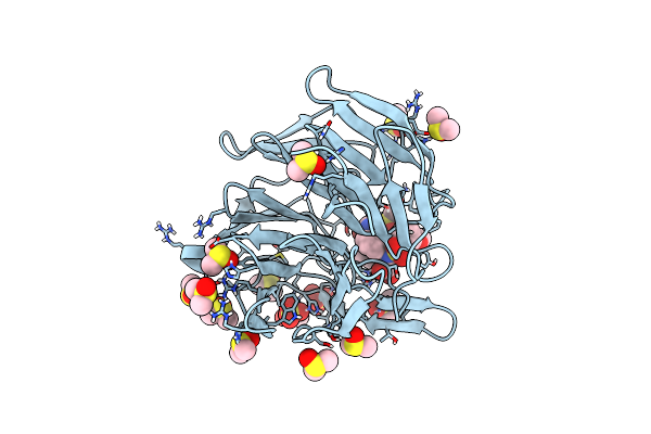
Keap1 Kelch Domain Bound To A Small Molecule Inhibitor Of The Keap1-Nrf2 Protein-Protein Interaction
Organism: Mus musculus
Method: X-RAY DIFFRACTION Resolution:1.37 Å Release Date: 2021-04-14 Classification: PEPTIDE BINDING PROTEIN Ligands: SO4, DMS, QHQ |
Organism: Mus musculus
Method: X-RAY DIFFRACTION
Release Date: 2021-04-14
Ligands: SO4, DMS, QHQ
 |
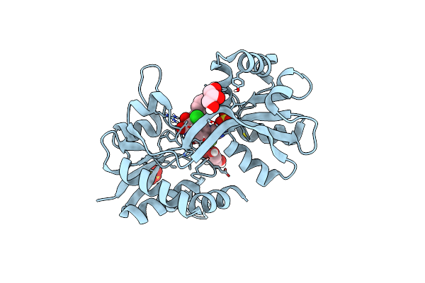
Keap1 Kelch Domain Bound To A Small Molecule Inhibitor Of The Keap1-Nrf2 Protein-Protein Interaction
Organism: Mus musculus
Method: X-RAY DIFFRACTION Resolution:1.75 Å Release Date: 2021-04-14 Classification: PEPTIDE BINDING PROTEIN Ligands: SO4, DMS, QHZ |
Organism: Mus musculus
Method: X-RAY DIFFRACTION
Release Date: 2021-04-14
Ligands: SO4, DMS, QHZ
 |
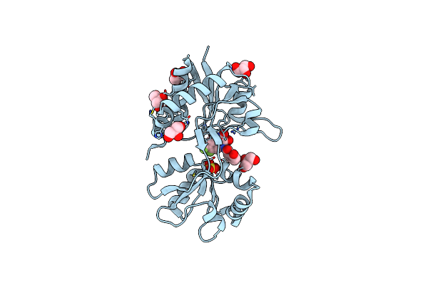
Structure Of Gluk1 Ligand-Binding Domain (S1S2) In Complex With N-(7-(1H-Imidazol-1-Yl)-2,3-Dioxo-6-(Trifluoromethyl)-3,4-Dihydroquinoxalin-1(2H)-Yl Benzamide At 2.3 A Resolution
Organism: Rattus norvegicus
Method: X-RAY DIFFRACTION Resolution:2.30 Å Release Date: 2019-10-30 Classification: MEMBRANE PROTEIN Ligands: SO4, L5H, CL, GOL |
Organism: Rattus norvegicus
Method: X-RAY DIFFRACTION
Release Date: 2019-10-30
Ligands: SO4, L5H, CL, GOL
 |
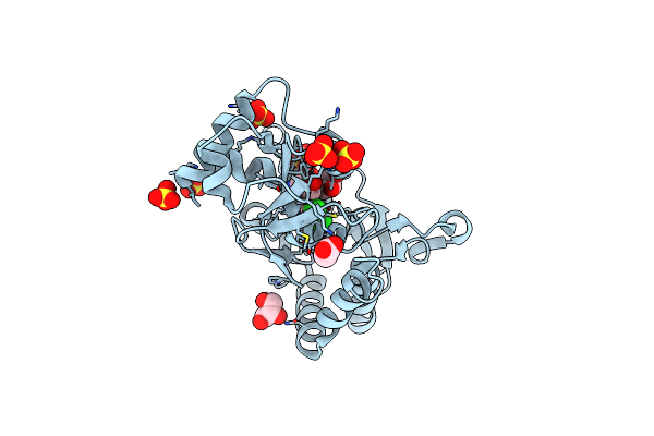
Structure Of Gluk1 Ligand-Binding Domain In Complex With N-(7-Fluoro-2,3-Dioxo-6-(Trifluoromethyl)-3,4-Dihydroquinoxalin-1(2H)-Yl)-2-Hydroxybenzamide At 1.85 A Resolution
Organism: Rattus norvegicus
Method: X-RAY DIFFRACTION Resolution:1.85 Å Release Date: 2019-01-23 Classification: MEMBRANE PROTEIN Ligands: GOL, SO4, EC8, CL |
Organism: Rattus norvegicus
Method: X-RAY DIFFRACTION
Release Date: 2019-01-23
Ligands: GOL, SO4, EC8, CL
 |
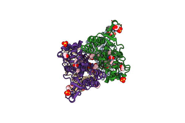
Crystal Structure Of The Glua2 Ligand-Binding Domain (S1S2J) In Complex With The Antagonist (S)-2-Amino-3-(3,4-Dichloro-5-(5-Hydroxypyridin-3-Yl)Phenyl)Propanoic Acid At 2.0A Resolution
Organism: Rattus norvegicus
Method: X-RAY DIFFRACTION Resolution:2.00 Å Release Date: 2015-12-30 Classification: MEMBRANE PROTEIN Ligands: 4ZK, GOL, SO4, ACT |
Organism: Rattus norvegicus
Method: X-RAY DIFFRACTION
Release Date: 2015-12-30
Ligands: 4ZK, GOL, SO4, ACT
 |
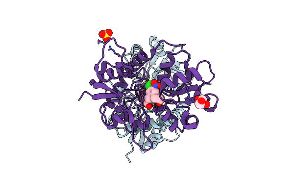
Crystal Structure Of The Glua2 Ligand-Binding Domain (S1S2J) In Complex With The Antagonist (R)-2-Amino-3-(3'-Hydroxybiphenyl-3-Yl)Propanoic Acid At 1.8A Resolution
Organism: Rattus norvegicus
Method: X-RAY DIFFRACTION Resolution:1.80 Å Release Date: 2015-12-30 Classification: MEMBRANE PROTEIN Ligands: E42, SO4, EDO, GOL, CL |
Organism: Rattus norvegicus
Method: X-RAY DIFFRACTION
Release Date: 2015-12-30
Ligands: E42, SO4, EDO, GOL, CL
 |
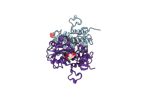
Crystal Structure Of The Gluk1 Ligand-Binding Domain (S1S2) In Complex With The Antagonist (S)-2-Amino-3-(2-(2-Carboxyethyl)-5-Chloro-4-Nitrophenyl)Propionic Acid At 2.0 A Resolution
Organism: Rattus norvegicus
Method: X-RAY DIFFRACTION Resolution:2.00 Å Release Date: 2012-10-10 Classification: MEMBRANE PROTEIN Ligands: TZG, CL, SO4, GOL |
Organism: Rattus norvegicus
Method: X-RAY DIFFRACTION
Release Date: 2012-10-10
Ligands: TZG, CL, SO4, GOL
 |
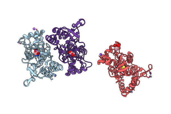
Crystal Structure Of The Glua2 Ligand-Binding Domain (S1S2J) In Complex With The Antagonist (S)-2-Amino-3-(2-(2-Carboxyethyl)-5-Chloro-4-Nitrophenyl)Propionic Acid At 1.9A Resolution
Organism: Rattus norvegicus
Method: X-RAY DIFFRACTION Resolution:1.90 Å Release Date: 2011-10-26 Classification: MEMBRANE PROTEIN Ligands: TZG, SO4 |
Organism: Rattus norvegicus
Method: X-RAY DIFFRACTION
Release Date: 2011-10-26
Ligands: TZG, SO4
 |
Structure Of The Ligand-Binding Core Of Glur2 In Complex With The Agonist (S)-Tdpa At 2.25 A Resolution
Organism: Rattus norvegicus
Method: X-RAY DIFFRACTION Resolution:2.27 Å Release Date: 2008-10-28 Classification: MEMBRANE PROTEIN Ligands: S2P, ZN, NA, CL, CAC |
Organism: Rattus norvegicus
Method: X-RAY DIFFRACTION
Release Date: 2008-10-28
Ligands: S2P, ZN, NA, CL, CAC