

Search Count: 20
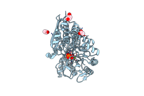 |
Mycobacterium Tuberculosis Enolase Mutant - E204A Complex With Phosphoenolpyruvate
Organism: Mycobacterium tuberculosis (strain atcc 25618 / h37rv)
Method: X-RAY DIFFRACTION Resolution:2.30 Å Release Date: 2022-02-16 Classification: HYDROLASE Ligands: PEP, PEG, EDO, ACT, MG |
Organism: Mycobacterium tuberculosis (strain atcc 25618 / h37rv)
Method: X-RAY DIFFRACTION
Release Date: 2022-02-16
Ligands: PEP, PEG, EDO, ACT, MG
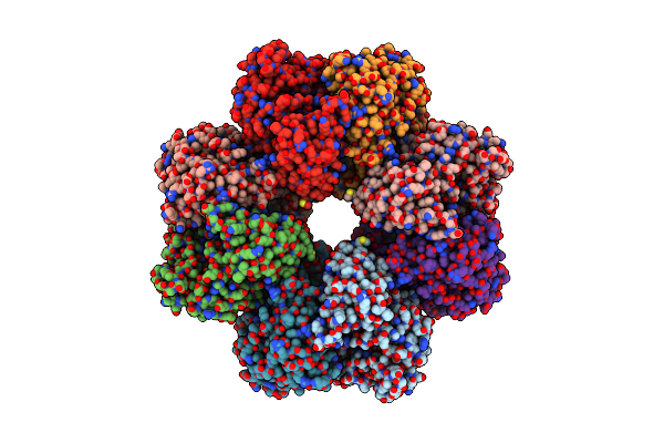 |
Structure Of Enolase From Mycobacterium Tuberculosis
Organism: Mycobacterium tuberculosis
Method: ELECTRON MICROSCOPY Release Date: 2022-02-16 Classification: METAL BINDING PROTEIN |
Organism: Mycobacterium tuberculosis
Method: ELECTRON MICROSCOPY
Release Date: 2022-02-16
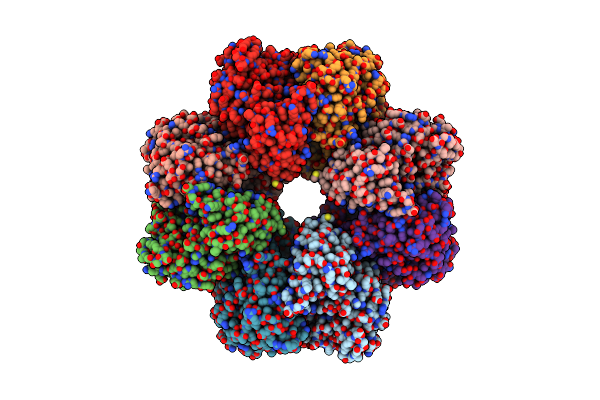 |
Structure Of Pep Bound Enolase From Mycobacterium Tuberculosis
Organism: Mycobacterium tuberculosis
Method: ELECTRON MICROSCOPY Release Date: 2022-02-16 Classification: METAL BINDING PROTEIN Ligands: PEP, MG |
Organism: Mycobacterium tuberculosis
Method: ELECTRON MICROSCOPY
Release Date: 2022-02-16
Ligands: PEP, MG
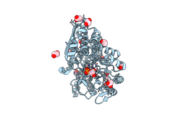 |
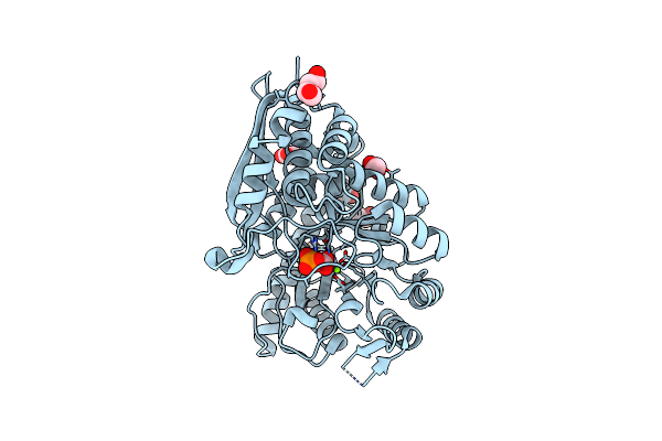
Mycobacterium Tuberculosis Enolase In Complex With Alternate 2-Phosphoglycerate
Organism: Mycobacterium tuberculosis
Method: X-RAY DIFFRACTION Resolution:2.15 Å Release Date: 2022-01-26 Classification: LYASE Ligands: EDO, ACT, MG, 2PG |
Organism: Mycobacterium tuberculosis
Method: X-RAY DIFFRACTION
Release Date: 2022-01-26
Ligands: EDO, ACT, MG, 2PG
 |
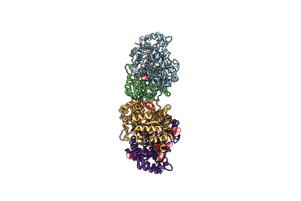
Mycobacterium Tuberculosis Enolase Mutant - E163A
Organism: Mycobacterium tuberculosis
Method: X-RAY DIFFRACTION Resolution:2.25 Å Release Date: 2021-12-01 Classification: LYASE Ligands: PEP, PEG, EDO, ACT, MG, CL |
Organism: Mycobacterium tuberculosis
Method: X-RAY DIFFRACTION
Release Date: 2021-12-01
Ligands: PEP, PEG, EDO, ACT, MG, CL
 |
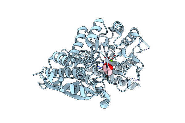
Mycobacterium Tubeculosis Enolase In Complex With 2-Phosphoglycerate
Organism: Mycobacterium tuberculosis
Method: X-RAY DIFFRACTION Resolution:1.99 Å Release Date: 2021-07-28 Classification: LYASE Ligands: MG, 2PG, GOL, ACT, PEG, CL |
Organism: Mycobacterium tuberculosis
Method: X-RAY DIFFRACTION
Release Date: 2021-07-28
Ligands: MG, 2PG, GOL, ACT, PEG, CL
 |
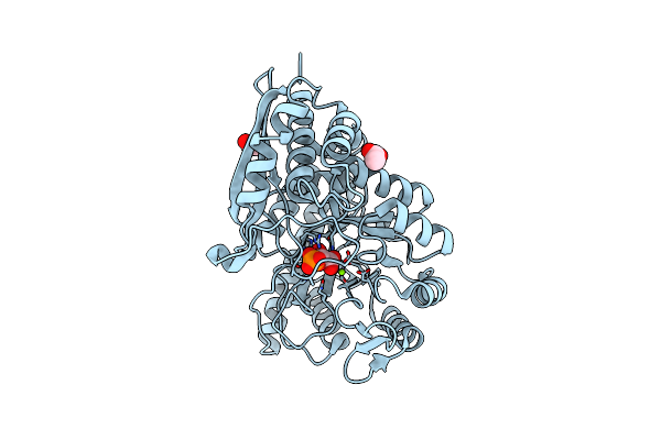
Mycobacterium Tuberculosis Enolase
Organism: Mycobacterium tuberculosis (strain atcc 25618 / h37rv)
Method: X-RAY DIFFRACTION Resolution:2.90 Å Release Date: 2021-07-21 Classification: LYASE Ligands: MG, MPD |
Organism: Mycobacterium tuberculosis (strain atcc 25618 / h37rv)
Method: X-RAY DIFFRACTION
Release Date: 2021-07-21
Ligands: MG, MPD
 |
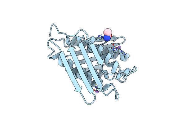
Mycobacterium Tuberculosis Enolase Mutant - S42A
Organism: Mycobacterium tuberculosis h37rv
Method: X-RAY DIFFRACTION Resolution:3.00 Å Release Date: 2020-11-04 Classification: HYDROLASE Ligands: EDO, 2PG, ACT, MG |
Organism: Mycobacterium tuberculosis h37rv
Method: X-RAY DIFFRACTION
Release Date: 2020-11-04
Ligands: EDO, 2PG, ACT, MG
 |
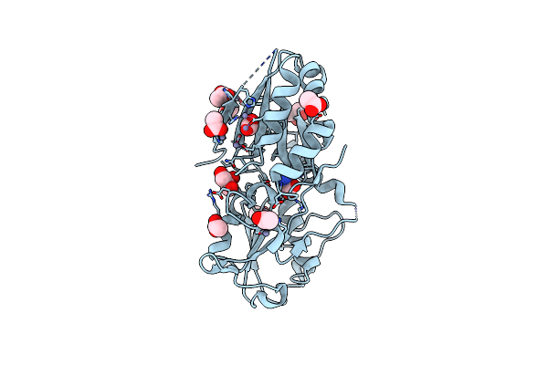
Crystal Structure Of Hisb From Mycobacterium Tuberculosis
Organism: Mycobacterium tuberculosis
Method: X-RAY DIFFRACTION Resolution:1.65 Å Release Date: 2020-07-15 Classification: LYASE Ligands: MN, CL, ACM |
Organism: Mycobacterium tuberculosis
Method: X-RAY DIFFRACTION
Release Date: 2020-07-15
Ligands: MN, CL, ACM
 |
Crystal Structure Of The Substrate Binding Protein Of A Methionine Transporter From Streptococcus Pneumoniae
Organism: Streptococcus pneumoniae serotype 4 (strain atcc baa-334 / tigr4)
Method: X-RAY DIFFRACTION Resolution:1.95 Å Release Date: 2019-07-10 Classification: TRANSPORT PROTEIN Ligands: MET, ZN, CL, ACT, GOL |
Organism: Streptococcus pneumoniae serotype 4 (strain atcc baa-334 / tigr4)
Method: X-RAY DIFFRACTION
Release Date: 2019-07-10
Ligands: MET, ZN, CL, ACT, GOL
 |
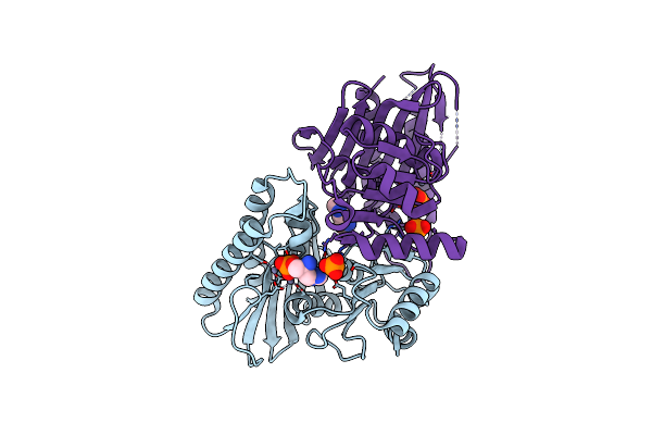
Achromobacter Dh1F Bacterioferritin
Organism: Achromobacter sp. dh1f
Method: X-RAY DIFFRACTION Resolution:1.95 Å Release Date: 2018-09-05 Classification: METAL BINDING PROTEIN Ligands: HEM, FE2, PEG, BA, CL |
Organism: Achromobacter sp. dh1f
Method: X-RAY DIFFRACTION
Release Date: 2018-09-05
Ligands: HEM, FE2, PEG, BA, CL
 |
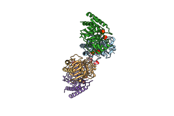
Crystal Structure Of A Phosphatase From Mycobacterium Tuberculosis In Complex With Its Substrate
Organism: Mycobacterium tuberculosis h37rv
Method: X-RAY DIFFRACTION Resolution:2.87 Å Release Date: 2018-05-23 Classification: HYDROLASE Ligands: ZN, HSA, PO4, HSO |
Organism: Mycobacterium tuberculosis h37rv
Method: X-RAY DIFFRACTION
Release Date: 2018-05-23
Ligands: ZN, HSA, PO4, HSO
 |
Histidinol Phosphate Phosphatase From Mycobacterium Tuberculosis
Organism: Mycobacterium tuberculosis
Method: X-RAY DIFFRACTION Resolution:1.94 Å Release Date: 2018-05-23 Classification: HYDROLASE Ligands: ZN, PO4, GOL |
Organism: Mycobacterium tuberculosis
Method: X-RAY DIFFRACTION
Release Date: 2018-05-23
Ligands: ZN, PO4, GOL
 |
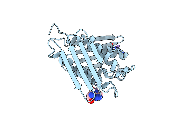
Crystal Structure Of Mycobacterium Tuberculosis Hisb In Complex With A Ligand
Organism: Mycobacterium tuberculosis h37rv
Method: X-RAY DIFFRACTION Resolution:1.80 Å Release Date: 2018-05-16 Classification: LYASE Ligands: MN, CL, IYP |
Organism: Mycobacterium tuberculosis h37rv
Method: X-RAY DIFFRACTION
Release Date: 2018-05-16
Ligands: MN, CL, IYP
 |
Crystal Structure Of Mycobacterium Tuberculosis Hisb Bound With An Inhibitor
Organism: Mycobacterium tuberculosis (strain atcc 25618 / h37rv)
Method: X-RAY DIFFRACTION Resolution:1.75 Å Release Date: 2018-04-04 Classification: LYASE/LYASE INHIBITOR Ligands: MN, CL, 83O |
Organism: Mycobacterium tuberculosis (strain atcc 25618 / h37rv)
Method: X-RAY DIFFRACTION
Release Date: 2018-04-04
Ligands: MN, CL, 83O
 |
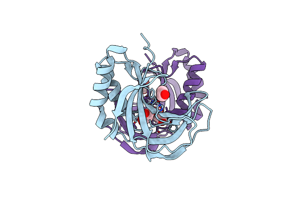
Crystal Structure Of Mhv Ns2 Pde Domain
Organism: Murine coronavirus (strain a59)
Method: X-RAY DIFFRACTION Resolution:3.05 Å Release Date: 2016-02-10 Classification: VIRAL PROTEIN |
Organism: Murine coronavirus (strain a59)
Method: X-RAY DIFFRACTION
Release Date: 2016-02-10
 |
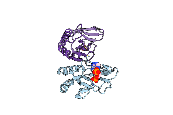
The 1.35A Structure Of A Viral Rnase L Antagonist Reveals Basis For The 2'-5'-Oligoadenylate Binding And Enzyme Activity.
Organism: Rotavirus a
Method: X-RAY DIFFRACTION Resolution:1.35 Å Release Date: 2015-04-29 Classification: VIRAL PROTEIN Ligands: MLI, EDO |
Organism: Rotavirus a
Method: X-RAY DIFFRACTION
Release Date: 2015-04-29
Ligands: MLI, EDO
 |
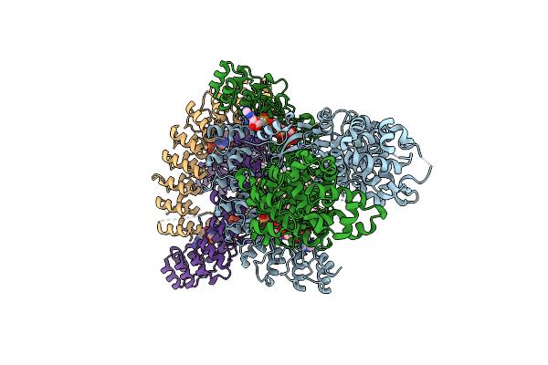
The 1.35 Structure Of A Viral Rnase L Antagonist Reveals Basis For The 2'-5'-Oligoadenylate Binding And Enzyme Activity.
Organism: Rotavirus a
Method: X-RAY DIFFRACTION Resolution:3.10 Å Release Date: 2015-04-29 Classification: VIRAL PROTEIN Ligands: A2P, SO4 |
Organism: Rotavirus a
Method: X-RAY DIFFRACTION
Release Date: 2015-04-29
Ligands: A2P, SO4
 |
Crystal Structure Of Rnase L In Complex With 2-5A
Organism: Sus scrofa
Method: X-RAY DIFFRACTION Resolution:3.27 Å Release Date: 2014-02-05 Classification: TRANSFERASE,HYDROLASE Ligands: 25L |
Organism: Sus scrofa
Method: X-RAY DIFFRACTION
Release Date: 2014-02-05
Ligands: 25L
 |
Crystal Structure Of Rnase L In Complex With 2-5A And Amp-Pnp
Organism: Sus scrofa
Method: X-RAY DIFFRACTION Resolution:2.50 Å Release Date: 2014-02-05 Classification: TRANSFERASE, HYDROLASE Ligands: ANP, MG, 25L |
Organism: Sus scrofa
Method: X-RAY DIFFRACTION
Release Date: 2014-02-05
Ligands: ANP, MG, 25L