

Search Count: 9
 |
Nmr Structure Of Repeat Domain 13 Of The Fibrillar Adhesin Csha From Streptococcus Gordonii.
Organism: Streptococcus gordonii (strain challis / atcc 35105 / bcrc 15272 / ch1 / dl1 / v288)
Method: SOLUTION NMR Release Date: 2020-04-08 Classification: CELL ADHESION |
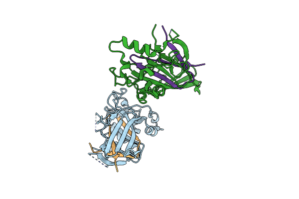 |
Streptococcal Surface Adhesin - Csha Nr2
Organism: Streptococcus gordonii (strain challis / atcc 35105 / bcrc 15272 / ch1 / dl1 / v288)
Method: X-RAY DIFFRACTION Resolution:2.66 Å Release Date: 2016-12-14 Classification: CELL ADHESION |
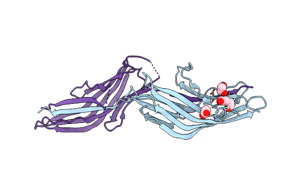 |
Streptococcus Agalactiae Agi/Ii Polypeptide Bspa Variable (V) Domain
Organism: Streptococcus agalactiae serotype iii (strain nem316)
Method: X-RAY DIFFRACTION Resolution:2.41 Å Release Date: 2016-06-22 Classification: CELL ADHESION Ligands: ACT, PEG, EDO |
Organism: Streptococcus agalactiae serotype iii (strain nem316)
Method: X-RAY DIFFRACTION
Release Date: 2016-06-22
Ligands: ACT, PEG, EDO
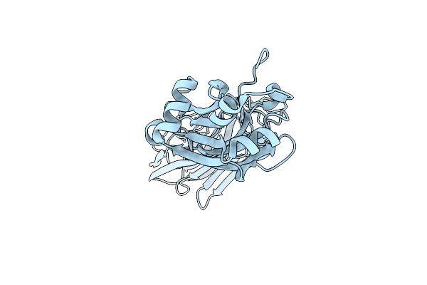 |
Streptococcus Agalactiae Agi/Ii Polypeptide Bspa C-Terminal Domain (Mut)
Organism: Streptococcus agalactiae serotype iii (strain nem316)
Method: X-RAY DIFFRACTION Resolution:1.89 Å Release Date: 2016-06-22 Classification: CELL ADHESION |
Organism: Streptococcus agalactiae serotype iii (strain nem316)
Method: X-RAY DIFFRACTION
Release Date: 2016-06-22
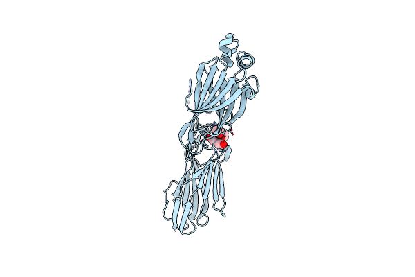 |
Streptococcus Agalactiae Agi/Ii Polypeptide Bspa C Terminal Domain (Wt)
Organism: Streptococcus agalactiae serotype iii (strain nem316)
Method: X-RAY DIFFRACTION Resolution:2.19 Å Release Date: 2016-06-22 Classification: CELL ADHESION Ligands: EDO, PEG |
Organism: Streptococcus agalactiae serotype iii (strain nem316)
Method: X-RAY DIFFRACTION
Release Date: 2016-06-22
Ligands: EDO, PEG
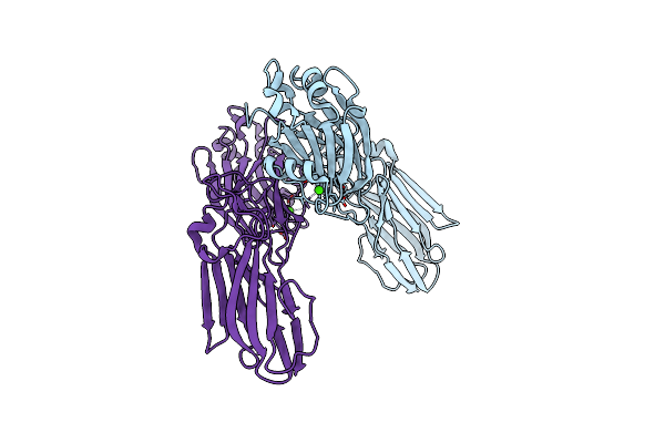 |
Structure Of The C-Terminal Domain Of The Streptococcus Pyogenes Antigen I/Ii-Family Protein Aspa
Organism: Streptococcus pyogenes
Method: X-RAY DIFFRACTION Resolution:1.80 Å Release Date: 2014-03-12 Classification: CELL ADHESION Ligands: CA |
Organism: Streptococcus pyogenes
Method: X-RAY DIFFRACTION
Release Date: 2014-03-12
Ligands: CA
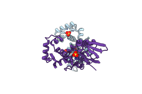 |
Crystal Structure Of The Manganese-Activated Repressor Scar: Apo Form
Organism: Streptococcus gordonii
Method: X-RAY DIFFRACTION Resolution:2.70 Å Release Date: 2009-06-23 Classification: TRANSCRIPTION Ligands: SO4 |
Organism: Streptococcus gordonii
Method: X-RAY DIFFRACTION
Release Date: 2009-06-23
Ligands: SO4
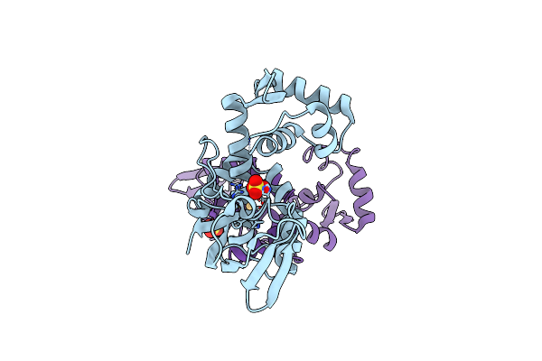 |
Crystal Structure Of Scar With Bound Cd2+
Organism: Streptococcus gordonii
Method: X-RAY DIFFRACTION Resolution:2.80 Å Release Date: 2009-06-23 Classification: TRANSCRIPTION Ligands: SO4, CD |
Organism: Streptococcus gordonii
Method: X-RAY DIFFRACTION
Release Date: 2009-06-23
Ligands: SO4, CD
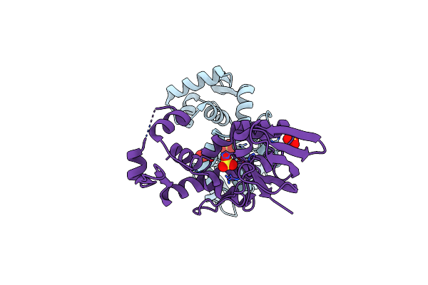 |
Crystal Structure Of Scar With Bound Zn2+
Organism: Streptococcus gordonii
Method: X-RAY DIFFRACTION Resolution:2.90 Å Release Date: 2009-06-23 Classification: TRANSCRIPTION Ligands: ZN, SO4 |
Organism: Streptococcus gordonii
Method: X-RAY DIFFRACTION
Release Date: 2009-06-23
Ligands: ZN, SO4