

Search Count: 43
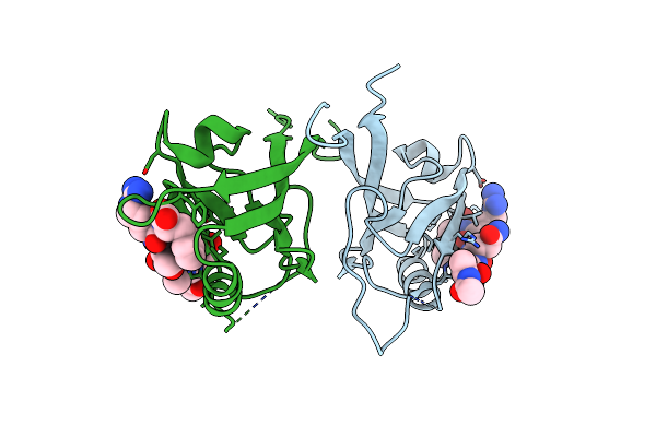 |
Crystal Structure Of Shank1 Pdz In Complex With L6F Mutant Of The C-Terminal Hexapeptide From Gkap
Organism: Homo sapiens
Method: X-RAY DIFFRACTION Resolution:1.78 Å Release Date: 2021-07-07 Classification: PEPTIDE BINDING PROTEIN |
Organism: Homo sapiens
Method: X-RAY DIFFRACTION
Release Date: 2021-07-07
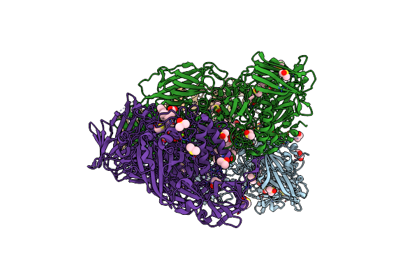 |
E.Coli Beta-Galactosidase (E537Q) In Complex With Fluorescent Probe Ksa02
Organism: Escherichia coli k-12
Method: X-RAY DIFFRACTION Resolution:2.67 Å Release Date: 2021-03-03 Classification: HYDROLASE Ligands: F4X, MG, NA, DMS, GOL |
Organism: Escherichia coli k-12
Method: X-RAY DIFFRACTION
Release Date: 2021-03-03
Ligands: F4X, MG, NA, DMS, GOL
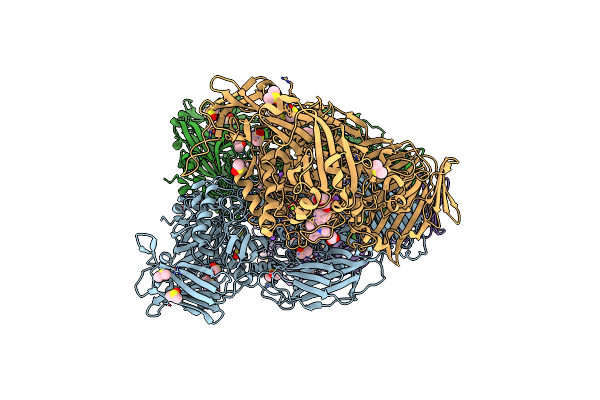 |
E.Coli Beta-Galactosidase (E537Q) In Complex With Fluorescent Probe Ksa01
Organism: Escherichia coli k-12
Method: X-RAY DIFFRACTION Resolution:2.70 Å Release Date: 2021-03-03 Classification: HYDROLASE Ligands: F6L, MG, NA, DMS, GOL |
Organism: Escherichia coli k-12
Method: X-RAY DIFFRACTION
Release Date: 2021-03-03
Ligands: F6L, MG, NA, DMS, GOL
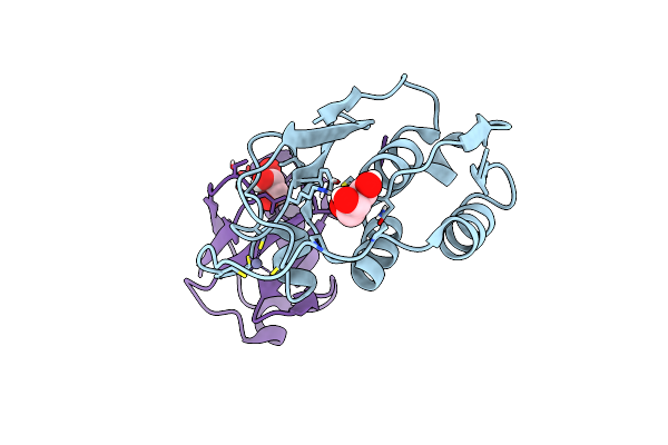 |
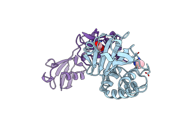
Equine Arteritis Virus Papain-Like Protease 2 (Plp2) Covalently Bound To Ubiquitin
Organism: Equine arteritis virus, Homo sapiens
Method: X-RAY DIFFRACTION Resolution:1.45 Å Release Date: 2013-02-13 Classification: HYDROLASE/PROTEIN BINDING Ligands: ZN, GOL |
Organism: Equine arteritis virus, Homo sapiens
Method: X-RAY DIFFRACTION
Release Date: 2013-02-13
Ligands: ZN, GOL
 |
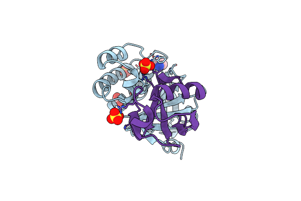
Structure Of A Viral Otu Domain Protease Bound To Interferon-Stimulated Gene 15 (Isg15)
Organism: Crimean-congo hemorrhagic fever virus, Homo sapiens
Method: X-RAY DIFFRACTION Resolution:2.30 Å Release Date: 2011-01-19 Classification: HYDROLASE/PROTEIN BINDING Ligands: 4LJ, GOL |
Organism: Crimean-congo hemorrhagic fever virus, Homo sapiens
Method: X-RAY DIFFRACTION
Release Date: 2011-01-19
Ligands: 4LJ, GOL
 |
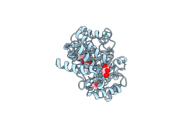
Structure Of A Viral Otu Domain Protease Bound To Ubiquitin
Organism: Crimean-congo hemorrhagic fever virus, Homo sapiens
Method: X-RAY DIFFRACTION Resolution:2.50 Å Release Date: 2011-01-19 Classification: HYDROLASE/PROTEIN BINDING Ligands: SO4, ACT, 4LJ |
Organism: Crimean-congo hemorrhagic fever virus, Homo sapiens
Method: X-RAY DIFFRACTION
Release Date: 2011-01-19
Ligands: SO4, ACT, 4LJ
 |
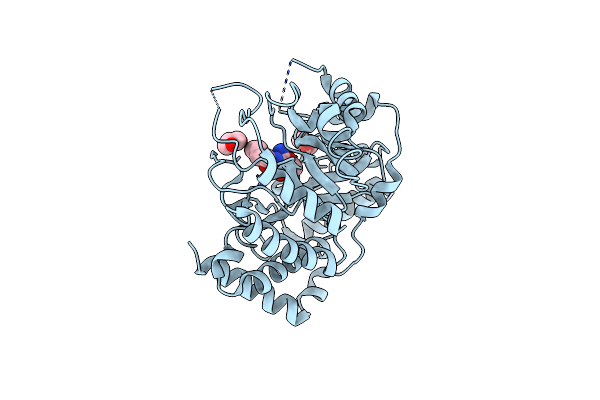
Vibrio Cholerea Family 3 Glycoside Hydrolase (Nagz)In Complex With N-Butyryl-Pugnac
Organism: Vibrio cholerae
Method: X-RAY DIFFRACTION Resolution:2.30 Å Release Date: 2009-06-30 Classification: HYDROLASE Ligands: NP6, GOL |
Organism: Vibrio cholerae
Method: X-RAY DIFFRACTION
Release Date: 2009-06-30
Ligands: NP6, GOL
 |
Vibrio Cholerae Family 3 Glycoside Hydrolase (Nagz) Bound To N-Valeryl-Pugnac
Organism: Vibrio cholerae
Method: X-RAY DIFFRACTION Resolution:2.40 Å Release Date: 2009-06-30 Classification: HYDROLASE Ligands: VPU, GOL |
Organism: Vibrio cholerae
Method: X-RAY DIFFRACTION
Release Date: 2009-06-30
Ligands: VPU, GOL
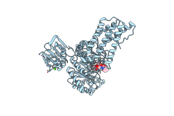 |
Btgh84 In Complex With N-Butyl Pugnac
Organism: Bacteroides thetaiotaomicron vpi-5482
Method: X-RAY DIFFRACTION Resolution:2.30 Å Release Date: 2009-06-16 Classification: HYDROLASE Ligands: NP6, CA |
Organism: Bacteroides thetaiotaomicron vpi-5482
Method: X-RAY DIFFRACTION
Release Date: 2009-06-16
Ligands: NP6, CA
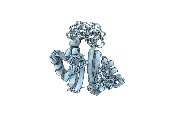 |
Pcbp2 Kh1-Kh2 Domains
Organism: Homo sapiens
Method: SOLUTION NMR Release Date: 2008-08-12 Classification: RNA BINDING PROTEIN |
Organism: Homo sapiens
Method: SOLUTION NMR
Release Date: 2008-08-12
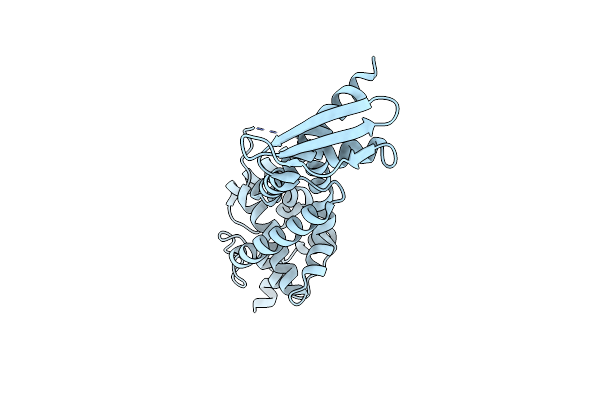 |
Structure Of Rnaseiiib And Dsrna Binding Domains Of Mouse Dicer
Organism: Mus musculus
Method: X-RAY DIFFRACTION Resolution:1.68 Å Release Date: 2008-02-19 Classification: HYDROLASE |
Organism: Mus musculus
Method: X-RAY DIFFRACTION
Release Date: 2008-02-19
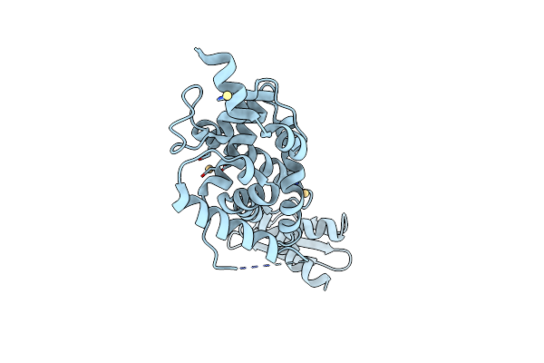 |
Structure Of Rnaseiiib And Dsrna Binding Domains Of Mouse Dicer
Organism: Mus musculus
Method: X-RAY DIFFRACTION Resolution:2.80 Å Release Date: 2008-02-19 Classification: HYDROLASE Ligands: CD |
Organism: Mus musculus
Method: X-RAY DIFFRACTION
Release Date: 2008-02-19
Ligands: CD
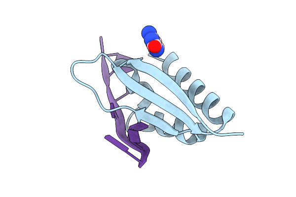 |
Crystal Structure Of The Third Kh Domain Of Human Poly(C)-Binding Protein-2 In Complex With C-Rich Strand Of Human Telomeric Dna
Organism: Homo sapiens
Method: X-RAY DIFFRACTION Resolution:1.60 Å Release Date: 2007-06-12 Classification: RNA AND DNA BINDING PROTEIN/DNA Ligands: CYT |
Organism: Homo sapiens
Method: X-RAY DIFFRACTION
Release Date: 2007-06-12
Ligands: CYT
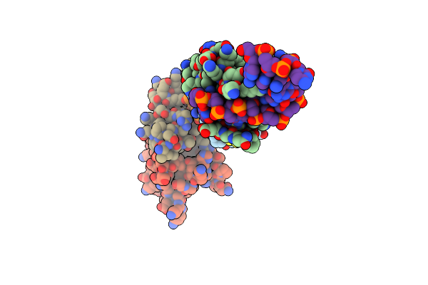 |
Crystal Structure Of Kh1 Domain Of Human Pcbp2 Complexed To Single-Stranded 12-Mer Telomeric Dna
Organism: Homo sapiens
Method: X-RAY DIFFRACTION Resolution:2.12 Å Release Date: 2007-06-12 Classification: RNA AND DNA Binding protein/DNA |
Organism: Homo sapiens
Method: X-RAY DIFFRACTION
Release Date: 2007-06-12
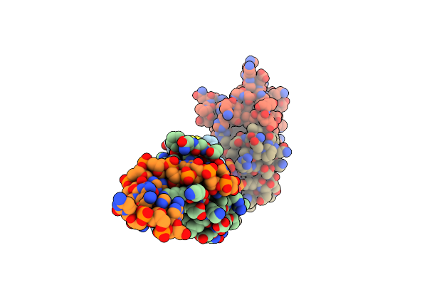 |
Protein-Rna Interaction Involving Kh1 Domain From Human Poly(C)-Binding Protein-2
Organism: Homo sapiens
Method: X-RAY DIFFRACTION Resolution:2.56 Å Release Date: 2007-06-12 Classification: RNA AND DNA binding protein/RNA |
Organism: Homo sapiens
Method: X-RAY DIFFRACTION
Release Date: 2007-06-12
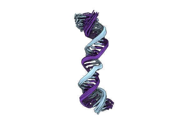 |
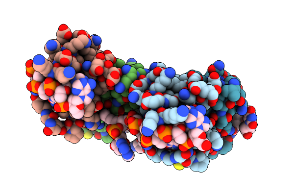
Linear Dimer Of Stemloop Sl1 From Hiv-1
|
Method: SOLUTION NMR
Release Date: 2006-04-25
 |
Crystal Structure Of Kh1 Domain Of Human Poly(C)-Binding Protein-2 With C-Rich Strand Of Human Telomeric Dna
Organism: Homo sapiens
Method: X-RAY DIFFRACTION Resolution:1.70 Å Release Date: 2005-09-27 Classification: DNA BINDING PROTEIN/DNA |
Organism: Homo sapiens
Method: X-RAY DIFFRACTION
Release Date: 2005-09-27
 |
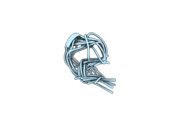
12-Mer From Site Ii Calbindin D9K (Dkngdgevsfee) Coordination Pb(Ii)
|
Method: SOLUTION NMR
Release Date: 2005-07-26
 |
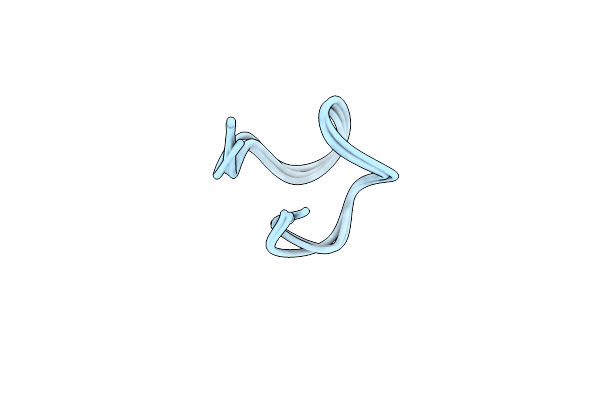
12-Mer From Site Ii Calbindin D9K (Dkngdgevsfee) Coordinating Cd(Ii)
|
Method: SOLUTION NMR
Release Date: 2005-07-26
 |
12-Mer From Site Ii Calbindin D9K (Dkngdgevsfee) Coordinating Zn(Ii)
|
Method: SOLUTION NMR
Release Date: 2005-07-26