

Search Count: 46
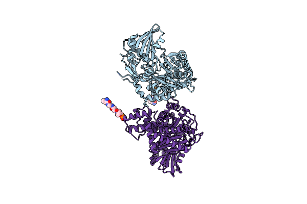 |
Non-Ribosomal Pcp-C Didomain (Amide Stabilised Leucine) Acceptor Bound State
Organism: Thermobifida fusca
Method: X-RAY DIFFRACTION Resolution:2.20 Å Release Date: 2023-06-21 Classification: BIOSYNTHETIC PROTEIN Ligands: YCK |
Organism: Thermobifida fusca
Method: X-RAY DIFFRACTION
Release Date: 2023-06-21
Ligands: YCK
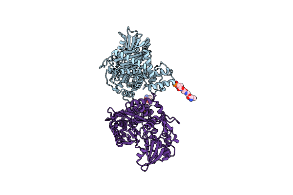 |
Non-Ribosomal Pcp-C Didomain (Ester Stabilised Leucine) Acceptor Bound State
Organism: Thermobifida fusca yx
Method: X-RAY DIFFRACTION Resolution:2.20 Å Release Date: 2023-06-21 Classification: BIOSYNTHETIC PROTEIN Ligands: YCT |
Organism: Thermobifida fusca yx
Method: X-RAY DIFFRACTION
Release Date: 2023-06-21
Ligands: YCT
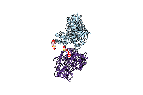 |
Non-Ribosomal Pcp-C Didomain (Thioether Stabilised Glycolic Acid) Acceptor Bound State
Organism: Thermobifida fusca yx
Method: X-RAY DIFFRACTION Resolution:3.02 Å Release Date: 2023-06-14 Classification: BIOSYNTHETIC PROTEIN Ligands: YJI |
Organism: Thermobifida fusca yx
Method: X-RAY DIFFRACTION
Release Date: 2023-06-14
Ligands: YJI
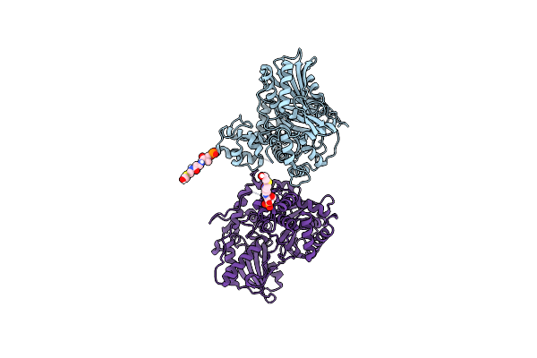 |
Non-Ribosomal Pcp-C Didomain R2577G (Thioether Stabilised Glycolic Acid) Acceptor Bound State
Organism: Thermobifida fusca yx
Method: X-RAY DIFFRACTION Resolution:2.10 Å Release Date: 2023-06-14 Classification: BIOSYNTHETIC PROTEIN Ligands: YJI |
Organism: Thermobifida fusca yx
Method: X-RAY DIFFRACTION
Release Date: 2023-06-14
Ligands: YJI
 |
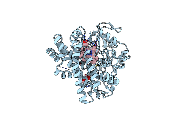
P450 (Oxya) From Kistamicin Biosynthesis, Mixed Heme Conformation, Attenuated Beam
Organism: Actinomadura parvosata subsp. kistnae
Method: X-RAY DIFFRACTION Resolution:2.00 Å Release Date: 2022-05-11 Classification: OXIDOREDUCTASE Ligands: GOL, HEM |
Organism: Actinomadura parvosata subsp. kistnae
Method: X-RAY DIFFRACTION
Release Date: 2022-05-11
Ligands: GOL, HEM
 |
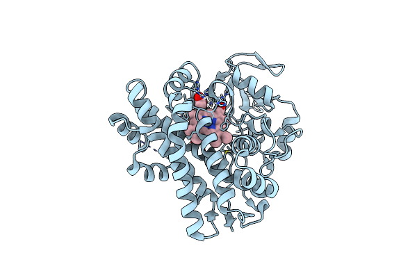
P450 (Oxya) From Kistamicin Biosynthesis, Y99F Mutant
Organism: Actinomadura parvosata subsp. kistnae
Method: X-RAY DIFFRACTION Resolution:1.80 Å Release Date: 2022-05-11 Classification: OXIDOREDUCTASE Ligands: HEM, GOL |
Organism: Actinomadura parvosata subsp. kistnae
Method: X-RAY DIFFRACTION
Release Date: 2022-05-11
Ligands: HEM, GOL
 |
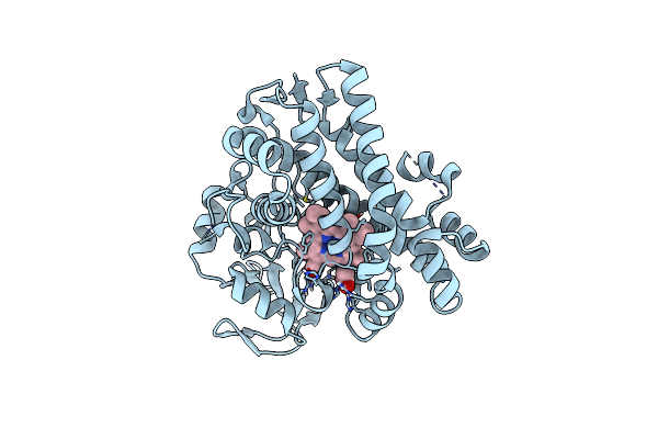
P450 (Oxya) From Kistamicin Biosynthesis, Mixed Heme Conformation
Organism: Actinomadura parvosata subsp. kistnae
Method: X-RAY DIFFRACTION Resolution:1.60 Å Release Date: 2022-05-11 Classification: OXIDOREDUCTASE Ligands: HEM |
Organism: Actinomadura parvosata subsp. kistnae
Method: X-RAY DIFFRACTION
Release Date: 2022-05-11
Ligands: HEM
 |
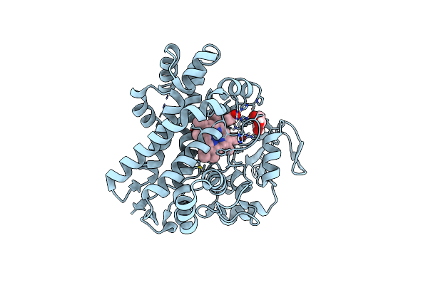
P450 (Oxya) From Kistamicin Biosynthesis, Mixed Heme Conformation
Organism: Actinomadura parvosata subsp. kistnae
Method: X-RAY DIFFRACTION Resolution:1.80 Å Release Date: 2022-05-11 Classification: OXIDOREDUCTASE Ligands: HEM, GOL |
Organism: Actinomadura parvosata subsp. kistnae
Method: X-RAY DIFFRACTION
Release Date: 2022-05-11
Ligands: HEM, GOL
 |
P450 (Oxya) From Kistamicin Biosynthesis, Imidazole Complex
Organism: Actinomadura parvosata subsp. kistnae
Method: X-RAY DIFFRACTION Resolution:1.62 Å Release Date: 2022-05-11 Classification: OXIDOREDUCTASE Ligands: IMD, HEM, GOL |
Organism: Actinomadura parvosata subsp. kistnae
Method: X-RAY DIFFRACTION
Release Date: 2022-05-11
Ligands: IMD, HEM, GOL
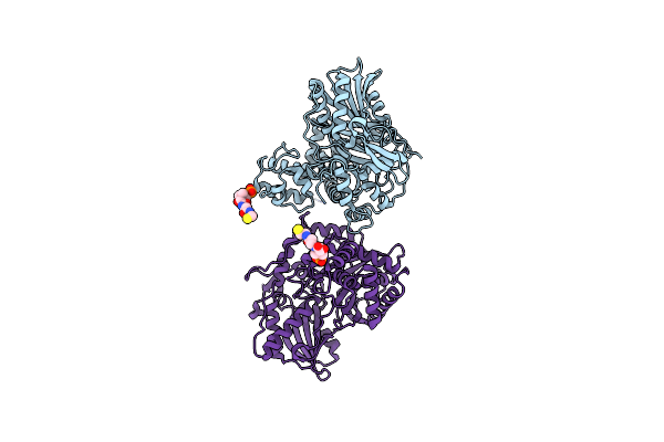 |
Non-Ribosomal Didomain (Holo-Pcp-C) Acceptor Bound State
Organism: Thermobifida fusca
Method: X-RAY DIFFRACTION Resolution:2.18 Å Release Date: 2021-03-24 Classification: BIOSYNTHETIC PROTEIN Ligands: PNS |
Organism: Thermobifida fusca
Method: X-RAY DIFFRACTION
Release Date: 2021-03-24
Ligands: PNS
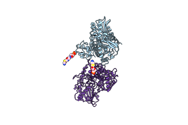 |
Non-Ribosomal Didomain (Stabilised Glycine-Pcp-C) Acceptor Bound State
Organism: Thermobifida fusca
Method: X-RAY DIFFRACTION Resolution:1.90 Å Release Date: 2021-03-24 Classification: BIOSYNTHETIC PROTEIN Ligands: XAG |
Organism: Thermobifida fusca
Method: X-RAY DIFFRACTION
Release Date: 2021-03-24
Ligands: XAG
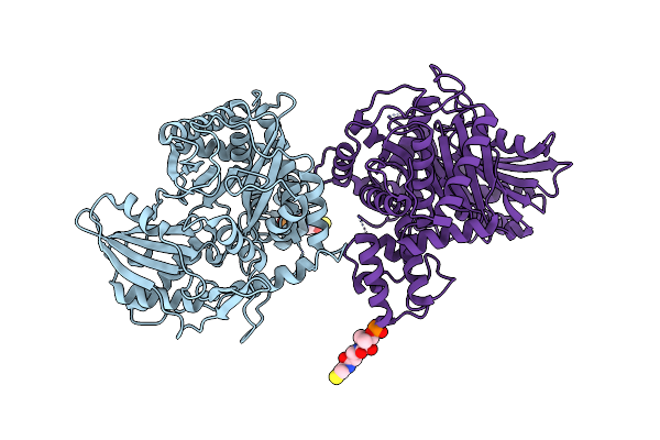 |
Non-Ribosomal Didomain (Holo-Pcp-C) Acceptor Bound State, R2577G
Organism: Thermobifida fusca
Method: X-RAY DIFFRACTION Resolution:2.00 Å Release Date: 2021-03-24 Classification: BIOSYNTHETIC PROTEIN Ligands: PNS |
Organism: Thermobifida fusca
Method: X-RAY DIFFRACTION
Release Date: 2021-03-24
Ligands: PNS
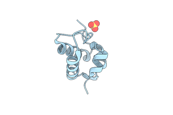 |
Non Ribosomal Pcp Domain
Organism: Thermobifida fusca
Method: X-RAY DIFFRACTION Resolution:2.30 Å Release Date: 2021-03-24 Classification: BIOSYNTHETIC PROTEIN Ligands: SO4 |
Organism: Thermobifida fusca
Method: X-RAY DIFFRACTION
Release Date: 2021-03-24
Ligands: SO4
 |
The Crystal Structure Of Fbia From Mycobacterium Smegmatis, Apo State
Organism: Mycolicibacterium smegmatis (strain atcc 700084 / mc(2)155)
Method: X-RAY DIFFRACTION Resolution:2.30 Å Release Date: 2020-05-13 Classification: BIOSYNTHETIC PROTEIN Ligands: CA |
Organism: Mycolicibacterium smegmatis (strain atcc 700084 / mc(2)155)
Method: X-RAY DIFFRACTION
Release Date: 2020-05-13
Ligands: CA
 |
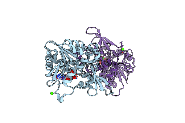
The Crystal Structure Of Fbia From Mycobacterium Smegmatis, Fo Bound Form
Organism: Mycolicibacterium smegmatis (strain atcc 700084 / mc(2)155)
Method: X-RAY DIFFRACTION Resolution:2.21 Å Release Date: 2020-05-13 Classification: BIOSYNTHETIC PROTEIN Ligands: FO1, CA |
Organism: Mycolicibacterium smegmatis (strain atcc 700084 / mc(2)155)
Method: X-RAY DIFFRACTION
Release Date: 2020-05-13
Ligands: FO1, CA
 |
The Crystal Structure Of Fbia From Mycobacterium Smegmatis, Gdp Bound Form
Organism: Mycolicibacterium smegmatis (strain atcc 700084 / mc(2)155)
Method: X-RAY DIFFRACTION Resolution:2.40 Å Release Date: 2020-05-13 Classification: BIOSYNTHETIC PROTEIN Ligands: GDP, GOL, CA |
Organism: Mycolicibacterium smegmatis (strain atcc 700084 / mc(2)155)
Method: X-RAY DIFFRACTION
Release Date: 2020-05-13
Ligands: GDP, GOL, CA
 |
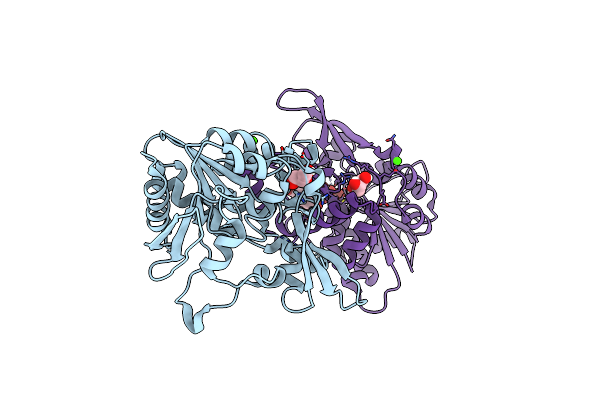
The Crystal Structure Of Fbia From Mycobacterium Smegmatis, Gdp And Fo Bound Form
Organism: Mycolicibacterium smegmatis (strain atcc 700084 / mc(2)155)
Method: X-RAY DIFFRACTION Resolution:2.20 Å Release Date: 2020-05-13 Classification: BIOSYNTHETIC PROTEIN Ligands: GDP, FO1, CA |
Organism: Mycolicibacterium smegmatis (strain atcc 700084 / mc(2)155)
Method: X-RAY DIFFRACTION
Release Date: 2020-05-13
Ligands: GDP, FO1, CA
 |
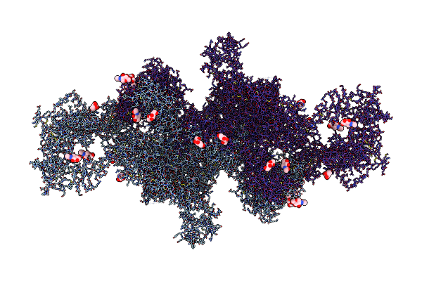
The Crystal Structure Of Fbia From Mycobacterium Smegmatis, Dehydro-F420-0 Bound Form
Organism: Mycolicibacterium smegmatis (strain atcc 700084 / mc(2)155)
Method: X-RAY DIFFRACTION Resolution:2.34 Å Release Date: 2020-05-13 Classification: BIOSYNTHETIC PROTEIN Ligands: GOL, QMD, CA |
Organism: Mycolicibacterium smegmatis (strain atcc 700084 / mc(2)155)
Method: X-RAY DIFFRACTION
Release Date: 2020-05-13
Ligands: GOL, QMD, CA
 |
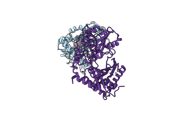
The Structure Of Human Thyroglobulin
Organism: Homo sapiens
Method: ELECTRON MICROSCOPY Release Date: 2020-02-12 Classification: HORMONE Ligands: NAG |
Organism: Homo sapiens
Method: ELECTRON MICROSCOPY
Release Date: 2020-02-12
Ligands: NAG
 |
Complex Of Oxya With The X-Domain From Gpa Biosynthesis
Organism: Actinomadura parvosata subsp. kistnae
Method: X-RAY DIFFRACTION Resolution:2.65 Å Release Date: 2019-05-22 Classification: BIOSYNTHETIC PROTEIN Ligands: HEM |
Organism: Actinomadura parvosata subsp. kistnae
Method: X-RAY DIFFRACTION
Release Date: 2019-05-22
Ligands: HEM