

Search Count: 37
 |
Crystal Structure Of Apkc Iota Kinase Domain With Llgl2 Peptide
Organism: Homo sapiens
Method: X-RAY DIFFRACTION Resolution:2.59 Å Release Date: 2024-11-20 Classification: CYTOSOLIC PROTEIN |
Organism: Homo sapiens
Method: X-RAY DIFFRACTION
Release Date: 2024-11-20
 |
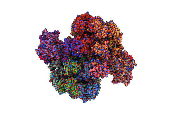
Cryo Em Structure Of A Stable Lgl/Apkc Iota/Par-6 Complex
Organism: Homo sapiens
Method: ELECTRON MICROSCOPY Release Date: 2024-11-20 Classification: CYTOSOLIC PROTEIN Ligands: ANP |
Organism: Homo sapiens
Method: ELECTRON MICROSCOPY
Release Date: 2024-11-20
Ligands: ANP
 |
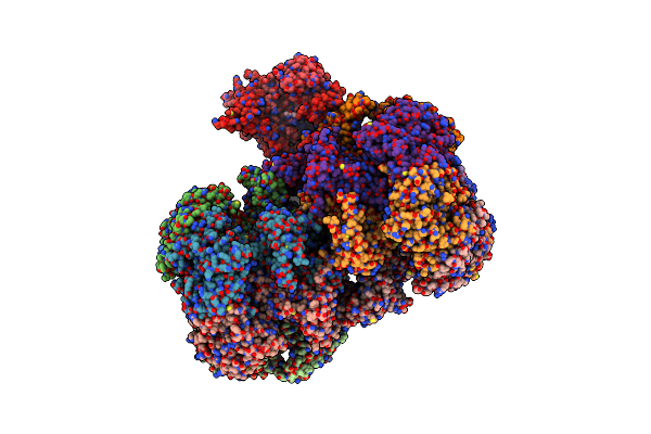
Structure Of Mcm2-7 Dh Complexed With Cdc7-Dbf4 In The Presence Of Atpgs, State Iii
Organism: Saccharomyces cerevisiae (strain atcc 204508 / s288c)
Method: ELECTRON MICROSCOPY Release Date: 2022-06-08 Classification: REPLICATION Ligands: AGS, MG, ZN, ADP |
Organism: Saccharomyces cerevisiae (strain atcc 204508 / s288c)
Method: ELECTRON MICROSCOPY
Release Date: 2022-06-08
Ligands: AGS, MG, ZN, ADP
 |
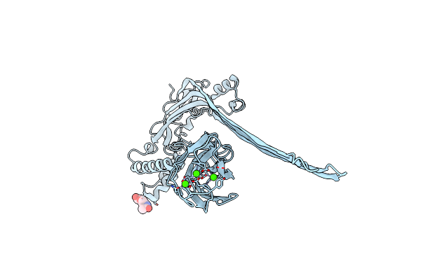
Structure Of Mcm2-7 Dh Complexed With Cdc7-Dbf4 In The Presence Of Adp:Bef3, State I
Organism: Saccharomyces cerevisiae (strain atcc 204508 / s288c)
Method: ELECTRON MICROSCOPY Release Date: 2022-06-08 Classification: REPLICATION Ligands: ADP, MG, ZN, BEF |
Organism: Saccharomyces cerevisiae (strain atcc 204508 / s288c)
Method: ELECTRON MICROSCOPY
Release Date: 2022-06-08
Ligands: ADP, MG, ZN, BEF
 |
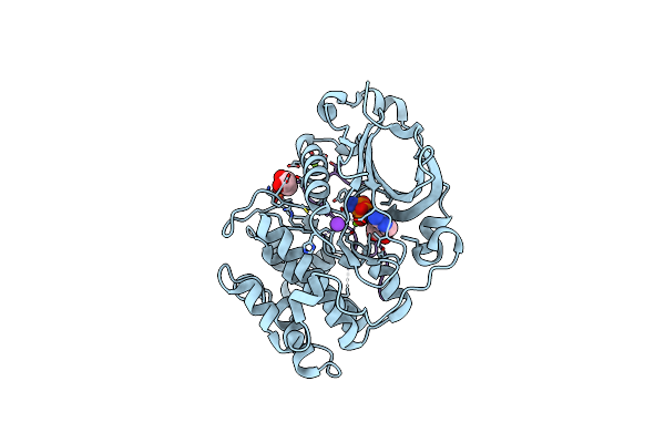
The Pore Conformation Of Lymphocyte Perforin
Organism: Mus musculus
Method: ELECTRON MICROSCOPY Release Date: 2022-02-16 Classification: IMMUNE SYSTEM Ligands: CA, NAG |
Organism: Mus musculus
Method: ELECTRON MICROSCOPY
Release Date: 2022-02-16
Ligands: CA, NAG
 |
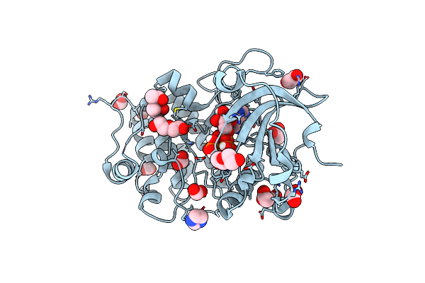
Structure Of A Par3-Inhibitory Peptide Bound To Pkciota Core Kinase Domain
Organism: Homo sapiens, Xenopus tropicalis
Method: X-RAY DIFFRACTION Resolution:2.00 Å Release Date: 2016-09-14 Classification: TRANSFERASE Ligands: ANP, GOL, K, MG |
Organism: Homo sapiens, Xenopus tropicalis
Method: X-RAY DIFFRACTION
Release Date: 2016-09-14
Ligands: ANP, GOL, K, MG
 |
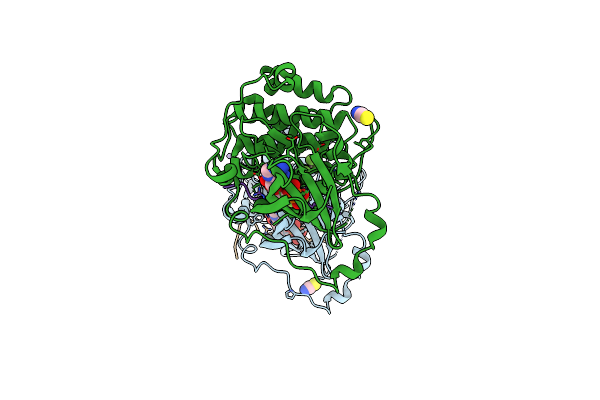
Structure Of A Nucleotide-Bound Form Of Pkciota Core Kinase Domain
Organism: Homo sapiens
Method: X-RAY DIFFRACTION Resolution:1.79 Å Release Date: 2016-09-14 Classification: TRANSFERASE Ligands: ACP, FMT, IMD, MRD, PEG, ACT |
Organism: Homo sapiens
Method: X-RAY DIFFRACTION
Release Date: 2016-09-14
Ligands: ACP, FMT, IMD, MRD, PEG, ACT
 |
Structure Of A Peptide-Substrate Bound To Pkciota Core Kinase Domain
Organism: Homo sapiens
Method: X-RAY DIFFRACTION Resolution:3.25 Å Release Date: 2016-09-14 Classification: TRANSFERASE Ligands: ADP, AF3, MN, SCN |
Organism: Homo sapiens
Method: X-RAY DIFFRACTION
Release Date: 2016-09-14
Ligands: ADP, AF3, MN, SCN
 |
Microed Structure Of The Segment, Gvvhgvttva, From The A53T Familial Mutant Of Parkinson'S Disease Protein, Alpha-Synuclein Residues 47-56
Organism: Homo sapiens
Method: ELECTRON CRYSTALLOGRAPHY Resolution:1.41 Å Release Date: 2015-09-09 Classification: LIPID BINDING PROTEIN |
Organism: Homo sapiens
Method: ELECTRON CRYSTALLOGRAPHY
Release Date: 2015-09-09
 |
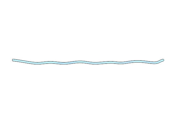
Amyloid Forming Segment, Avvtgvtav, From The Nac Domain Of Parkinson'S Disease Protein Alpha-Synuclein, Residues 69-77
Organism: Homo sapiens
Method: X-RAY DIFFRACTION Resolution:1.85 Å Release Date: 2015-08-26 Classification: LIPID BINDING PROTEIN |
Organism: Homo sapiens
Method: X-RAY DIFFRACTION
Release Date: 2015-08-26
 |
Structure Of The Amyloid Forming Segment, Gavvtgvtava, From The Nac Domain Of Parkinson'S Disease Protein Alpha-Synuclein, Residues 68-78, Determined By Electron Diffraction
Organism: Homo sapiens
Method: ELECTRON CRYSTALLOGRAPHY Resolution:1.43 Å Release Date: 2015-08-26 Classification: LIPID BINDING PROTEIN |
Organism: Homo sapiens
Method: ELECTRON CRYSTALLOGRAPHY
Release Date: 2015-08-26
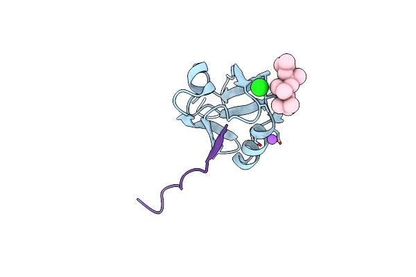 |
Crystal Structure Of The Pdz Domain Of Pals1 In Complex With The Crb Peptide
Organism: Homo sapiens
Method: X-RAY DIFFRACTION Resolution:1.23 Å Release Date: 2015-01-14 Classification: CELL ADHESION Ligands: CL, NA, PUN |
Organism: Homo sapiens
Method: X-RAY DIFFRACTION
Release Date: 2015-01-14
Ligands: CL, NA, PUN
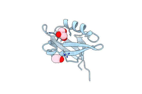 |
Crystal Structure Of The Pdz Domain Of Pals1
Organism: Homo sapiens
Method: X-RAY DIFFRACTION Resolution:1.80 Å Release Date: 2015-01-14 Classification: STRUCTURAL PROTEIN Ligands: GOL, CL, ACT |
Organism: Homo sapiens
Method: X-RAY DIFFRACTION
Release Date: 2015-01-14
Ligands: GOL, CL, ACT
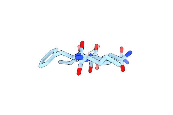 |
Ifqins, An Amyloid Forming Segment From Human Lysozyme Spanning Residues 56-61
Organism: Homo sapiens
Method: X-RAY DIFFRACTION Resolution:1.52 Å Release Date: 2014-12-17 Classification: PROTEIN FIBRIL |
Organism: Homo sapiens
Method: X-RAY DIFFRACTION
Release Date: 2014-12-17
 |
Tgvtava, An Amyloid Forming Segment From Alpha Synuclein, Residues 72-78
Organism: Homo sapiens
Method: X-RAY DIFFRACTION Resolution:1.38 Å Release Date: 2014-12-17 Classification: PROTEIN FIBRIL |
Organism: Homo sapiens
Method: X-RAY DIFFRACTION
Release Date: 2014-12-17
 |
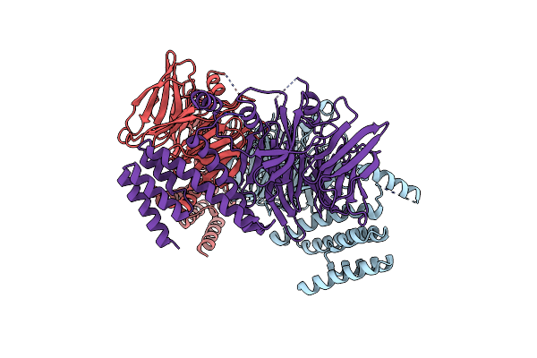
Vvtgvta, An Amyloid Forming Segment From Alpha Synuclein, Residues 70-76
Organism: Homo sapiens
Method: X-RAY DIFFRACTION Resolution:1.50 Å Release Date: 2014-12-17 Classification: PROTEIN FIBRIL Ligands: HOH |
Organism: Homo sapiens
Method: X-RAY DIFFRACTION
Release Date: 2014-12-17
Ligands: HOH
 |
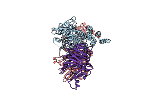
Crystal Structure Of The C-Terminal Region Of Yeast Ctf4, Selenomethionine Protein.
Organism: Saccharomyces cerevisiae
Method: X-RAY DIFFRACTION Resolution:2.69 Å Release Date: 2014-04-30 Classification: DNA REPLICATION |
Organism: Saccharomyces cerevisiae
Method: X-RAY DIFFRACTION
Release Date: 2014-04-30
 |
Crystal Structure Of The C-Terminal Region Of Yeast Ctf4
Organism: Saccharomyces cerevisiae
Method: X-RAY DIFFRACTION Resolution:3.00 Å Release Date: 2014-04-30 Classification: DNA REPLICATION |
Organism: Saccharomyces cerevisiae
Method: X-RAY DIFFRACTION
Release Date: 2014-04-30
 |
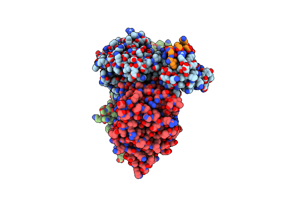
Crystal Structure Of The Carboxy-Terminal Domain Of Yeast Ctf4 Bound To Pol Alpha.
Organism: Saccharomyces cerevisiae
Method: X-RAY DIFFRACTION Resolution:2.69 Å Release Date: 2014-04-30 Classification: DNA REPLICATION |
Organism: Saccharomyces cerevisiae
Method: X-RAY DIFFRACTION
Release Date: 2014-04-30
 |
Crystal Structure Of The Carboxy-Terminal Domain Of Yeast Ctf4 Bound To Sld5
Organism: Saccharomyces cerevisiae
Method: X-RAY DIFFRACTION Resolution:2.69 Å Release Date: 2014-04-30 Classification: DNA REPLICATION |
Organism: Saccharomyces cerevisiae
Method: X-RAY DIFFRACTION
Release Date: 2014-04-30