

Search Count: 77
 |
Crystal Structure Of Htead4 Yap Binding Domain (Htead4-Ybd) In Complex With Peptide 6
Organism: Homo sapiens
Method: X-RAY DIFFRACTION Resolution:1.74 Å Release Date: 2026-04-08 Classification: TRANSCRIPTION Ligands: EDO, MYR, PO4 |
Organism: Homo sapiens
Method: X-RAY DIFFRACTION
Release Date: 2026-04-08
Ligands: EDO, MYR, PO4
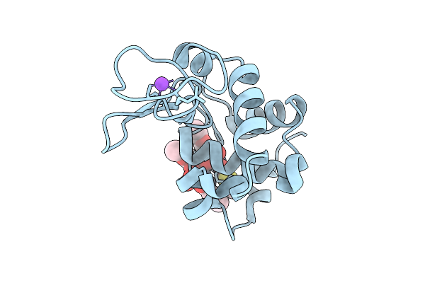 |
X-Ray Structure Of Lysozyme Obtained Upon Reaction With The Dioxidovanadium(V) Complex With Thiophene-2-Carboxylic Acid (3-Ethoxy-2-Hydroxybenzylidene)Hydrazide
Organism: Gallus gallus
Method: X-RAY DIFFRACTION Resolution:1.37 Å Release Date: 2026-04-01 Classification: HYDROLASE |
Organism: Gallus gallus
Method: X-RAY DIFFRACTION
Release Date: 2026-04-01
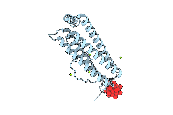 |
X-Ray Structure Of A Polyoxidovanadate/Human H-Ferritin Adduct Obtained When The Protein Is Treated Overnight With [Vivo(Acac)2]
Organism: Homo sapiens
Method: X-RAY DIFFRACTION Resolution:1.38 Å Release Date: 2025-12-03 Classification: METAL TRANSPORT Ligands: CL, MG, A1JGJ |
Organism: Homo sapiens
Method: X-RAY DIFFRACTION
Release Date: 2025-12-03
Ligands: CL, MG, A1JGJ
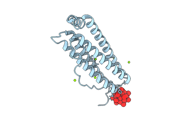 |
X-Ray Structure Of A Polyoxidovanadate/Human H-Ferritin Adduct Obtained When The Protein Is Treated 6 Days With [Vivo(Acac)2]
Organism: Homo sapiens
Method: X-RAY DIFFRACTION Resolution:1.54 Å Release Date: 2025-12-03 Classification: METAL TRANSPORT Ligands: CL, MG, A1JGJ |
Organism: Homo sapiens
Method: X-RAY DIFFRACTION
Release Date: 2025-12-03
Ligands: CL, MG, A1JGJ
 |
X-Ray Structure Of A Polyoxidovanadate/Human H-Ferritin Adduct Obtained When The Protein Is Treated 24 H With [Vivo(Acac)2]
Organism: Homo sapiens
Method: X-RAY DIFFRACTION Resolution:1.50 Å Release Date: 2025-12-03 Classification: METAL TRANSPORT Ligands: CL, MG, A1JGJ |
Organism: Homo sapiens
Method: X-RAY DIFFRACTION
Release Date: 2025-12-03
Ligands: CL, MG, A1JGJ
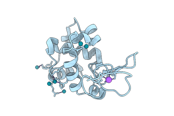 |
X-Ray Structure Of The Adduct Formed Upon Reaction Of Dirhodium-Tetraacetate With Lysozyme At Body Temperature
Organism: Gallus gallus
Method: X-RAY DIFFRACTION Resolution:2.12 Å Release Date: 2025-08-13 Classification: HYDROLASE Ligands: RH, NA |
Organism: Gallus gallus
Method: X-RAY DIFFRACTION
Release Date: 2025-08-13
Ligands: RH, NA
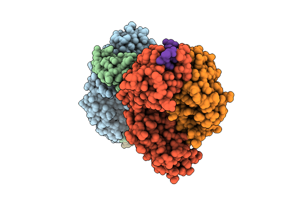 |
Crystal Structure Of Ma6 Fab In Complex With Pfcsp Repeat Region Peptide Npna3
Organism: Homo sapiens, Plasmodium falciparum
Method: X-RAY DIFFRACTION Resolution:1.80 Å Release Date: 2025-07-09 Classification: IMMUNE SYSTEM |
Organism: Homo sapiens, Plasmodium falciparum
Method: X-RAY DIFFRACTION
Release Date: 2025-07-09
 |
X-Ray Structure Of A Polyoxidovanadate/Lysozyme Adduct Obtained When The Protein Is Treated With [Vivo(Acac)2</Sub>] At 310 K
Organism: Gallus gallus
Method: X-RAY DIFFRACTION Resolution:1.83 Å Release Date: 2025-05-14 Classification: HYDROLASE Ligands: CL, NA, A1ICR, A1H8D |
Organism: Gallus gallus
Method: X-RAY DIFFRACTION
Release Date: 2025-05-14
Ligands: CL, NA, A1ICR, A1H8D
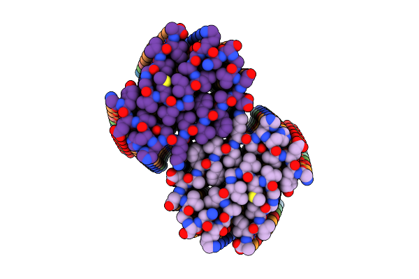 |
Cryo-Em Structure Of The Class I Amyloid-Beta 42 Fibril Containing A D-Asp At Position 23
Organism: Homo sapiens
Method: ELECTRON MICROSCOPY Release Date: 2025-03-26 Classification: PROTEIN FIBRIL |
Organism: Homo sapiens
Method: ELECTRON MICROSCOPY
Release Date: 2025-03-26
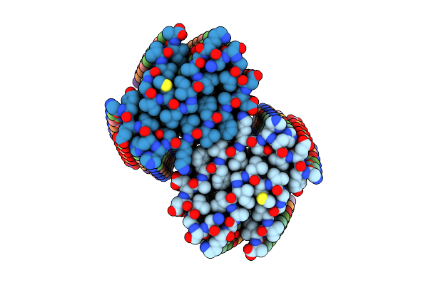 |
Cryo-Em Structure Of The Class Ii Amyloid-Beta 42 Fibril Containing A D-Asp At Position 23
Organism: Homo sapiens
Method: ELECTRON MICROSCOPY Release Date: 2025-03-26 Classification: PROTEIN FIBRIL |
Organism: Homo sapiens
Method: ELECTRON MICROSCOPY
Release Date: 2025-03-26
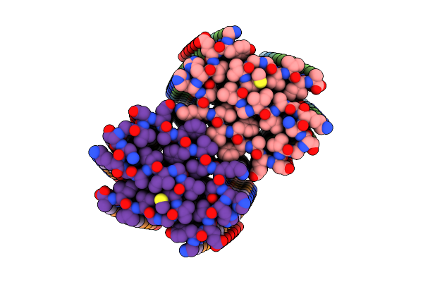 |
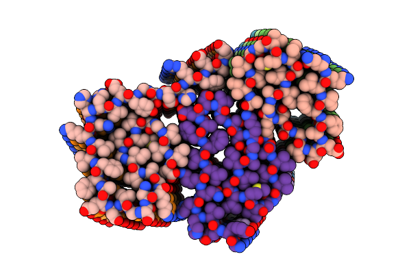
Cryo-Em Structure Of The Type I Amyloid-Beta 42 Fibril Containing A D-Asp At Positions 7 And 23
Organism: Homo sapiens
Method: ELECTRON MICROSCOPY Release Date: 2025-03-26 Classification: PROTEIN FIBRIL |
Organism: Homo sapiens
Method: ELECTRON MICROSCOPY
Release Date: 2025-03-26
 |
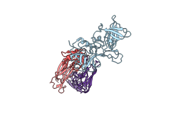
Cryo-Em Structure Of The Type Ii Amyloid-Beta 42 Fibril Containing A D-Asp At Positions 7 And 23
Organism: Homo sapiens
Method: ELECTRON MICROSCOPY Release Date: 2025-03-26 Classification: PROTEIN FIBRIL |
Organism: Homo sapiens
Method: ELECTRON MICROSCOPY
Release Date: 2025-03-26
 |
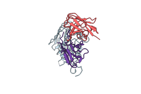
Vx22 Bound To Gii.4 P Domain
Organism: Homo sapiens, Norovirus hu/gii.4/sydney/nsw0514/2012/au
Method: X-RAY DIFFRACTION Resolution:3.35 Å Release Date: 2025-02-19 Classification: VIRAL PROTEIN/IMMUNE SYSTEM |
Organism: Homo sapiens, Norovirus hu/gii.4/sydney/nsw0514/2012/au
Method: X-RAY DIFFRACTION
Release Date: 2025-02-19
 |
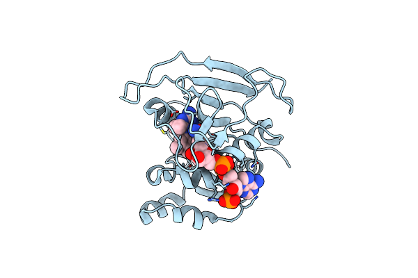
Structure Of Ut14 Fab In Complex With The Head Domain Of H3 (A/Singapore/Infimh-16-0019/2016)
Organism: Homo sapiens, Influenza a virus
Method: ELECTRON MICROSCOPY Release Date: 2024-11-27 Classification: IMMUNE SYSTEM |
Organism: Homo sapiens, Influenza a virus
Method: ELECTRON MICROSCOPY
Release Date: 2024-11-27
 |
Crystal Structure Of Trypanosoma Brucei Dhfr In Complex With The Cofactor And Inhibitor P25
Organism: Trypanosoma brucei brucei
Method: X-RAY DIFFRACTION Resolution:2.90 Å Release Date: 2024-11-20 Classification: OXIDOREDUCTASE Ligands: NDP, A1H0S |
Organism: Trypanosoma brucei brucei
Method: X-RAY DIFFRACTION
Release Date: 2024-11-20
Ligands: NDP, A1H0S
 |
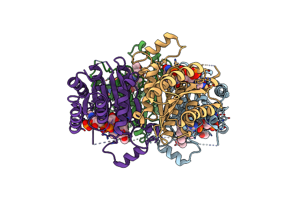
Crystal Structure Of Trypanosoma Brucei Ptr1 In Complex With The Cofactor And Inhibitor P25
Organism: Trypanosoma brucei brucei
Method: X-RAY DIFFRACTION Resolution:1.71 Å Release Date: 2024-11-20 Classification: OXIDOREDUCTASE Ligands: NDP, A1H0S, ACT |
Organism: Trypanosoma brucei brucei
Method: X-RAY DIFFRACTION
Release Date: 2024-11-20
Ligands: NDP, A1H0S, ACT
 |
Crystal Structure Of Trypanosoma Brucei Ptr1 In Complex With The Cofactor And Inhibitor P30
Organism: Trypanosoma brucei brucei
Method: X-RAY DIFFRACTION Resolution:1.70 Å Release Date: 2024-11-20 Classification: OXIDOREDUCTASE Ligands: NDP, ACT, A1H0U |
Organism: Trypanosoma brucei brucei
Method: X-RAY DIFFRACTION
Release Date: 2024-11-20
Ligands: NDP, ACT, A1H0U
 |
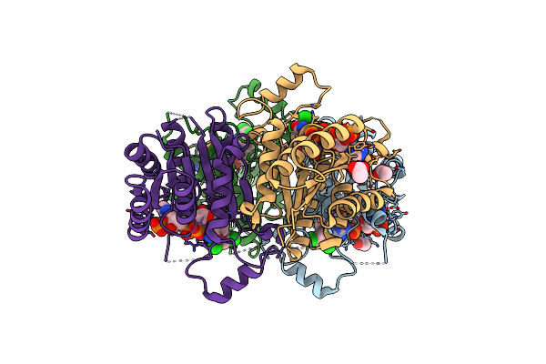
Crystal Structure Of Trypanosoma Brucei Ptr1 In Complex With The Cofactor And Inhibitor P31
Organism: Trypanosoma brucei brucei
Method: X-RAY DIFFRACTION Resolution:1.70 Å Release Date: 2024-11-20 Classification: OXIDOREDUCTASE Ligands: NDP, A1H0W, ACT |
Organism: Trypanosoma brucei brucei
Method: X-RAY DIFFRACTION
Release Date: 2024-11-20
Ligands: NDP, A1H0W, ACT
 |
Crystal Structure Of Trypanosoma Brucei Ptr1 In Complex With The Cofactor And Inhibitor P32
Organism: Trypanosoma brucei brucei
Method: X-RAY DIFFRACTION Resolution:1.48 Å Release Date: 2024-11-20 Classification: OXIDOREDUCTASE Ligands: NDP, A1H0T, ACT |
Organism: Trypanosoma brucei brucei
Method: X-RAY DIFFRACTION
Release Date: 2024-11-20
Ligands: NDP, A1H0T, ACT
 |
Crystal Structure Of Trypanosoma Brucei Ptr1 In Complex With The Cofactor And Inhibitor P34
Organism: Trypanosoma brucei brucei
Method: X-RAY DIFFRACTION Resolution:1.86 Å Release Date: 2024-11-20 Classification: OXIDOREDUCTASE Ligands: NDP, A1H0V |
Organism: Trypanosoma brucei brucei
Method: X-RAY DIFFRACTION
Release Date: 2024-11-20
Ligands: NDP, A1H0V