

Search Count: 27
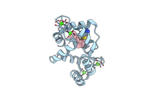 |
Calmodulin In Complex With Farnesyl Cysteine Methyl Ester
Organism: Homo sapiens
Method: X-RAY DIFFRACTION Resolution:2.05 Å Release Date: 2020-04-08 Classification: METAL BINDING PROTEIN Ligands: CA, 5U0 |
Organism: Homo sapiens
Method: X-RAY DIFFRACTION
Release Date: 2020-04-08
Ligands: CA, 5U0
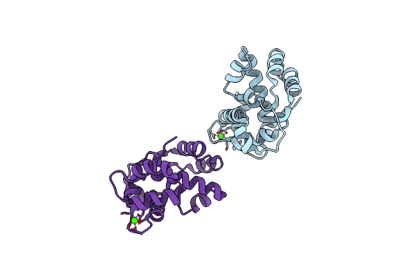 |
X-Ray Crystal Structure Of C. Elegans Stim Ef-Sam Domain
Organism: Caenorhabditis elegans
Method: X-RAY DIFFRACTION Resolution:1.89 Å Release Date: 2019-11-13 Classification: SIGNALING PROTEIN Ligands: CA |
Organism: Caenorhabditis elegans
Method: X-RAY DIFFRACTION
Release Date: 2019-11-13
Ligands: CA
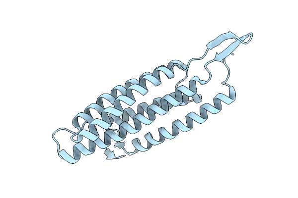 |
Crystal Structure Of The Alpha-N-Catenin Actin-Binding Domain H1 Mutant
Organism: Homo sapiens
Method: X-RAY DIFFRACTION Resolution:2.20 Å Release Date: 2018-12-19 Classification: CELL ADHESION |
Organism: Homo sapiens
Method: X-RAY DIFFRACTION
Release Date: 2018-12-19
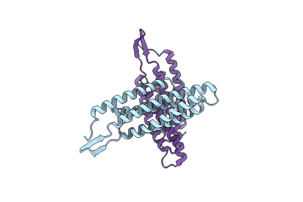 |
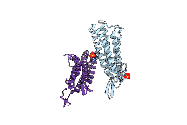
Crystal Structure Of The Alpha-N-Catenin Actin-Binding Domain H1 Mutant
Organism: Mus musculus
Method: X-RAY DIFFRACTION Resolution:2.81 Å Release Date: 2018-12-19 Classification: CELL ADHESION |
Organism: Mus musculus
Method: X-RAY DIFFRACTION
Release Date: 2018-12-19
 |
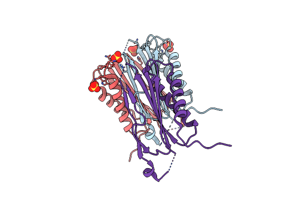
Crystal Structure Of The Alpha-E-Catenin Actin-Binding Domain
Organism: Mus musculus
Method: X-RAY DIFFRACTION Resolution:2.20 Å Release Date: 2018-12-19 Classification: CELL ADHESION Ligands: BR, SO4 |
Organism: Mus musculus
Method: X-RAY DIFFRACTION
Release Date: 2018-12-19
Ligands: BR, SO4
 |
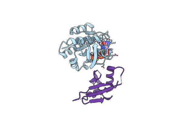
Crystal Structure Of Dynlt1/Tctex-1 In Complex With Arhgef2
Organism: Mus musculus
Method: X-RAY DIFFRACTION Resolution:2.00 Å Release Date: 2017-11-15 Classification: MOTOR PROTEIN Ligands: SO4 |
Organism: Mus musculus
Method: X-RAY DIFFRACTION
Release Date: 2017-11-15
Ligands: SO4
 |
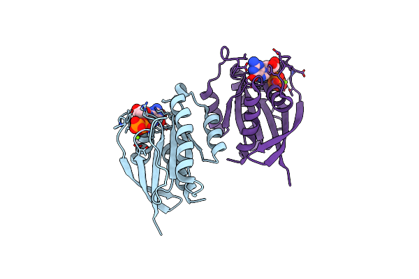
Crystal Structure Of The Afadin Ra1 Domain In Complex With Hras
Organism: Homo sapiens, Mus musculus
Method: X-RAY DIFFRACTION Resolution:2.50 Å Release Date: 2017-11-01 Classification: SIGNALING PROTEIN Ligands: GNP, MG |
Organism: Homo sapiens, Mus musculus
Method: X-RAY DIFFRACTION
Release Date: 2017-11-01
Ligands: GNP, MG
 |
Structure Of Wild Type Mus Musculus Rheb Bound To Gtp
Organism: Mus musculus
Method: X-RAY DIFFRACTION Resolution:2.20 Å Release Date: 2014-03-26 Classification: HYDROLASE Ligands: GTP, MG |
Organism: Mus musculus
Method: X-RAY DIFFRACTION
Release Date: 2014-03-26
Ligands: GTP, MG
 |
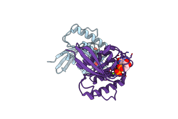
Structure Of Mus Musculus Rheb G63A Mutant Bound To Gtp
Organism: Mus musculus
Method: X-RAY DIFFRACTION Resolution:2.40 Å Release Date: 2014-03-26 Classification: HYDROLASE Ligands: GTP, MG, TRS, ACT |
Organism: Mus musculus
Method: X-RAY DIFFRACTION
Release Date: 2014-03-26
Ligands: GTP, MG, TRS, ACT
 |
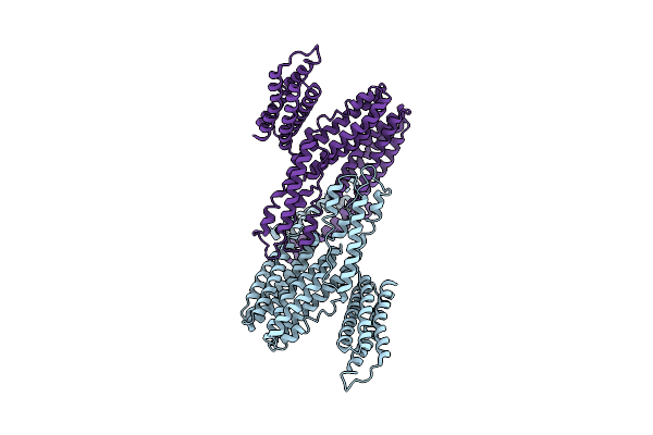
Structure Of Mus Musculus Rheb G63V Mutant Bound To Gdp
Organism: Mus musculus
Method: X-RAY DIFFRACTION Resolution:2.25 Å Release Date: 2014-03-26 Classification: HYDROLASE Ligands: GDP, MG |
Organism: Mus musculus
Method: X-RAY DIFFRACTION
Release Date: 2014-03-26
Ligands: GDP, MG
 |
Crystal Structure Of Full-Length Mouse Alphae-Catenin
Organism: Mus musculus
Method: X-RAY DIFFRACTION Resolution:6.50 Å Release Date: 2013-05-01 Classification: CELL ADHESION |
Organism: Mus musculus
Method: X-RAY DIFFRACTION
Release Date: 2013-05-01
 |
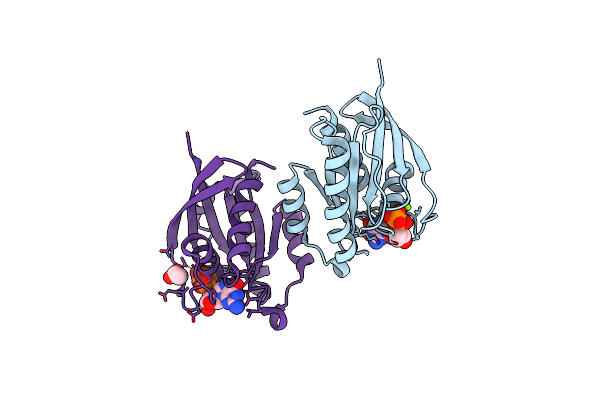
Crystal Structure Of The Alphan-Catenin Actin-Binding Domain
Organism: Mus musculus
Method: X-RAY DIFFRACTION Resolution:2.60 Å Release Date: 2013-05-01 Classification: CELL ADHESION Ligands: SO4 |
Organism: Mus musculus
Method: X-RAY DIFFRACTION
Release Date: 2013-05-01
Ligands: SO4
 |
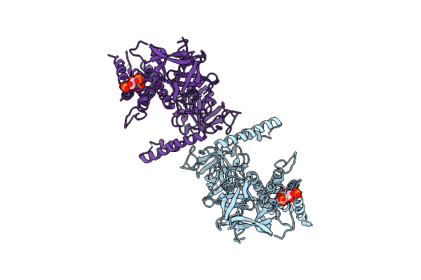
Structure Of Rheb-Y35A Mutant In Gdp- And Gmppnp-Bound Forms
Organism: Homo sapiens
Method: X-RAY DIFFRACTION Resolution:2.00 Å Release Date: 2012-06-20 Classification: HYDROLASE Ligands: MG, GDP, GNP, ACT |
Organism: Homo sapiens
Method: X-RAY DIFFRACTION
Release Date: 2012-06-20
Ligands: MG, GDP, GNP, ACT
 |
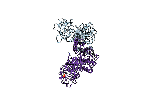
Crystal Structure Of The Inositol 1,4,5-Trisphosphate Receptor With Ligand Bound Form.
Organism: Rattus norvegicus
Method: X-RAY DIFFRACTION Resolution:3.60 Å Release Date: 2012-02-15 Classification: SIGNALING PROTEIN Ligands: I3P |
Organism: Rattus norvegicus
Method: X-RAY DIFFRACTION
Release Date: 2012-02-15
Ligands: I3P
 |
Crystal Structure Of The Apo-Inositol 1,4,5-Trisphosphate Receptor
Organism: Rattus norvegicus
Method: X-RAY DIFFRACTION Resolution:3.00 Å Release Date: 2012-02-15 Classification: SIGNALING PROTEIN Ligands: SO4 |
Organism: Rattus norvegicus
Method: X-RAY DIFFRACTION
Release Date: 2012-02-15
Ligands: SO4
 |
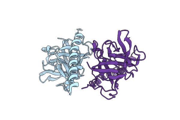
Crystal Structure Of The Ryr Domain Of The Rabbit Ryanodine Receptor
Organism: Oryctolagus cuniculus
Method: X-RAY DIFFRACTION Resolution:2.16 Å Release Date: 2011-06-22 Classification: METAL TRANSPORT |
Organism: Oryctolagus cuniculus
Method: X-RAY DIFFRACTION
Release Date: 2011-06-22
 |
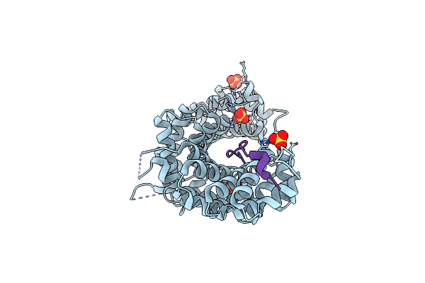
Crystal Structure Of The Ligand Binding Suppressor Domain Of Type 3 Inositol 1,4,5-Trisphosphate Receptor
Organism: Mus musculus
Method: X-RAY DIFFRACTION Resolution:1.90 Å Release Date: 2010-09-15 Classification: TRANSPORT PROTEIN |
Organism: Mus musculus
Method: X-RAY DIFFRACTION
Release Date: 2010-09-15
 |
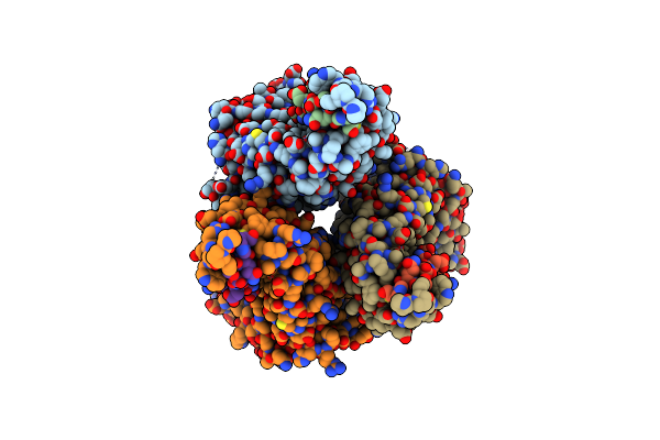
Crystal Structure Of P120 Catenin In Complex With E-Cadherin
Organism: Homo sapiens
Method: X-RAY DIFFRACTION Resolution:2.40 Å Release Date: 2010-04-21 Classification: CELL ADHESION Ligands: SO4 |
Organism: Homo sapiens
Method: X-RAY DIFFRACTION
Release Date: 2010-04-21
Ligands: SO4
 |
Crystal Structure Of P120 Catenin In Complex With E-Cadherin
Organism: Homo sapiens
Method: X-RAY DIFFRACTION Resolution:3.00 Å Release Date: 2010-04-21 Classification: CELL ADHESION |
Organism: Homo sapiens
Method: X-RAY DIFFRACTION
Release Date: 2010-04-21
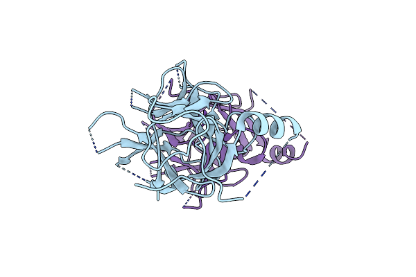 |
Crystal Structure Of Distal N-Terminal Beta-Trefoil Domain Of Ryanodine Receptor Type 1
Organism: Oryctolagus cuniculus
Method: X-RAY DIFFRACTION Resolution:2.50 Å Release Date: 2009-07-28 Classification: SIGNALING PROTEIN |
Organism: Oryctolagus cuniculus
Method: X-RAY DIFFRACTION
Release Date: 2009-07-28