

Search Count: 71
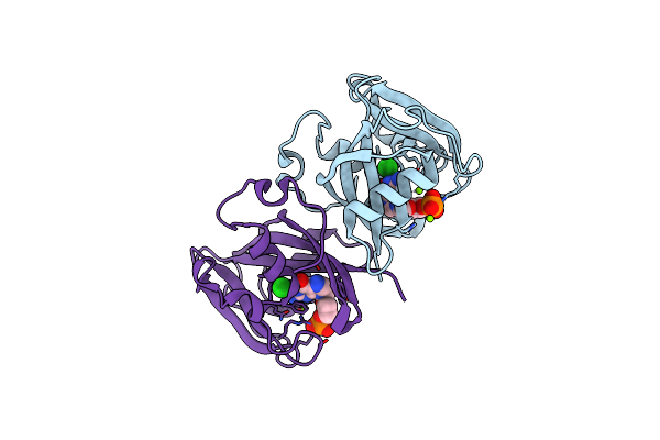 |
Structure Of Nudt15 In Complex With Acyclovir Monophosphate
Organism: Homo sapiens
Method: X-RAY DIFFRACTION Resolution:1.60 Å Release Date: 2021-05-19 Classification: HYDROLASE Ligands: 4DG, MG, CL |
Organism: Homo sapiens
Method: X-RAY DIFFRACTION
Release Date: 2021-05-19
Ligands: 4DG, MG, CL
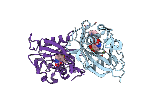 |
Structure Of Nudt15 In Complex With Th7755
Organism: Homo sapiens
Method: X-RAY DIFFRACTION Resolution:1.60 Å Release Date: 2021-03-24 Classification: HYDROLASE Ligands: SYW, MG |
Organism: Homo sapiens
Method: X-RAY DIFFRACTION
Release Date: 2021-03-24
Ligands: SYW, MG
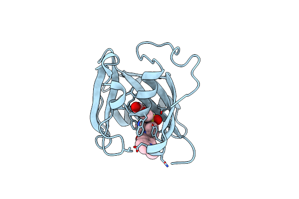 |
Structure Of Nudt15 V18I Mutant In Complex With Th7755
Organism: Homo sapiens
Method: X-RAY DIFFRACTION Resolution:1.50 Å Release Date: 2021-03-24 Classification: HYDROLASE Ligands: SYW |
Organism: Homo sapiens
Method: X-RAY DIFFRACTION
Release Date: 2021-03-24
Ligands: SYW
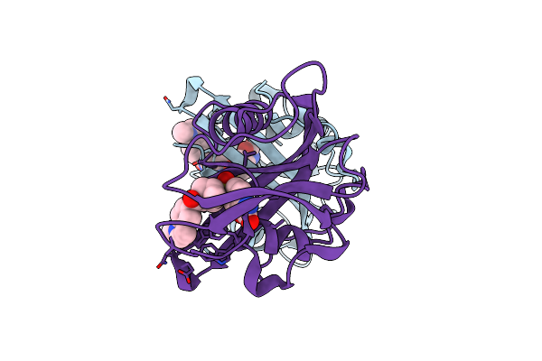 |
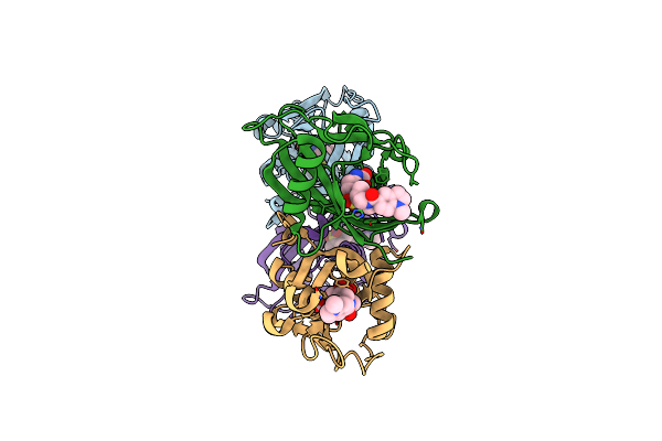
Structure Of Nudt15 R139C Mutant In Complex With Th7755
Organism: Homo sapiens
Method: X-RAY DIFFRACTION Resolution:1.60 Å Release Date: 2021-03-24 Classification: HYDROLASE Ligands: SYW |
Organism: Homo sapiens
Method: X-RAY DIFFRACTION
Release Date: 2021-03-24
Ligands: SYW
 |
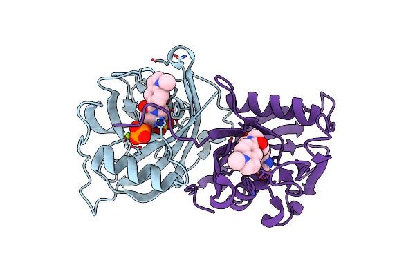
Structure Of Nudt15 R139H Mutant In Complex With Th7755
Organism: Homo sapiens
Method: X-RAY DIFFRACTION Resolution:1.60 Å Release Date: 2021-03-24 Classification: HYDROLASE Ligands: SYW |
Organism: Homo sapiens
Method: X-RAY DIFFRACTION
Release Date: 2021-03-24
Ligands: SYW
 |
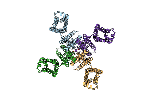
Structure Of Nudt15 V18_V19Insgv Mutant In Complex With Th7755
Organism: Homo sapiens
Method: X-RAY DIFFRACTION Resolution:1.45 Å Release Date: 2021-03-24 Classification: HYDROLASE Ligands: SYW, PO4, MG |
Organism: Homo sapiens
Method: X-RAY DIFFRACTION
Release Date: 2021-03-24
Ligands: SYW, PO4, MG
 |
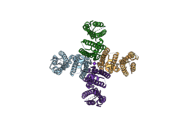
Cryo-Em Structure Of K+-Bound Herg Channel
Organism: Homo sapiens
Method: ELECTRON MICROSCOPY Release Date: 2021-01-20 Classification: TRANSPORT PROTEIN Ligands: K |
Organism: Homo sapiens
Method: ELECTRON MICROSCOPY
Release Date: 2021-01-20
Ligands: K
 |
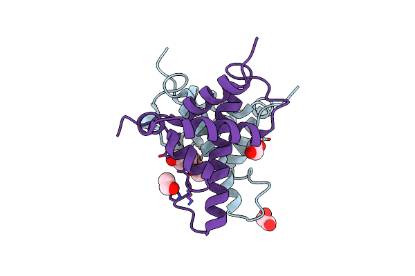
Cryo-Em Structure Of K+-Bound Herg Channel In The Presence Of Astemizole
Organism: Homo sapiens
Method: ELECTRON MICROSCOPY Release Date: 2021-01-20 Classification: TRANSPORT PROTEIN Ligands: K |
Organism: Homo sapiens
Method: ELECTRON MICROSCOPY
Release Date: 2021-01-20
Ligands: K
 |
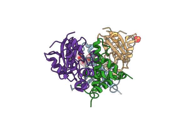
Crystal Structure Of The Type Vi Amidase Immunity (Tai4) From Agrobacterium Tumefaciens
Organism: Rhizobium radiobacter
Method: X-RAY DIFFRACTION Resolution:1.55 Å Release Date: 2018-12-19 Classification: ANTITOXIN Ligands: EDO |
Organism: Rhizobium radiobacter
Method: X-RAY DIFFRACTION
Release Date: 2018-12-19
Ligands: EDO
 |
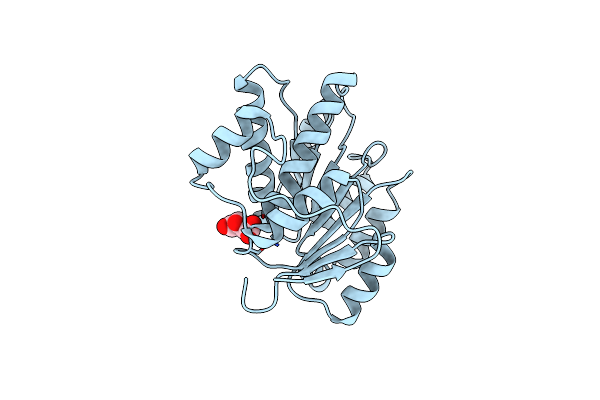
Crystal Structure Of The Type Vi Effector-Immunity Complex (Tae4-Tai4) From Agrobacterium Tumefaciens
Organism: Agrobacterium tumefaciens
Method: X-RAY DIFFRACTION Resolution:1.90 Å Release Date: 2018-12-19 Classification: ANTITOXIN Ligands: 1PE, SO4 |
Organism: Agrobacterium tumefaciens
Method: X-RAY DIFFRACTION
Release Date: 2018-12-19
Ligands: 1PE, SO4
 |
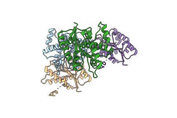
Crystal Structure Of The Ddx41 Dead Domain In An Apo Closed Form
Organism: Homo sapiens
Method: X-RAY DIFFRACTION Resolution:1.50 Å Release Date: 2016-10-19 Classification: HYDROLASE Ligands: LMR |
Organism: Homo sapiens
Method: X-RAY DIFFRACTION
Release Date: 2016-10-19
Ligands: LMR
 |
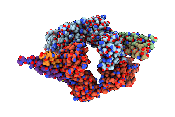
Crystal Structure Of The Ddx41 Dead Domain In An Apo Open Form
Organism: Homo sapiens
Method: X-RAY DIFFRACTION Resolution:2.20 Å Release Date: 2016-10-19 Classification: HYDROLASE |
Organism: Homo sapiens
Method: X-RAY DIFFRACTION
Release Date: 2016-10-19
 |
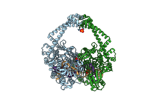
Crystal Structure Of Optineurin Uban In Complex With Linear Ubiquitin
Organism: Homo sapiens
Method: X-RAY DIFFRACTION Resolution:2.69 Å Release Date: 2016-09-07 Classification: SIGNALING PROTEIN |
Organism: Homo sapiens
Method: X-RAY DIFFRACTION
Release Date: 2016-09-07
 |
Crystal Structure Of S.A. Gyrase In Complex With Compound 7
Organism: Staphylococcus aureus, Staphylococcus aureus subsp. aureus ed98
Method: X-RAY DIFFRACTION Resolution:2.65 Å Release Date: 2015-06-17 Classification: Isomerase/DNA/RNA Ligands: MN, SO4, WCP |
Organism: Staphylococcus aureus, Staphylococcus aureus subsp. aureus ed98
Method: X-RAY DIFFRACTION
Release Date: 2015-06-17
Ligands: MN, SO4, WCP
 |
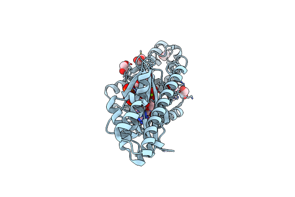
Crystal Structure Of Vibrio Cholerae Dncv Apo Form
Organism: Vibrio cholerae o1 biovar el tor str. n16961
Method: X-RAY DIFFRACTION Resolution:1.77 Å Release Date: 2015-04-29 Classification: TRANSFERASE Ligands: EDO |
Organism: Vibrio cholerae o1 biovar el tor str. n16961
Method: X-RAY DIFFRACTION
Release Date: 2015-04-29
Ligands: EDO
 |
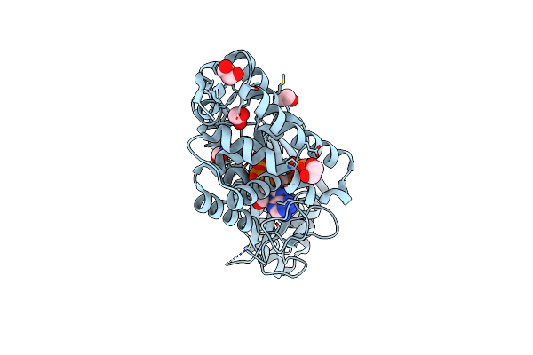
Crystal Structure Of Vibrio Cholerae Dncv Gtp Bound Form
Organism: Vibrio cholerae o1 biovar el tor str. n16961
Method: X-RAY DIFFRACTION Resolution:1.65 Å Release Date: 2015-04-29 Classification: TRANSFERASE Ligands: MG, EDO, GTP |
Organism: Vibrio cholerae o1 biovar el tor str. n16961
Method: X-RAY DIFFRACTION
Release Date: 2015-04-29
Ligands: MG, EDO, GTP
 |
Crystal Structure Of Vibrio Cholerae Dncv 3'-Deoxy Atp Bound Form
Organism: Vibrio cholerae o1 biovar el tor str. n16961
Method: X-RAY DIFFRACTION Resolution:1.60 Å Release Date: 2015-04-29 Classification: TRANSFERASE Ligands: EDO, 3AT, MG |
Organism: Vibrio cholerae o1 biovar el tor str. n16961
Method: X-RAY DIFFRACTION
Release Date: 2015-04-29
Ligands: EDO, 3AT, MG
 |
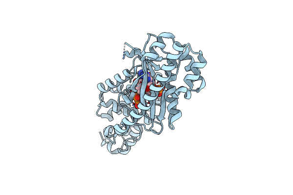
Crystal Structure Of Vibrio Cholerae Dncv 3'-Deoxy Gtp Bound Form
Organism: Vibrio cholerae o1 biovar el tor str. n16961
Method: X-RAY DIFFRACTION Resolution:1.55 Å Release Date: 2015-04-29 Classification: TRANSFERASE Ligands: MG, EDO, GH3, NA |
Organism: Vibrio cholerae o1 biovar el tor str. n16961
Method: X-RAY DIFFRACTION
Release Date: 2015-04-29
Ligands: MG, EDO, GH3, NA
 |
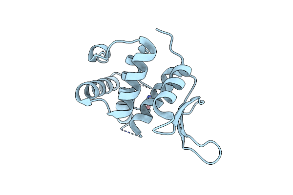
Crystal Structure Of Escherichia Coli Dncv 3'-Deoxy Gtp Bound Form
Organism: Escherichia coli
Method: X-RAY DIFFRACTION Resolution:2.31 Å Release Date: 2015-04-29 Classification: TRANSFERASE Ligands: MG, GH3 |
Organism: Escherichia coli
Method: X-RAY DIFFRACTION
Release Date: 2015-04-29
Ligands: MG, GH3
 |
Crystal Structure Of E. Coli Yfcm Bound To Co(Ii)
Organism: Escherichia coli
Method: X-RAY DIFFRACTION Resolution:1.96 Å Release Date: 2015-04-01 Classification: TRANSLATION Ligands: CO |
Organism: Escherichia coli
Method: X-RAY DIFFRACTION
Release Date: 2015-04-01
Ligands: CO