

Search Count: 34
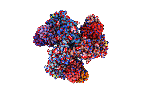 |
G002-293-0536 Fab In Complex With 001428_T278M_L14 Sosip And Rm20A3 Fab
Organism: Human immunodeficiency virus 1, Homo sapiens, Macaca mulatta
Method: ELECTRON MICROSCOPY Resolution:3.20 Å Release Date: 2025-05-28 Classification: VIRAL PROTEIN/IMMUNE SYSTEM Ligands: NAG |
Organism: Human immunodeficiency virus 1, Homo sapiens, Macaca mulatta
Method: ELECTRON MICROSCOPY
Release Date: 2025-05-28
Ligands: NAG
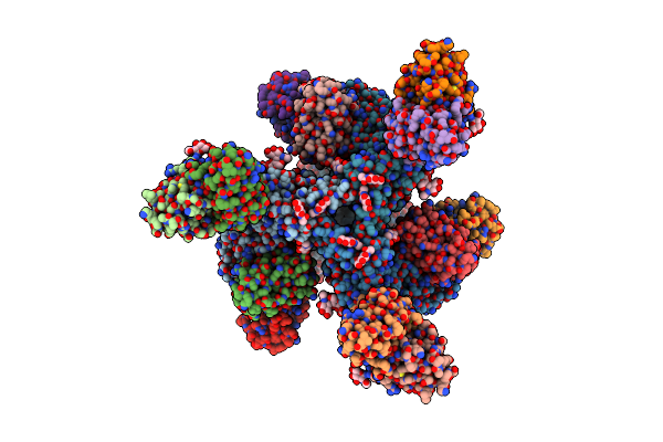 |
G002-480-0546 Fab In Complex With V703-0537_T278M_L14 Sosip And Bg18 Fab
Organism: Human immunodeficiency virus 1, Homo sapiens
Method: ELECTRON MICROSCOPY Resolution:3.40 Å Release Date: 2025-05-28 Classification: VIRAL PROTEIN/IMMUNE SYSTEM Ligands: NAG |
Organism: Human immunodeficiency virus 1, Homo sapiens
Method: ELECTRON MICROSCOPY
Release Date: 2025-05-28
Ligands: NAG
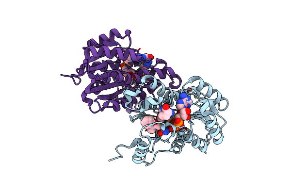 |
Crystal Structure Of E. Coli Fabi In Complex With Nad And (R,E)-3-(7-Amino-8-Oxo-6,7,8,9-Tetrahydro-5H-Pyrido[2,3-B]Azepin-3-Yl)-N-Methyl-N-((3-Methylbenzofuran-2-Yl)Methyl)Acrylamide
Organism: Escherichia coli str. k-12 substr. mg1655
Method: X-RAY DIFFRACTION Resolution:1.70 Å Release Date: 2023-02-15 Classification: ANTIBIOTIC Ligands: NAD, NQF |
Organism: Escherichia coli str. k-12 substr. mg1655
Method: X-RAY DIFFRACTION
Release Date: 2023-02-15
Ligands: NAD, NQF
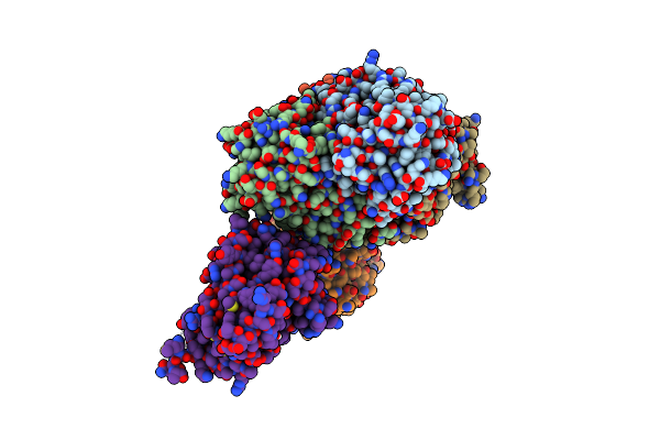 |
Crystal Structure Of Acinetobacter Baumannii Fabi In Complex With Nad And (R,E)-3-(7-Amino-8-Oxo-6,7,8,9-Tetrahydro-5H-Pyrido[2,3-B]Azepin-3-Yl)-N-Methyl-N-((3-Methylbenzofuran-2-Yl)Methyl)Acrylamide
Organism: Acinetobacter baumannii atcc 19606 = cip 70.34 = jcm 6841
Method: X-RAY DIFFRACTION Resolution:2.39 Å Release Date: 2023-02-15 Classification: ANTIBIOTIC Ligands: NQF, NAD |
Organism: Acinetobacter baumannii atcc 19606 = cip 70.34 = jcm 6841
Method: X-RAY DIFFRACTION
Release Date: 2023-02-15
Ligands: NQF, NAD
 |
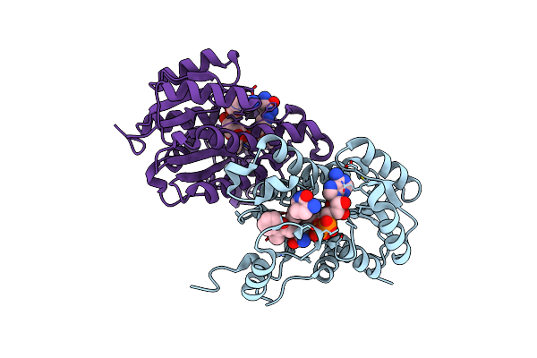
Crystal Structure Of Acinetobacter Baumannii Fabi In Complex With Nad And Fabimycin ((S,E)-3-(7-Amino-8-Oxo-6,7,8,9-Tetrahydro-5H-Pyrido[2,3-B]Azepin-3-Yl)-N-Methyl-N-((3-Methylbenzofuran-2-Yl)Methyl)Acrylamide)
Organism: Acinetobacter baumannii atcc 19606 = cip 70.34 = jcm 6841
Method: X-RAY DIFFRACTION Resolution:2.74 Å Release Date: 2023-02-15 Classification: ANTIBIOTIC Ligands: NAD, NUC |
Organism: Acinetobacter baumannii atcc 19606 = cip 70.34 = jcm 6841
Method: X-RAY DIFFRACTION
Release Date: 2023-02-15
Ligands: NAD, NUC
 |
Crystal Structure Of E. Coli Fabi In Complex With Nad And Fabimycin ((S,E)-3-(7-Amino-8-Oxo-6,7,8,9-Tetrahydro-5H-Pyrido[2,3-B]Azepin-3-Yl)-N-Methyl-N-((3-Methylbenzofuran-2-Yl)Methyl)Acrylamide)
Organism: Escherichia coli str. k-12 substr. mg1655
Method: X-RAY DIFFRACTION Resolution:1.54 Å Release Date: 2022-12-14 Classification: ANTIBIOTIC Ligands: NAD, NUC |
Organism: Escherichia coli str. k-12 substr. mg1655
Method: X-RAY DIFFRACTION
Release Date: 2022-12-14
Ligands: NAD, NUC
 |
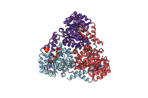
Crystal Structure Of Protein Cite From Mycobacterium Tuberculosis In Complex With Magnesium, Pyruvate And Citramalyl-Coa
Organism: Mycobacterium tuberculosis h37rv
Method: X-RAY DIFFRACTION Resolution:1.83 Å Release Date: 2018-08-01 Classification: LYASE Ligands: MG, CQM, PO4, GOL, CL, PYR |
Organism: Mycobacterium tuberculosis h37rv
Method: X-RAY DIFFRACTION
Release Date: 2018-08-01
Ligands: MG, CQM, PO4, GOL, CL, PYR
 |
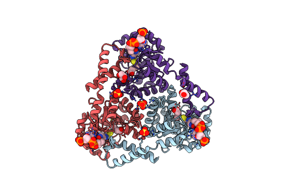
Crystal Structure Of Protein Cite From Mycobacterium Tuberculosis In Complex With Magnesium, Pyruvate And Coenzyme A
Organism: Mycobacterium tuberculosis h37rv
Method: X-RAY DIFFRACTION Resolution:1.72 Å Release Date: 2018-08-01 Classification: LYASE Ligands: MG, PYR, COA, PO4, GOL |
Organism: Mycobacterium tuberculosis h37rv
Method: X-RAY DIFFRACTION
Release Date: 2018-08-01
Ligands: MG, PYR, COA, PO4, GOL
 |
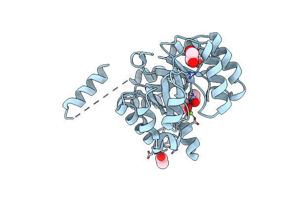
Crystal Structure Of Protein Cite From Mycobacterium Tuberculosis In Complex With Magnesium, Acetoacetate And Coenzyme A
Organism: Mycobacterium tuberculosis h37rv
Method: X-RAY DIFFRACTION Resolution:2.04 Å Release Date: 2018-08-01 Classification: LYASE Ligands: MG, COA, AAE, SO4, GOL |
Organism: Mycobacterium tuberculosis h37rv
Method: X-RAY DIFFRACTION
Release Date: 2018-08-01
Ligands: MG, COA, AAE, SO4, GOL
 |
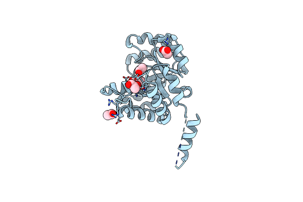
Crystal Structure Of Protein Cite From Mycobacterium Tuberculosis In Complex With Magnesium And Acetate
Organism: Mycobacterium tuberculosis h37rv
Method: X-RAY DIFFRACTION Resolution:1.61 Å Release Date: 2018-08-01 Classification: LYASE Ligands: MG, ACT, EDO |
Organism: Mycobacterium tuberculosis h37rv
Method: X-RAY DIFFRACTION
Release Date: 2018-08-01
Ligands: MG, ACT, EDO
 |
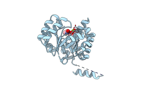
Crystal Structure Of Protein Cite From Mycobacterium Tuberculosis In Complex With Magnesium And Pyruvate
Organism: Mycobacterium tuberculosis h37rv
Method: X-RAY DIFFRACTION Resolution:1.73 Å Release Date: 2018-08-01 Classification: LYASE Ligands: MG, ACT, PYR |
Organism: Mycobacterium tuberculosis h37rv
Method: X-RAY DIFFRACTION
Release Date: 2018-08-01
Ligands: MG, ACT, PYR
 |
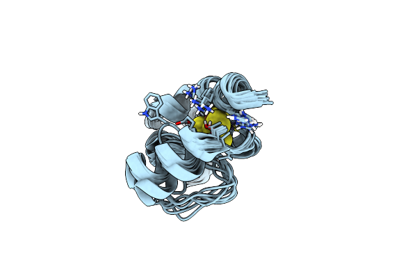
Crystal Structure Of Protein Cite From Mycobacterium Tuberculosis In Complex With Magnesium And Acetoacetate
Organism: Mycobacterium tuberculosis h37rv
Method: X-RAY DIFFRACTION Resolution:1.73 Å Release Date: 2018-08-01 Classification: LYASE Ligands: MG, ACT, AAE |
Organism: Mycobacterium tuberculosis h37rv
Method: X-RAY DIFFRACTION
Release Date: 2018-08-01
Ligands: MG, ACT, AAE
 |
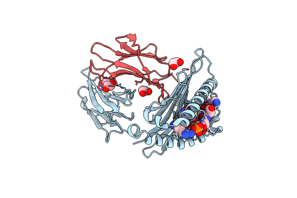
M. Tuberculosis [4Fe-4S] Protein Whib1 Is A Four-Helix Bundle That Forms A No-Sensitive Complex With Sigmaa And Regulates The Major Virulence Factor Esx-1
Organism: Mycobacterium tuberculosis (strain atcc 25618 / h37rv)
Method: SOLUTION NMR Release Date: 2018-01-03 Classification: SIGNALING PROTEIN Ligands: SF4 |
Organism: Mycobacterium tuberculosis (strain atcc 25618 / h37rv)
Method: SOLUTION NMR
Release Date: 2018-01-03
Ligands: SF4
 |
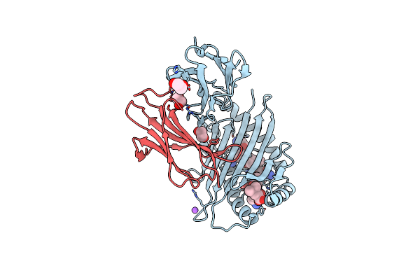
Crystal Structure Of Ampd2 Phosphopeptide Bound To Hla-A2
Organism: Homo sapiens
Method: X-RAY DIFFRACTION Resolution:1.70 Å Release Date: 2014-12-24 Classification: IMMUNE SYSTEM/ANTIGEN Ligands: EDO |
Organism: Homo sapiens
Method: X-RAY DIFFRACTION
Release Date: 2014-12-24
Ligands: EDO
 |
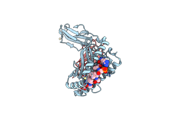
Crystal Structure Of Non-Phosphorylated Form Of Ampd2 Phosphopeptide Bound To Hla-A2
Organism: Homo sapiens
Method: X-RAY DIFFRACTION Resolution:2.10 Å Release Date: 2014-12-24 Classification: IMMUNE SYSTEM/ANTIGEN Ligands: EDO, NA |
Organism: Homo sapiens
Method: X-RAY DIFFRACTION
Release Date: 2014-12-24
Ligands: EDO, NA
 |
Crystal Structure Of Pkd2 Phosphopeptide Bound To Hla-A2
Organism: Homo sapiens
Method: X-RAY DIFFRACTION Resolution:2.10 Å Release Date: 2014-11-19 Classification: IMMUNE SYSTEM/ANTIGEN Ligands: CD, GOL |
Organism: Homo sapiens
Method: X-RAY DIFFRACTION
Release Date: 2014-11-19
Ligands: CD, GOL
 |
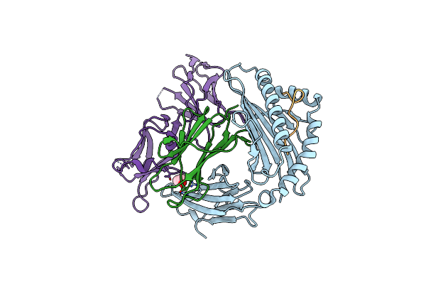
Crystal Structure Of Non-Phosphorylated Form Of Pkd2 Phosphopeptide Bound To Hla-A2
Organism: Homo sapiens
Method: X-RAY DIFFRACTION Resolution:1.90 Å Release Date: 2014-11-19 Classification: IMMUNE SYSTEM/ANTIGEN Ligands: CD, GOL |
Organism: Homo sapiens
Method: X-RAY DIFFRACTION
Release Date: 2014-11-19
Ligands: CD, GOL
 |
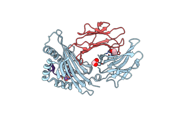
Crystal Structure Of Non-Phosphorylated Form Of Rqa_V Phosphopeptide Bound To Hla-A2 In Complex With Lilrb1
Organism: Homo sapiens
Method: X-RAY DIFFRACTION Resolution:2.70 Å Release Date: 2014-11-19 Classification: IMMUNE SYSTEM/ANTIGEN Ligands: EDO |
Organism: Homo sapiens
Method: X-RAY DIFFRACTION
Release Date: 2014-11-19
Ligands: EDO
 |
Crystal Structure Of Rqa_V Phosphopeptide Bound To Hla-A2
Organism: Homo sapiens
Method: X-RAY DIFFRACTION Resolution:2.00 Å Release Date: 2014-11-19 Classification: IMMUNE SYSTEM/ANTIGEN Ligands: EDO, NA |
Organism: Homo sapiens
Method: X-RAY DIFFRACTION
Release Date: 2014-11-19
Ligands: EDO, NA
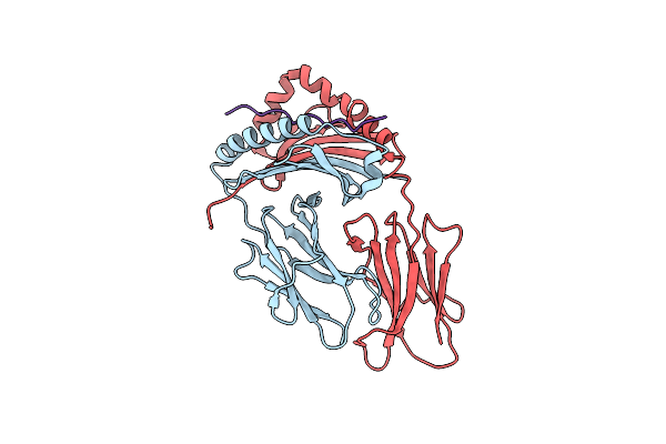 |
Crystal Structure Of Hla-Dr4 Bound To Gp100 Peptide
Organism: Homo sapiens
Method: X-RAY DIFFRACTION Resolution:2.50 Å Release Date: 2013-10-23 Classification: IMMUNE SYSTEM |
Organism: Homo sapiens
Method: X-RAY DIFFRACTION
Release Date: 2013-10-23