

Search Count: 25
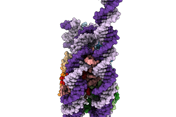 |
Structural Characterisation Of Chromatin Remodelling Intermediates Supports Linker Dna Dependent Product Inhibition As A Mechanism For Nucleosome Spacing.
Organism: Xenopus laevis, Synthetic construct
Method: ELECTRON MICROSCOPY Resolution:3.80 Å Release Date: 2026-02-04 Classification: GENE REGULATION |
Organism: Xenopus laevis, Synthetic construct
Method: ELECTRON MICROSCOPY
Release Date: 2026-02-04
 |
Structural Characterisation Of Chromatin Remodelling Intermediates Supports Linker Dna Dependent Product Inhibition As A Mechanism For Nucleosome Spacing.
Organism: Xenopus laevis, Synthetic construct
Method: ELECTRON MICROSCOPY Resolution:4.20 Å Release Date: 2026-01-28 Classification: GENE REGULATION |
Organism: Xenopus laevis, Synthetic construct
Method: ELECTRON MICROSCOPY
Release Date: 2026-01-28
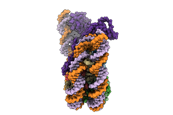 |
Structural Characterisation Of Chromatin Remodelling Intermediates Supports Linker Dna Dependent Product Inhibition As A Mechanism For Nucleosome Spacing.
Organism: Xenopus laevis, Saccharomyces cerevisiae, Synthetic construct
Method: ELECTRON MICROSCOPY Resolution:3.80 Å Release Date: 2026-01-28 Classification: GENE REGULATION |
Organism: Xenopus laevis, Saccharomyces cerevisiae, Synthetic construct
Method: ELECTRON MICROSCOPY
Release Date: 2026-01-28
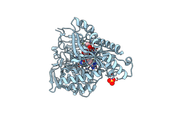 |
Crystal Structure Of The Peptide Binding Protein, Oppa, From Bacillus Subtilis In Complex With A Phre-Derived Pentapeptide
Organism: Bacillus subtilis subsp. subtilis str. 168
Method: X-RAY DIFFRACTION Resolution:1.90 Å Release Date: 2023-02-22 Classification: TRANSPORT PROTEIN Ligands: SO4 |
Organism: Bacillus subtilis subsp. subtilis str. 168
Method: X-RAY DIFFRACTION
Release Date: 2023-02-22
Ligands: SO4
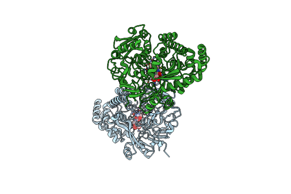 |
Crystal Structure Of The Peptide Binding Protein, Oppa, From Bacillus Subtilis In Complex With An Endogenous Tetrapeptide
Organism: Bacillus subtilis subsp. subtilis str. 168, Escherichia coli
Method: X-RAY DIFFRACTION Resolution:1.50 Å Release Date: 2023-02-22 Classification: TRANSPORT PROTEIN |
Organism: Bacillus subtilis subsp. subtilis str. 168, Escherichia coli
Method: X-RAY DIFFRACTION
Release Date: 2023-02-22
 |
Crystal Structure Of The Peptide Binding Protein Dppe From Bacillus Subtilis In Complex With Murein Tripeptide
Organism: Bacillus subtilis
Method: X-RAY DIFFRACTION Resolution:1.51 Å Release Date: 2023-02-22 Classification: TRANSPORT PROTEIN Ligands: MHI, MG, EDO |
Organism: Bacillus subtilis
Method: X-RAY DIFFRACTION
Release Date: 2023-02-22
Ligands: MHI, MG, EDO
 |
Crystal Structure Of The Peptide Binding Protein Dppe From Bacillus Subtilis In The Unliganded State
Organism: Bacillus subtilis subsp. subtilis str. 168
Method: X-RAY DIFFRACTION Resolution:1.40 Å Release Date: 2023-02-22 Classification: TRANSPORT PROTEIN |
Organism: Bacillus subtilis subsp. subtilis str. 168
Method: X-RAY DIFFRACTION
Release Date: 2023-02-22
 |
Crystal Structure Of Shiga Toxin 2K
Organism: Escherichia coli
Method: X-RAY DIFFRACTION Resolution:2.29 Å Release Date: 2020-07-01 Classification: TOXIN Ligands: EPE, EDO, ZN |
Organism: Escherichia coli
Method: X-RAY DIFFRACTION
Release Date: 2020-07-01
Ligands: EPE, EDO, ZN
 |
Crystal Structure Of A Putative Peptide Binding Protein Appa From Clostridium Difficile
Organism: Clostridioides difficile
Method: X-RAY DIFFRACTION Resolution:2.00 Å Release Date: 2019-04-10 Classification: PEPTIDE BINDING PROTEIN Ligands: IOD, SO4 |
Organism: Clostridioides difficile
Method: X-RAY DIFFRACTION
Release Date: 2019-04-10
Ligands: IOD, SO4
 |
Structure Of Two Molecules Of The Chromatin Remodelling Enzyme Chd1 Bound To A Nucleosome
Organism: Xenopus laevis, Saccharomyces cerevisiae s288c, Synthetic construct
Method: ELECTRON MICROSCOPY Release Date: 2018-08-22 Classification: MOTOR PROTEIN Ligands: ADP, BEF |
Organism: Xenopus laevis, Saccharomyces cerevisiae s288c, Synthetic construct
Method: ELECTRON MICROSCOPY
Release Date: 2018-08-22
Ligands: ADP, BEF
 |
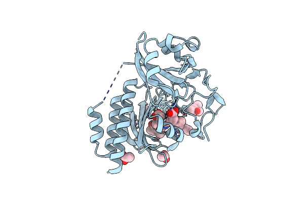
Structure Of The Chromatin Remodelling Enzyme Chd1 Bound To A Ubiquitinylated Nucleosome
Organism: Petromyzon marinus, Xenopus laevis, Xenopus tropicalis, Homo sapiens, Saccharomyces cerevisiae, Synthetic construct
Method: ELECTRON MICROSCOPY Resolution:4.50 Å Release Date: 2018-08-08 Classification: MOTOR PROTEIN Ligands: BEF, ADP |
Organism: Petromyzon marinus, Xenopus laevis, Xenopus tropicalis, Homo sapiens, Saccharomyces cerevisiae, Synthetic construct
Method: ELECTRON MICROSCOPY
Release Date: 2018-08-08
Ligands: BEF, ADP
 |
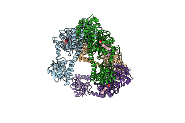
Deinococcus Radiodurans Bphp Pas-Gaf Y263F Mutant
Organism: Deinococcus radiodurans r1
Method: X-RAY DIFFRACTION Resolution:1.34 Å Release Date: 2018-04-18 Classification: TRANSFERASE Ligands: ACT, MPD, LBV |
Organism: Deinococcus radiodurans r1
Method: X-RAY DIFFRACTION
Release Date: 2018-04-18
Ligands: ACT, MPD, LBV
 |
Deinococcus Radiodurans Bphp Pas-Gaf-Phy Y263F Mutant, Pre-Illuminated
Organism: Deinococcus radiodurans (strain atcc 13939 / dsm 20539 / jcm 16871 / lmg 4051 / nbrc 15346 / ncimb 9279 / r1 / vkm b-1422)
Method: X-RAY DIFFRACTION Resolution:3.30 Å Release Date: 2018-04-18 Classification: TRANSFERASE Ligands: LBV |
 |
Deinococcus Radiodurans Bphp Pas-Gaf-Phy Y263F Mutant, Dark
Organism: Deinococcus radiodurans (strain atcc 13939 / dsm 20539 / jcm 16871 / lmg 4051 / nbrc 15346 / ncimb 9279 / r1 / vkm b-1422)
Method: X-RAY DIFFRACTION Resolution:3.60 Å Release Date: 2018-04-18 Classification: TRANSFERASE Ligands: LBV |
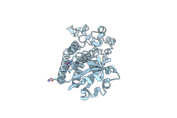 |
Ceue (Y288F Variant) A Periplasmic Protein From Campylobacter Jejuni.
Organism: Campylobacter jejuni
Method: X-RAY DIFFRACTION Resolution:1.47 Å Release Date: 2017-04-12 Classification: METAL TRANSPORT Ligands: ZN |
Organism: Campylobacter jejuni
Method: X-RAY DIFFRACTION
Release Date: 2017-04-12
Ligands: ZN
 |
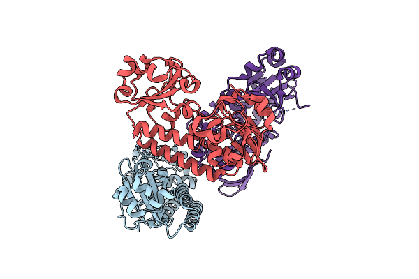
Ceue (H227L Variant) A Periplasmic Protein From Campylobacter Jejuni
Organism: Campylobacter jejuni bj-cjgb96299
Method: X-RAY DIFFRACTION Resolution:1.52 Å Release Date: 2017-04-12 Classification: METAL TRANSPORT Ligands: BR, NA |
Organism: Campylobacter jejuni bj-cjgb96299
Method: X-RAY DIFFRACTION
Release Date: 2017-04-12
Ligands: BR, NA
 |
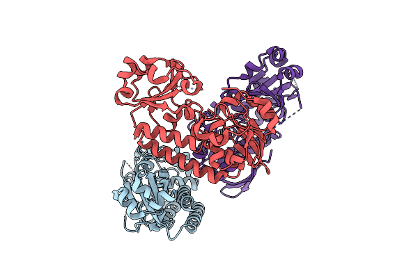
Ceue (H227A Variant) A Periplasmic Protein From Campylobacter Jejuni
Organism: Campylobacter jejuni
Method: X-RAY DIFFRACTION Resolution:1.33 Å Release Date: 2017-04-12 Classification: METAL TRANSPORT |
Organism: Campylobacter jejuni
Method: X-RAY DIFFRACTION
Release Date: 2017-04-12
 |
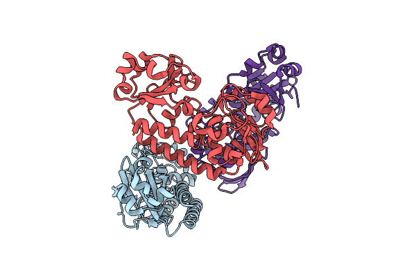
Ceue (H227L, Y288F Variant) A Periplasmic Protein From Campylobacter Jejuni
Organism: Campylobacter jejuni
Method: X-RAY DIFFRACTION Resolution:1.80 Å Release Date: 2017-04-12 Classification: METAL TRANSPORT |
Organism: Campylobacter jejuni
Method: X-RAY DIFFRACTION
Release Date: 2017-04-12
 |
Ceue (H227A, Y288F Variant) A Periplasmic Protein From Campylobacter Jejuni
Organism: Campylobacter jejuni
Method: X-RAY DIFFRACTION Resolution:1.81 Å Release Date: 2017-04-12 Classification: METAL TRANSPORT |
Organism: Campylobacter jejuni
Method: X-RAY DIFFRACTION
Release Date: 2017-04-12
 |
A Complex Of The Synthetic Siderophore Analogue Fe(Iii)-5-Licam With Ceue (H227L Variant), A Periplasmic Protein From Campylobacter Jejuni.
Organism: Campylobacter jejuni bj-cjgb96299
Method: X-RAY DIFFRACTION Resolution:1.90 Å Release Date: 2017-04-12 Classification: METAL TRANSPORT Ligands: FE, 5LC |
Organism: Campylobacter jejuni bj-cjgb96299
Method: X-RAY DIFFRACTION
Release Date: 2017-04-12
Ligands: FE, 5LC