
Search Count: 891
 |
Organism: Homo sapiens
Method: ELECTRON MICROSCOPY Resolution:2.30 Å Release Date: 2026-06-03 Classification: MEMBRANE PROTEIN Ligands: K |
 |
Organism: Homo sapiens
Method: ELECTRON MICROSCOPY Resolution:2.70 Å Release Date: 2026-06-03 Classification: MEMBRANE PROTEIN Ligands: K, A1E3Q |
 |
Organism: Homo sapiens
Method: ELECTRON MICROSCOPY Resolution:2.90 Å Release Date: 2026-06-03 Classification: MEMBRANE PROTEIN Ligands: K, A1E3Q |
 |
Organism: Homo sapiens
Method: ELECTRON MICROSCOPY Resolution:2.80 Å Release Date: 2026-06-03 Classification: MEMBRANE PROTEIN Ligands: K, A1E3Q |
 |
Organism: Homo sapiens
Method: ELECTRON MICROSCOPY Resolution:3.00 Å Release Date: 2026-06-03 Classification: MEMBRANE PROTEIN Ligands: K, A1E3Q |
 |
Organism: Homo sapiens
Method: ELECTRON MICROSCOPY Resolution:2.60 Å Release Date: 2026-06-03 Classification: MEMBRANE PROTEIN Ligands: K, A1EY8 |
 |
Organism: Homo sapiens
Method: ELECTRON MICROSCOPY Resolution:2.50 Å Release Date: 2026-06-03 Classification: MEMBRANE PROTEIN Ligands: K, A1EY8 |
 |
Organism: Homo sapiens
Method: ELECTRON MICROSCOPY Resolution:2.50 Å Release Date: 2026-06-03 Classification: MEMBRANE PROTEIN Ligands: K, A1EY8 |
 |
Organism: Homo sapiens
Method: ELECTRON MICROSCOPY Resolution:2.80 Å Release Date: 2026-06-03 Classification: MEMBRANE PROTEIN Ligands: A1EY8, K |
 |
Organism: Homo sapiens
Method: ELECTRON MICROSCOPY Resolution:2.60 Å Release Date: 2026-06-03 Classification: MEMBRANE PROTEIN Ligands: K, A1EY8 |
 |
Organism: Homo sapiens
Method: ELECTRON MICROSCOPY Resolution:2.40 Å Release Date: 2026-06-03 Classification: MEMBRANE PROTEIN Ligands: K |
 |
Organism: Homo sapiens
Method: ELECTRON MICROSCOPY Resolution:2.70 Å Release Date: 2026-06-03 Classification: MEMBRANE PROTEIN Ligands: K |
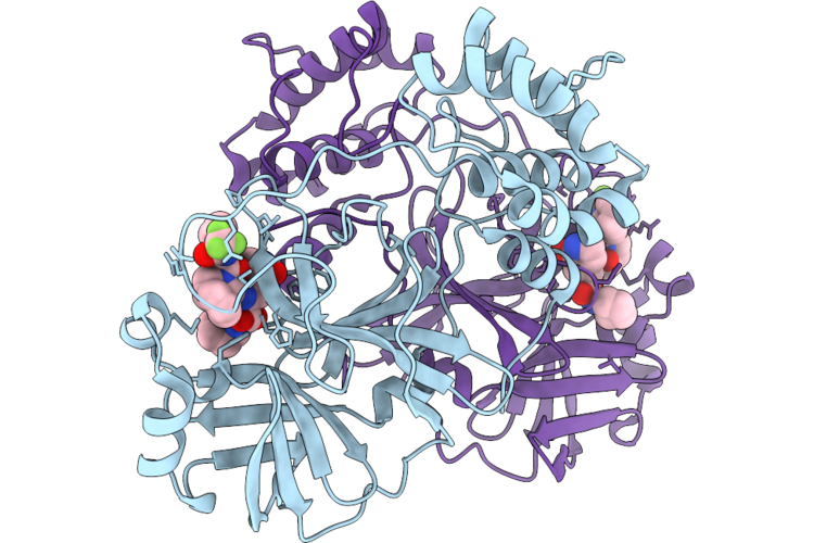 |
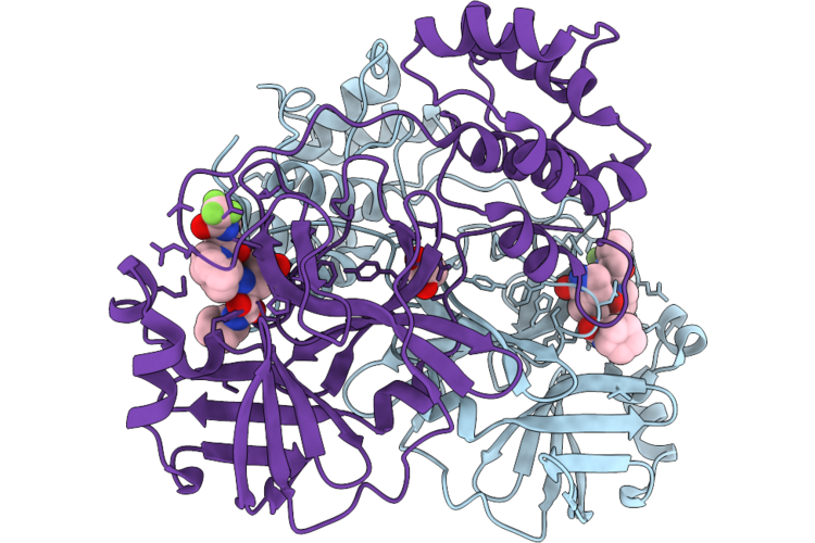
Crystal Structure Of Sars-Cov-2 Main Protease A173V Mutant In Complex With Leritrelvir
Organism: Severe acute respiratory syndrome coronavirus 2
Method: X-RAY DIFFRACTION Resolution:1.65 Å Release Date: 2026-06-03 Classification: VIRAL PROTEIN Ligands: 7ON |
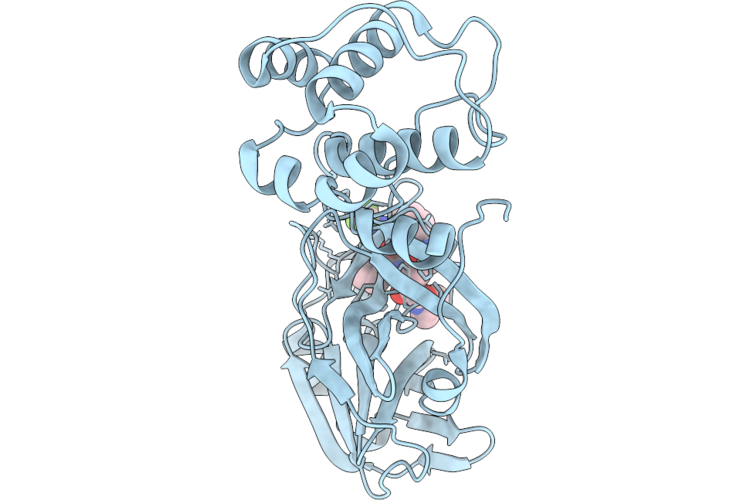 |
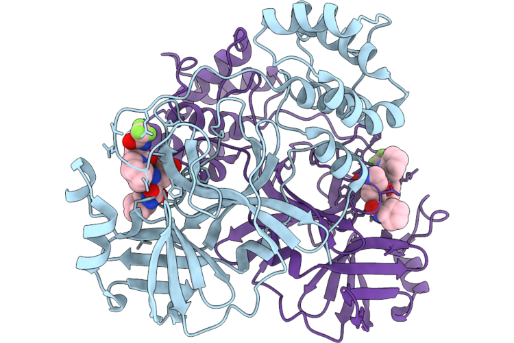
Crystal Structure Of Sars-Cov-2 Main Protease P168 Deletion Mutant In Complex With Leritrelvir
Organism: Severe acute respiratory syndrome coronavirus 2
Method: X-RAY DIFFRACTION Resolution:1.70 Å Release Date: 2026-06-03 Classification: VIRAL PROTEIN Ligands: 7ON |
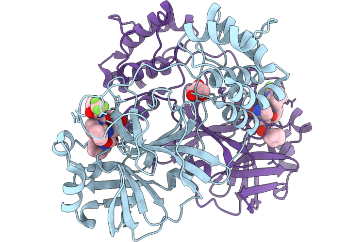 |
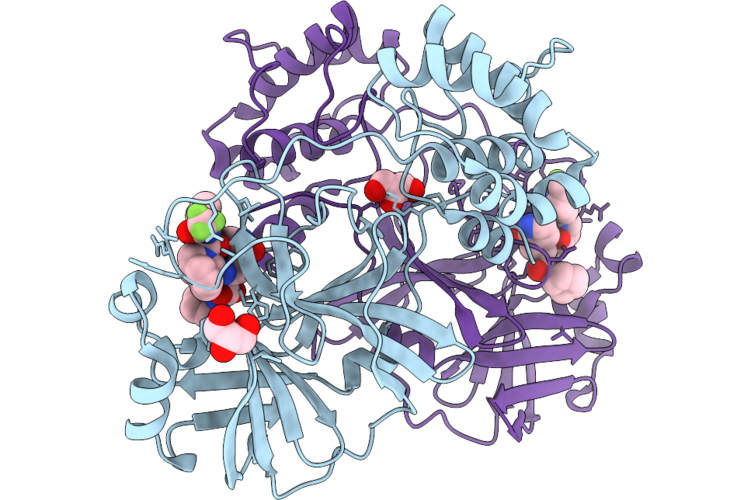
Crystal Structure Of Sars-Cov-2 Main Protease E166V Mutant In Complex With Leritrelvir
Organism: Severe acute respiratory syndrome coronavirus 2
Method: X-RAY DIFFRACTION Resolution:1.56 Å Release Date: 2026-06-03 Classification: VIRAL PROTEIN Ligands: 7ON, GOL |
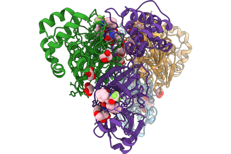 |
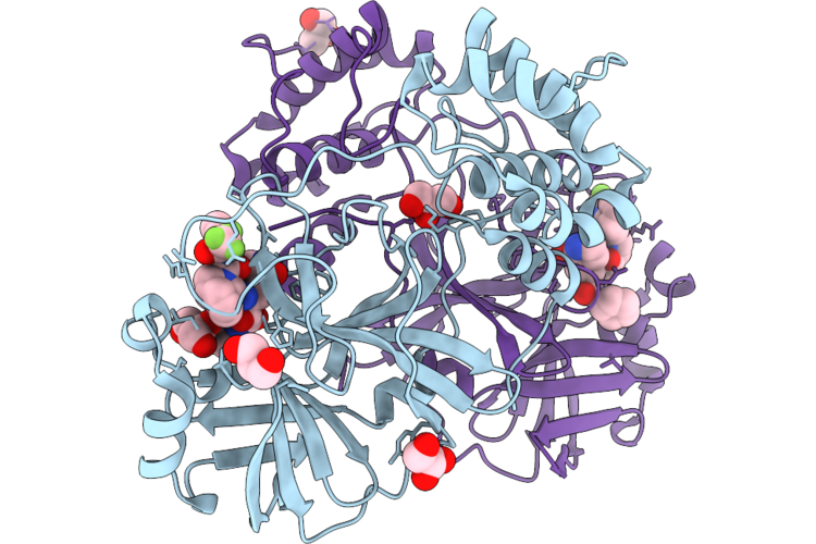
Crystal Structure Of Sars-Cov-2 Main Protease G143S Mutant In Complex With Leritrelvir
Organism: Severe acute respiratory syndrome coronavirus 2
Method: X-RAY DIFFRACTION Resolution:1.97 Å Release Date: 2026-06-03 Classification: VIRAL PROTEIN Ligands: GOL, 7ON |
 |
Crystal Structure Of Sars-Cov-2 Main Protease H172Y Mutant In Complex With Leritrelvir
Organism: Severe acute respiratory syndrome coronavirus 2
Method: X-RAY DIFFRACTION Resolution:2.27 Å Release Date: 2026-06-03 Classification: VIRAL PROTEIN Ligands: 7ON, GOL |
 |
Crystal Structure Of Sars-Cov-2 Main Protease L50F/E166V Mutant In Complex With Leritrelvir
Organism: Severe acute respiratory syndrome coronavirus 2
Method: X-RAY DIFFRACTION Resolution:2.50 Å Release Date: 2026-06-03 Classification: VIRAL PROTEIN Ligands: A1EN0 |
 |
Crystal Structure Of Sars-Cov-2 Main Protease M49I Mutant In Complex With Leritrelvir
Organism: Severe acute respiratory syndrome coronavirus 2
Method: X-RAY DIFFRACTION Resolution:1.87 Å Release Date: 2026-06-03 Classification: VIRAL PROTEIN Ligands: 7ON, GOL |
 |
Crystal Structure Of Sars-Cov-2 Main Protease M49I/M165I Mutant In Complex With Leritrelvir
Organism: Severe acute respiratory syndrome coronavirus 2
Method: X-RAY DIFFRACTION Resolution:1.80 Å Release Date: 2026-06-03 Classification: VIRAL PROTEIN Ligands: A1EN0, GOL |