

Search Count: 29
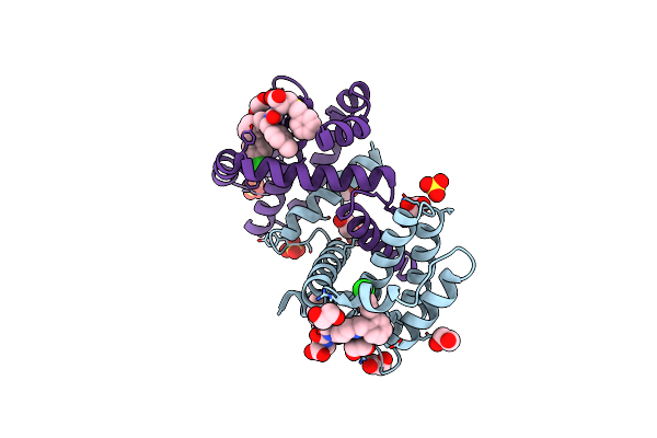 |
Crystal Structure Of Bcl-Xl Bound To Compound 8: (R)-3-(Benzylthio)-2-(3-(2-((4'-Chloro-[1,1'-Biphenyl]-2-Yl)Methyl)-1,2,3,4-Tetrahydroisoquinoline-6-Carbonyl)-3-(4-Methylbenzyl)Ureido)Propanoic Acid
Organism: Homo sapiens
Method: X-RAY DIFFRACTION Resolution:1.90 Å Release Date: 2021-05-05 Classification: Apoptosis/INHIBITOR Ligands: XOY, SO4, EDO |
Organism: Homo sapiens
Method: X-RAY DIFFRACTION
Release Date: 2021-05-05
Ligands: XOY, SO4, EDO
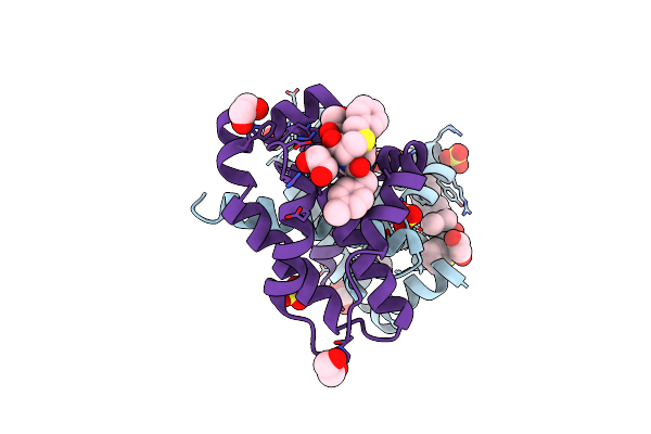 |
Crystal Structure Of Bcl-Xl Bound To Compound 2: (2R)-3-(Benzylsulfanyl)-2-({[(4-Methylphenyl)Methyl] [(4 Phenylphenyl)Carbonyl] Carbamoyl}Amino) Propanoic Acid
Organism: Homo sapiens
Method: X-RAY DIFFRACTION Resolution:2.15 Å Release Date: 2021-05-05 Classification: Apoptosis/INHIBITOR Ligands: XOU, SO4, EDO |
Organism: Homo sapiens
Method: X-RAY DIFFRACTION
Release Date: 2021-05-05
Ligands: XOU, SO4, EDO
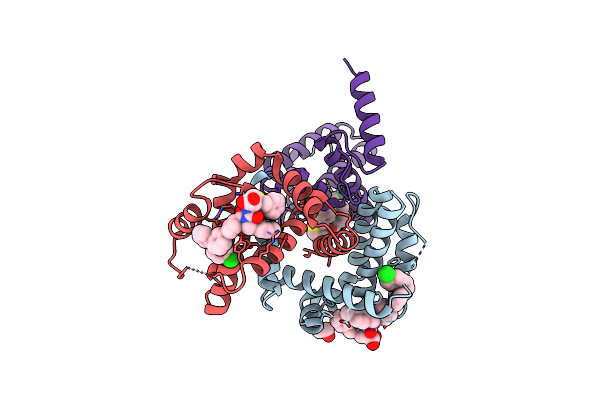 |
Crystal Structure Of Bcl-Xl Bound To Compound 7: (R)-3-(Benzylthio)-2-(3-(4-Chloro-[1,1':2',1'':3'',1'''-Quaterphenyl]-4'''-Carbonyl)-3-(4-Methylbenzyl)Ureido)Propanoic Acid
Organism: Homo sapiens
Method: X-RAY DIFFRACTION Resolution:2.87 Å Release Date: 2021-05-05 Classification: Apoptosis/INHIBITOR Ligands: QHV, EDO |
Organism: Homo sapiens
Method: X-RAY DIFFRACTION
Release Date: 2021-05-05
Ligands: QHV, EDO
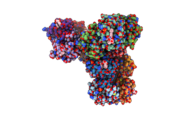 |
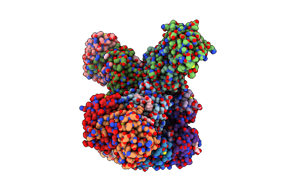
Crystal Structure Of Bcl-Xl Bound To Compound 12: (R)-2-(3-([1,1'-Biphenyl]-4-Carbonyl)-3-(4-Methylbenzyl)Ureido)-3-((Cyclohexylmethyl)Sulfonyl)Propanoic Acid
Organism: Homo sapiens
Method: X-RAY DIFFRACTION Resolution:2.24 Å Release Date: 2021-05-05 Classification: Apoptosis/INHIBITOR Ligands: SO4, EDO, QHS |
Organism: Homo sapiens
Method: X-RAY DIFFRACTION
Release Date: 2021-05-05
Ligands: SO4, EDO, QHS
 |
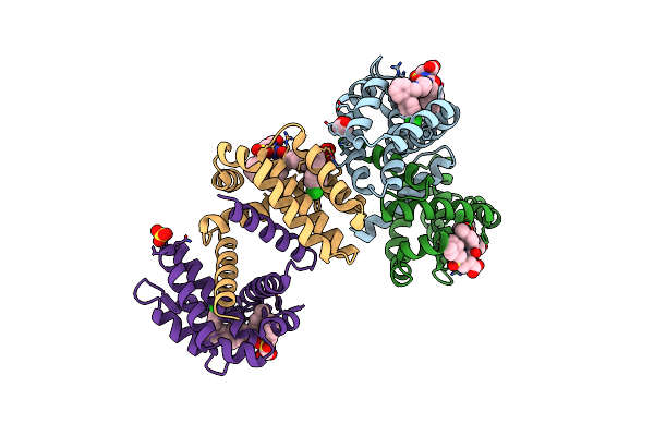
Crystal Structure Of Bcl-Xl Bound To Compound 13: (R)-2-(3-([1,1'-Biphenyl]-4-Carbonyl)-3-(4-Methylbenzyl)Ureido)-3-(((3R,5R,7R)-Adamantan-1-Ylmethyl)Sulfonyl)Propanoic Acid
Organism: Homo sapiens
Method: X-RAY DIFFRACTION Resolution:2.10 Å Release Date: 2021-05-05 Classification: Apoptosis/INHIBITOR Ligands: QHP, SO4, EDO |
Organism: Homo sapiens
Method: X-RAY DIFFRACTION
Release Date: 2021-05-05
Ligands: QHP, SO4, EDO
 |
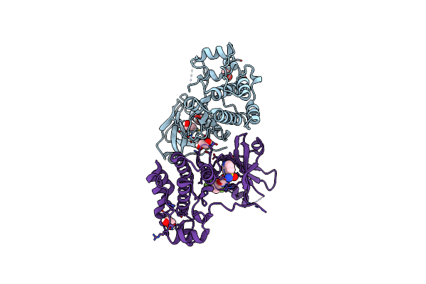
Crystal Structure Of Bcl-Xl Bound To Compound 15: (R)-2-(3-(2-((4'-Chloro-[1,1'-Biphenyl]-2-Yl)Methyl)-1,2,3,4-Tetrahydroisoquinoline-6-Carbonyl)-3-(4-Methylbenzyl)Ureido)-3-((Cyclohexylmethyl)Sulfonyl)Propanoic Acid
Organism: Homo sapiens
Method: X-RAY DIFFRACTION Resolution:2.19 Å Release Date: 2021-05-05 Classification: Apoptosis/INHIBITOR Ligands: EDO, SO4, QHJ |
Organism: Homo sapiens
Method: X-RAY DIFFRACTION
Release Date: 2021-05-05
Ligands: EDO, SO4, QHJ
 |
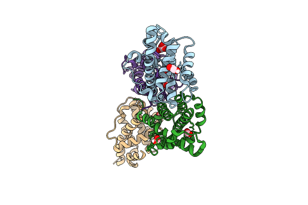
Crystal Structure Of The Human Mlkl Pseudokinase Domain Bound To Compound 2
Organism: Homo sapiens
Method: X-RAY DIFFRACTION Resolution:2.29 Å Release Date: 2020-09-16 Classification: transferase/transferase inhibitor Ligands: EDO, LN4 |
Organism: Homo sapiens
Method: X-RAY DIFFRACTION
Release Date: 2020-09-16
Ligands: EDO, LN4
 |
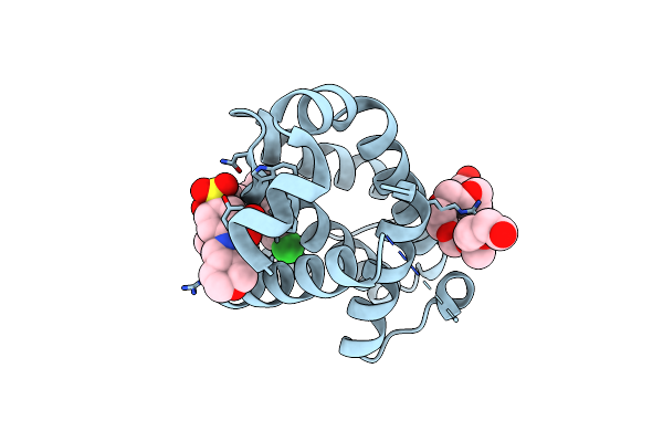
Crystal Structure Of Mouse Bak
Organism: Mus musculus
Method: X-RAY DIFFRACTION Resolution:1.75 Å Release Date: 2019-09-11 Classification: APOPTOSIS Ligands: FMT, EDO |
Organism: Mus musculus
Method: X-RAY DIFFRACTION
Release Date: 2019-09-11
Ligands: FMT, EDO
 |
Crystal Structure Of Bcl-2 With Venetoclax
Organism: Homo sapiens
Method: X-RAY DIFFRACTION Resolution:1.62 Å Release Date: 2019-05-22 Classification: APOPTOSIS Ligands: LBM, 2PE |
Organism: Homo sapiens
Method: X-RAY DIFFRACTION
Release Date: 2019-05-22
Ligands: LBM, 2PE
 |
Crystal Structure Of Bcl-2 G101V Mutation With Venetoclax
Organism: Homo sapiens
Method: X-RAY DIFFRACTION Resolution:2.20 Å Release Date: 2019-05-22 Classification: APOPTOSIS Ligands: LBM, PEG, CL |
Organism: Homo sapiens
Method: X-RAY DIFFRACTION
Release Date: 2019-05-22
Ligands: LBM, PEG, CL
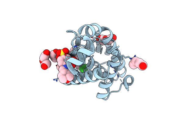 |
Crystal Structure Of Bcl-2 F104L Mutation With Venetoclax
Organism: Homo sapiens
Method: X-RAY DIFFRACTION Resolution:1.75 Å Release Date: 2019-05-22 Classification: APOPTOSIS Ligands: LBM, PEG |
Organism: Homo sapiens
Method: X-RAY DIFFRACTION
Release Date: 2019-05-22
Ligands: LBM, PEG
 |
Crystal Structure Of Bcl-2 G101V Mutation With S55746
Organism: Homo sapiens
Method: X-RAY DIFFRACTION Resolution:2.00 Å Release Date: 2019-05-22 Classification: APOPTOSIS Ligands: F3Q |
Organism: Homo sapiens
Method: X-RAY DIFFRACTION
Release Date: 2019-05-22
Ligands: F3Q
 |
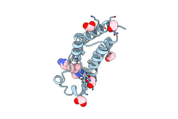
Crystal Structure Of Bcl-2 G101A Mutation With Venetoclax
Organism: Homo sapiens
Method: X-RAY DIFFRACTION Resolution:1.80 Å Release Date: 2019-05-22 Classification: APOPTOSIS Ligands: LBM, PEG |
Organism: Homo sapiens
Method: X-RAY DIFFRACTION
Release Date: 2019-05-22
Ligands: LBM, PEG
 |
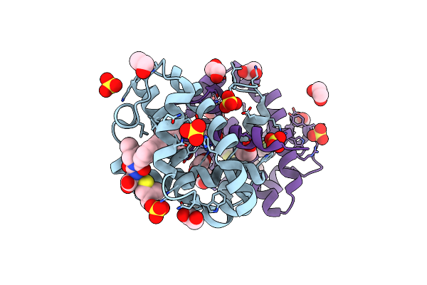
X-Ray Crystal Structure Of 1,2,3-Triazolobenzodiazepine In Complex With Brd2(D2)
Organism: Homo sapiens
Method: X-RAY DIFFRACTION Resolution:1.78 Å Release Date: 2017-12-13 Classification: transcription/transcription inhibitor Ligands: 7WY, EDO |
Organism: Homo sapiens
Method: X-RAY DIFFRACTION
Release Date: 2017-12-13
Ligands: 7WY, EDO
 |
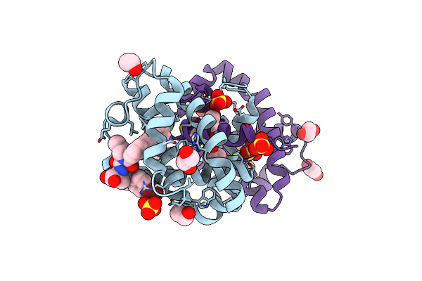
Crystal Structure Of Bcl-Xl In Complex With Benzoylurea Compound (39B)
Organism: Homo sapiens
Method: X-RAY DIFFRACTION Resolution:2.05 Å Release Date: 2014-02-05 Classification: APOPTOSIS Ligands: X0D, SO4, EDO |
Organism: Homo sapiens
Method: X-RAY DIFFRACTION
Release Date: 2014-02-05
Ligands: X0D, SO4, EDO
 |
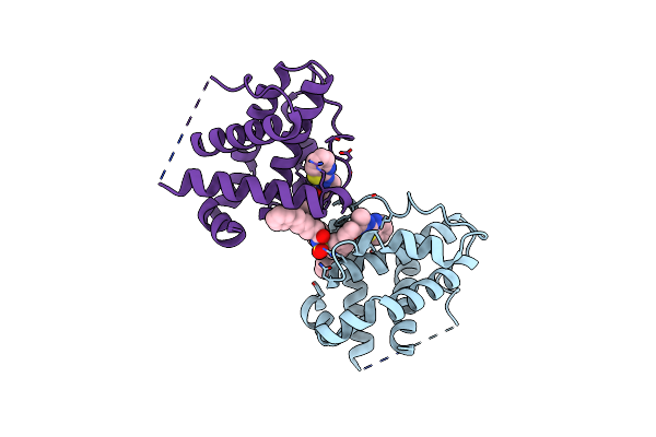
Crystal Structure Of Bcl-Xl In Complex With Benzoylurea Compound (42)
Organism: Homo sapiens
Method: X-RAY DIFFRACTION Resolution:2.30 Å Release Date: 2014-02-05 Classification: APOPTOSIS Ligands: X0R, SO4, EDO |
Organism: Homo sapiens
Method: X-RAY DIFFRACTION
Release Date: 2014-02-05
Ligands: X0R, SO4, EDO
 |
Crystal Structure Of Bcl-Xl In Complex With Inhibitor (Compound 2).
Organism: Homo sapiens
Method: X-RAY DIFFRACTION Resolution:2.48 Å Release Date: 2013-04-24 Classification: APOPTOSIS Ligands: H1I |
Organism: Homo sapiens
Method: X-RAY DIFFRACTION
Release Date: 2013-04-24
Ligands: H1I
 |
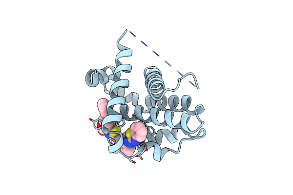
Crystal Structure Of Bcl-Xl In Complex With Inhibitor (Compound 3)
Organism: Homo sapiens
Method: X-RAY DIFFRACTION Resolution:2.29 Å Release Date: 2013-04-24 Classification: APOPTOSIS Ligands: H0Y, SO4, EDO |
Organism: Homo sapiens
Method: X-RAY DIFFRACTION
Release Date: 2013-04-24
Ligands: H0Y, SO4, EDO
 |
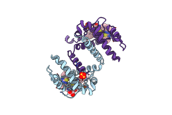
Crystal Structure Of Bcl-Xl In Complex With Inhibitor (Compound 6)
Organism: Homo sapiens
Method: X-RAY DIFFRACTION Resolution:2.60 Å Release Date: 2013-04-24 Classification: APOPTOSIS Ligands: X8U |
Organism: Homo sapiens
Method: X-RAY DIFFRACTION
Release Date: 2013-04-24
Ligands: X8U
 |
Crystal Structure Of Bcl-Xl In Complex With Inhibitor (Wehi-539)
Organism: Homo sapiens
Method: X-RAY DIFFRACTION Resolution:2.03 Å Release Date: 2013-04-24 Classification: APOPTOSIS Ligands: X0B, EDO, SO4 |
Organism: Homo sapiens
Method: X-RAY DIFFRACTION
Release Date: 2013-04-24
Ligands: X0B, EDO, SO4