
Search Count: 20
All
Selected
 |
Organism: Synthetic construct
Method: X-RAY DIFFRACTION Resolution:1.99 Å Release Date: 2026-04-29 Classification: DE NOVO PROTEIN |
 |
Organism: Synthetic construct
Method: X-RAY DIFFRACTION Resolution:1.66 Å Release Date: 2026-04-29 Classification: DE NOVO PROTEIN Ligands: A1DD0 |
 |
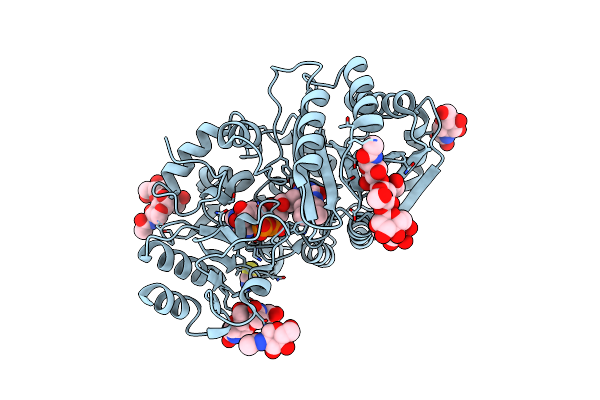
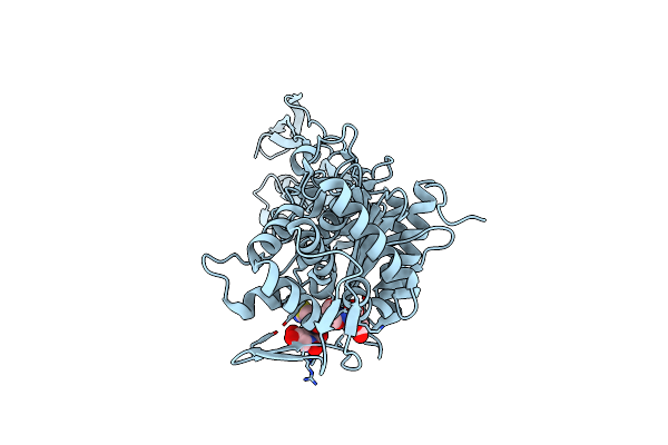
Organism: Penicillium rubens
Method: X-RAY DIFFRACTION Resolution:1.38 Å Release Date: 2025-03-19 Classification: OXIDOREDUCTASE Ligands: FAD, PEG |
 |
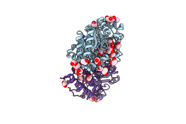
Organism: Penicillium rubens wisconsin 54-1255
Method: X-RAY DIFFRACTION Resolution:3.00 Å Release Date: 2025-03-19 Classification: OXIDOREDUCTASE Ligands: FAD, NAG, A1ITD |
 |
Organism: Penicillium rubens wisconsin 54-1255
Method: X-RAY DIFFRACTION Resolution:1.71 Å Release Date: 2025-03-19 Classification: OXIDOREDUCTASE Ligands: FAD, A1ITD |
 |
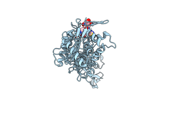
Organism: Penicillium rubens wisconsin 54-1255
Method: X-RAY DIFFRACTION Resolution:1.55 Å Release Date: 2025-03-19 Classification: OXIDOREDUCTASE Ligands: FAD, NAG, SCN |
 |
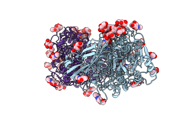
Organism: Penicillium expansum
Method: X-RAY DIFFRACTION Resolution:2.40 Å Release Date: 2023-09-06 Classification: FLAVOPROTEIN Ligands: FAD, NAG, PEG, GOL |
 |
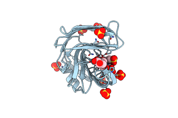
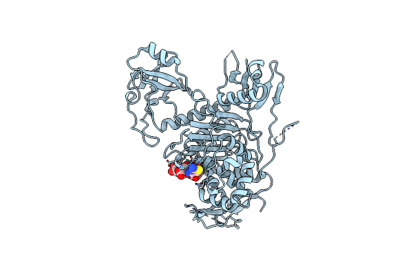
Organism: Emericella nidulans
Method: X-RAY DIFFRACTION Resolution:1.48 Å Release Date: 2019-06-05 Classification: HYDROLASE Ligands: NAG, EDO, ACT, GOL, K |
 |
Gh3 Exo-Beta-Xylosidase (Xlnd) In Complex With Xylobiose Aziridine Activity Based Probe
Organism: Emericella nidulans (strain fgsc a4 / atcc 38163 / cbs 112.46 / nrrl 194 / m139)
Method: X-RAY DIFFRACTION Resolution:2.14 Å Release Date: 2019-06-05 Classification: HYDROLASE Ligands: NAG, HLB, EDO |
 |
Organism: Aspergillus aculeatus atcc 16872
Method: X-RAY DIFFRACTION Resolution:1.42 Å Release Date: 2019-06-05 Classification: HYDROLASE Ligands: NAG, EDO, K |
 |
Organism: Aspergillus aculeatus atcc 16872
Method: X-RAY DIFFRACTION Resolution:1.76 Å Release Date: 2019-06-05 Classification: HYDROLASE Ligands: NAG, EDO, K, HQ8, XYP |
 |
Crystal Structure Of Aspergillus Niger Gh11 Endoxylanase Xyna In Complex With Xylobiose Epoxide Activity Based Probe
Organism: Aspergillus niger
Method: X-RAY DIFFRACTION Resolution:1.79 Å Release Date: 2019-06-05 Classification: HYDROLASE Ligands: MES, SO4, HZB |
 |
Crystal Structure Of Pbp3 In Complex With Compound 14 ((2E)-2-({[(2S)-2-{[(2Z)-2-(2-Amino-1,3-Thiazol-4-Yl)-2-{[(1,5-Dihydroxy-4-Oxo-1,4-Dihydropyridin-2-Yl)Methoxy]Imino}Acetyl]Amino}-3-Oxopropyl]Oxy}Imino)Pentanedioic Acid)
Organism: Pseudomonas aeruginosa pa1r
Method: X-RAY DIFFRACTION Resolution:2.30 Å Release Date: 2014-05-07 Classification: transferase/transferase inhibitor Ligands: 2U2 |
 |
Crystal Structure Of Pbp3 In Complex With Bal30072 ((2Z)-2-(2-Amino-1,3-Thiazol-4-Yl)-2-{[(1,5-Dihydroxy-4-Oxo-1,4-Dihydropyridin-2-Yl)Methoxy]Imino}-N-{(2S)-1-Hydroxy-3-Methyl-3-[(Sulfooxy)Amino]Butan-2-Yl}Ethanamide)
Organism: Pseudomonas aeruginosa pa1r
Method: X-RAY DIFFRACTION Resolution:2.00 Å Release Date: 2014-05-07 Classification: transferase/transferase inhibitor Ligands: 2U3 |
 |
Crystal Structure Of Pbp1A In Complex With Compound 17 ((4Z,8S,11E,14S)-5-(2-Amino-1,3-Thiazol-4-Yl)-14-(5,6-Dihydroxy-1,3-Dioxo-1,3-Dihydro-2H-Isoindol-2-Yl)-8-Formyl-2-Methyl-6-Oxo-3,10-Dioxa-4,7,11-Triazatetradeca-4,11-Diene-2,12,14-Tricarboxylic Acid)
Organism: Pseudomonas aeruginosa pao1
Method: X-RAY DIFFRACTION Resolution:3.20 Å Release Date: 2014-05-07 Classification: transferase/transferase inhibitor Ligands: 2U4 |
 |
Nmr Structure Of The Ground State Of The Photoactive Yellow Protein Lacking The N-Terminal Part
Organism: Halorhodospira halophila
Method: SOLUTION NMR Release Date: 2005-08-16 Classification: SIGNALING PROTEIN Ligands: HC4 |
 |
Structure Of The Blue Shifted Intermediate State Of The Photoactive Yellow Protein Lacking The N-Terminal Part
Organism: Halorhodospira halophila
Method: SOLUTION NMR Release Date: 2005-08-16 Classification: SIGNALING PROTEIN Ligands: HC4 |
 |
Organism: Ectothiorhodospira halophila
Method: X-RAY DIFFRACTION Resolution:1.14 Å Release Date: 2003-03-18 Classification: SIGNALLING Ligands: HC4 |
 |
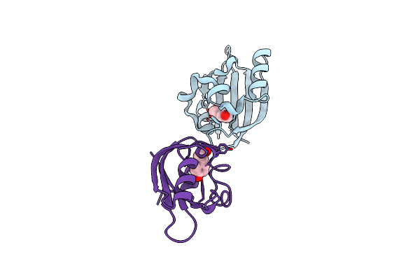
Crystal Structure Of The Von Willebrand Factor Binding Domain Of Glycoprotein Ib Alpha
Organism: Homo sapiens
Method: X-RAY DIFFRACTION Resolution:1.85 Å Release Date: 2002-08-28 Classification: BLOOD CLOTTING |
 |
Crystal Structure Of The Complex Of Glycoprotein Ib Alpha And The Von Willebrand Factor A1 Domain
Organism: Homo sapiens
Method: X-RAY DIFFRACTION Resolution:3.10 Å Release Date: 2002-08-28 Classification: BLOOD CLOTTING |