

Search Count: 23
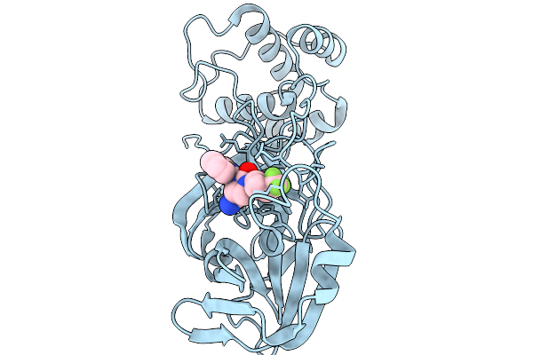 |
Co-Structure Of Sars-Cov-2 3C-Like Proteinase Nsp5 With Compound 34
Organism: Severe acute respiratory syndrome coronavirus 2
Method: X-RAY DIFFRACTION Resolution:1.87 Å Release Date: 2026-03-04 Classification: HYDROLASE Ligands: A1CAR |
Organism: Severe acute respiratory syndrome coronavirus 2
Method: X-RAY DIFFRACTION
Release Date: 2026-03-04
Ligands: A1CAR
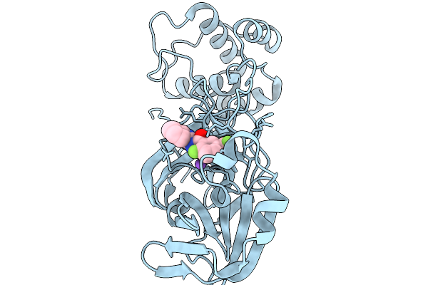 |
Co-Structure Of Main Protease Of Sars-Cov-2 With Compound 1
Organism: Severe acute respiratory syndrome coronavirus 2
Method: X-RAY DIFFRACTION Resolution:1.60 Å Release Date: 2026-02-18 Classification: HYDROLASE Ligands: A1CAS, NA |
Organism: Severe acute respiratory syndrome coronavirus 2
Method: X-RAY DIFFRACTION
Release Date: 2026-02-18
Ligands: A1CAS, NA
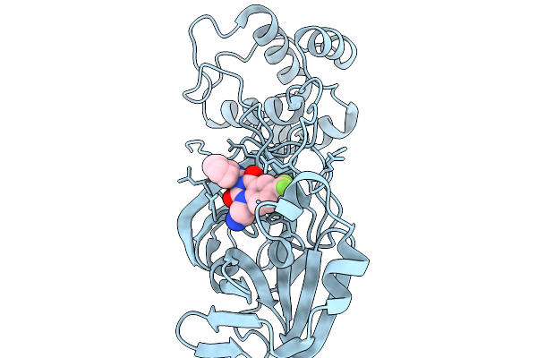 |
Co-Structure Of Main Protease Of Sars-Cov-2 With Compound 11
Organism: Severe acute respiratory syndrome coronavirus 2
Method: X-RAY DIFFRACTION Resolution:2.46 Å Release Date: 2026-02-18 Classification: HYDROLASE Ligands: A1CBN |
Organism: Severe acute respiratory syndrome coronavirus 2
Method: X-RAY DIFFRACTION
Release Date: 2026-02-18
Ligands: A1CBN
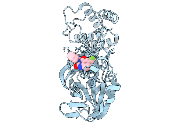 |
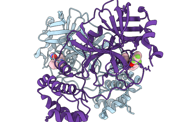
Co-Structure Of Main Protease Of Sars-Cov-2 With Compound 2
Organism: Severe acute respiratory syndrome coronavirus 2
Method: X-RAY DIFFRACTION Resolution:2.21 Å Release Date: 2026-02-18 Classification: HYDROLASE Ligands: A1CBY |
Organism: Severe acute respiratory syndrome coronavirus 2
Method: X-RAY DIFFRACTION
Release Date: 2026-02-18
Ligands: A1CBY
 |
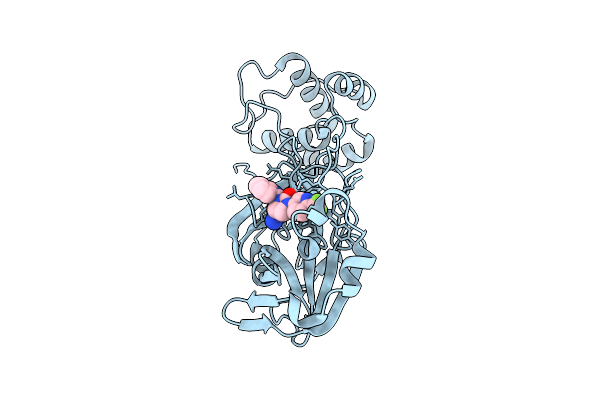
Co-Structure Of Main Protease Of Sars-Cov-2 With Compound 10
Organism: Severe acute respiratory syndrome coronavirus 2
Method: X-RAY DIFFRACTION Resolution:1.71 Å Release Date: 2026-02-18 Classification: HYDROLASE Ligands: A1CB2 |
Organism: Severe acute respiratory syndrome coronavirus 2
Method: X-RAY DIFFRACTION
Release Date: 2026-02-18
Ligands: A1CB2
 |
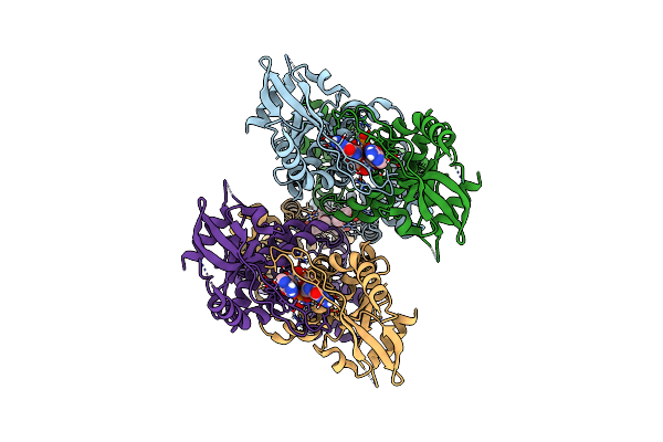
Co-Structure Of Main Protease Of Sars-Cov-2 With Nvp-Egt710
Organism: Severe acute respiratory syndrome coronavirus 2
Method: X-RAY DIFFRACTION Resolution:1.87 Å Release Date: 2025-12-24 Classification: HYDROLASE Ligands: A1CBU |
Organism: Severe acute respiratory syndrome coronavirus 2
Method: X-RAY DIFFRACTION
Release Date: 2025-12-24
Ligands: A1CBU
 |
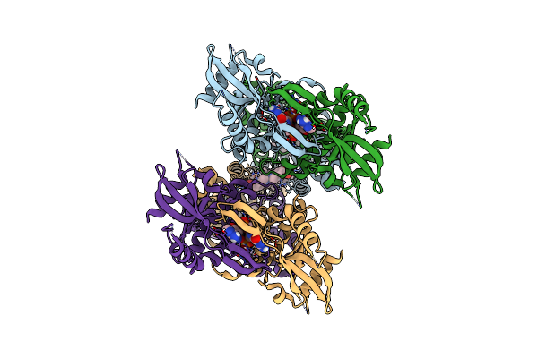
Cryo-Em Structure Of Sting Oligomer Bound To Cgamp And Nvs-Stg2
Organism: Homo sapiens
Method: ELECTRON MICROSCOPY Release Date: 2023-11-01 Classification: IMMUNE SYSTEM Ligands: 1SY, Y6H |
Organism: Homo sapiens
Method: ELECTRON MICROSCOPY
Release Date: 2023-11-01
Ligands: 1SY, Y6H
 |
Cryo-Em Structure Of Sting Oligomer Bound To Cgamp, Nvs-Stg2 And C53
Organism: Homo sapiens
Method: ELECTRON MICROSCOPY Release Date: 2023-11-01 Classification: IMMUNE SYSTEM Ligands: Y6H, 1SY, 9IM |
Organism: Homo sapiens
Method: ELECTRON MICROSCOPY
Release Date: 2023-11-01
Ligands: Y6H, 1SY, 9IM
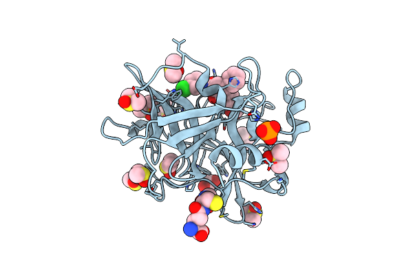 |
Human Plasmakallikrein Protease Domain In Complex With Active Site Directed Inhibitor
Organism: Homo sapiens
Method: X-RAY DIFFRACTION Resolution:1.42 Å Release Date: 2020-07-08 Classification: HYDROLASE Ligands: GSH, DMS, PO4, GOL, MU8 |
Organism: Homo sapiens
Method: X-RAY DIFFRACTION
Release Date: 2020-07-08
Ligands: GSH, DMS, PO4, GOL, MU8
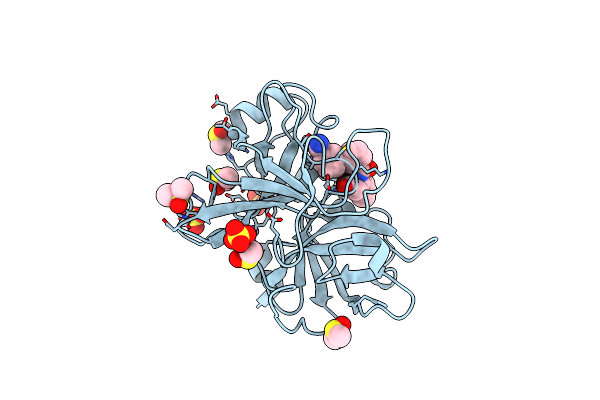 |
Coagulation Factor Xi Protease Domain In Complex With Active Site Inhibitor
Organism: Homo sapiens
Method: X-RAY DIFFRACTION Resolution:1.17 Å Release Date: 2020-07-08 Classification: HYDROLASE Ligands: SO4, DMS, NA, J7B |
Organism: Homo sapiens
Method: X-RAY DIFFRACTION
Release Date: 2020-07-08
Ligands: SO4, DMS, NA, J7B
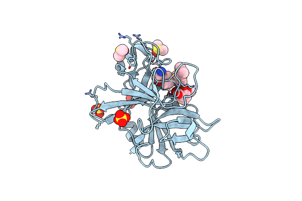 |
Coagulation Factor Xi Protease Domain In Complex With Active Site Inhibitor
Organism: Homo sapiens
Method: X-RAY DIFFRACTION Resolution:1.29 Å Release Date: 2020-07-08 Classification: HYDROLASE Ligands: NW2, SO4, DMS |
Organism: Homo sapiens
Method: X-RAY DIFFRACTION
Release Date: 2020-07-08
Ligands: NW2, SO4, DMS
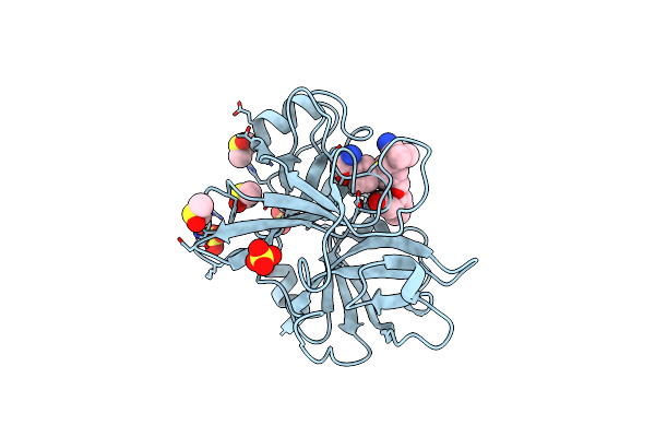 |
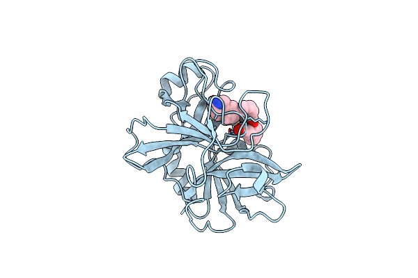
Coagulation Factor Xi Protease Domain In Complex With Active Site Inhibitor
Organism: Homo sapiens
Method: X-RAY DIFFRACTION Resolution:1.33 Å Release Date: 2020-07-08 Classification: HYDROLASE Ligands: SO4, DMS, NW5 |
Organism: Homo sapiens
Method: X-RAY DIFFRACTION
Release Date: 2020-07-08
Ligands: SO4, DMS, NW5
 |
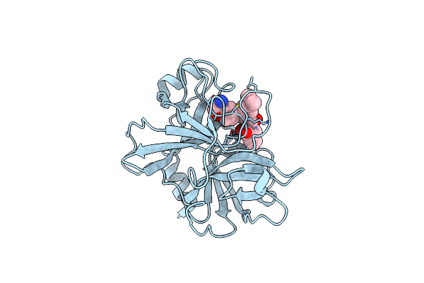
Coagulation Factor Xi Protease Domain In Complex With Active Site Inhibitor
Organism: Homo sapiens
Method: X-RAY DIFFRACTION Resolution:2.63 Å Release Date: 2020-07-08 Classification: HYDROLASE Ligands: NWE |
Organism: Homo sapiens
Method: X-RAY DIFFRACTION
Release Date: 2020-07-08
Ligands: NWE
 |
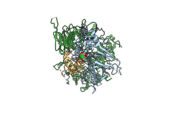
Coagulation Factor Xi Catalytic Domain (C123S) In Complex With Nvp-Xiv936
Organism: Homo sapiens
Method: X-RAY DIFFRACTION Resolution:1.26 Å Release Date: 2020-07-01 Classification: HYDROLASE Ligands: QGS |
Organism: Homo sapiens
Method: X-RAY DIFFRACTION
Release Date: 2020-07-01
Ligands: QGS
 |
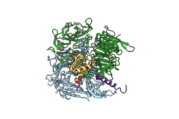
Structure Of The Human Ddb1-Dda1-Dcaf15 E3 Ubiquitin Ligase Bound To Rbm39 And Indisulam
Organism: Homo sapiens
Method: ELECTRON MICROSCOPY Release Date: 2019-12-18 Classification: ONCOPROTEIN Ligands: EF6 |
Organism: Homo sapiens
Method: ELECTRON MICROSCOPY
Release Date: 2019-12-18
Ligands: EF6
 |
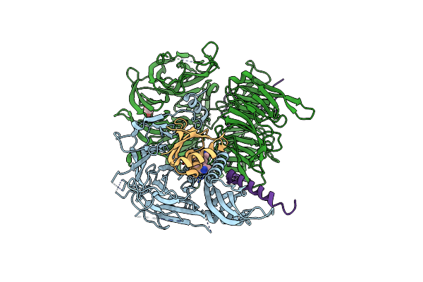
Crystal Structure Of Full-Length Human Dcaf15-Ddb1(Deltabpb)-Dda1-Rbm39 In Complex With Indisulam
Organism: Homo sapiens
Method: X-RAY DIFFRACTION Resolution:2.30 Å Release Date: 2019-12-18 Classification: LIGASE Ligands: GOL, EF6 |
Organism: Homo sapiens
Method: X-RAY DIFFRACTION
Release Date: 2019-12-18
Ligands: GOL, EF6
 |
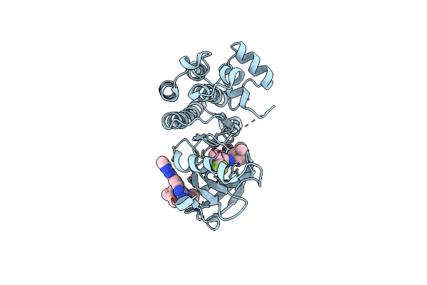
Crystal Structure Of Full-Length Human Dcaf15-Ddb1-Deltapbp-Dda1-Rbm39 In Complex With 4-(Aminomethyl)-N-(3-Cyano-4-Methyl-1H-Indol-7-Yl)Benzenesulfonamide
Organism: Homo sapiens
Method: X-RAY DIFFRACTION Resolution:2.61 Å Release Date: 2019-12-18 Classification: LIGASE Ligands: Q5J, GOL |
Organism: Homo sapiens
Method: X-RAY DIFFRACTION
Release Date: 2019-12-18
Ligands: Q5J, GOL
 |
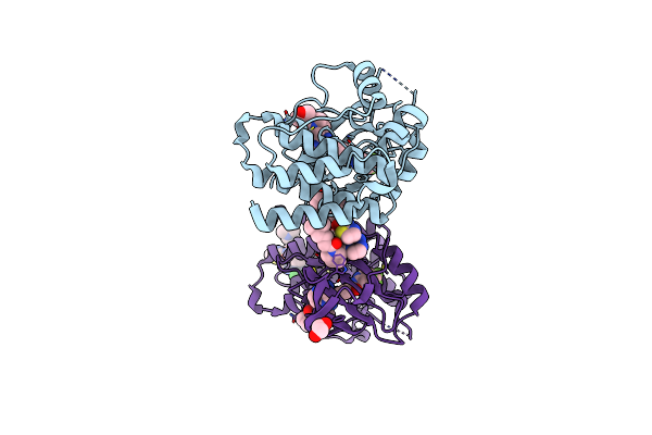
Btk-Inhibitor Co-Structure
Organism: Homo sapiens
Method: X-RAY DIFFRACTION Resolution:1.89 Å Release Date: 2016-03-23 Classification: TRANSFERASE/TRANSFERASE inhibitor Ligands: 5WH, 5WE |
Organism: Homo sapiens
Method: X-RAY DIFFRACTION
Release Date: 2016-03-23
Ligands: 5WH, 5WE
 |
Btk Kinase Domain With Inhibitor 1
Organism: Homo sapiens
Method: X-RAY DIFFRACTION Resolution:1.80 Å Release Date: 2016-02-03 Classification: TRANSFERASE Ligands: 5WF, 5WE, CL, EDO |
Organism: Homo sapiens
Method: X-RAY DIFFRACTION
Release Date: 2016-02-03
Ligands: 5WF, 5WE, CL, EDO
 |
Structure Of The Q61G Mutant Of Ras In The Gdp-Bound Form
Organism: Homo sapiens
Method: X-RAY DIFFRACTION Resolution:2.00 Å Release Date: 2006-03-14 Classification: ONCOPROTEIN Ligands: MG, GDP |
Organism: Homo sapiens
Method: X-RAY DIFFRACTION
Release Date: 2006-03-14
Ligands: MG, GDP