
Search Count: 34
All
Selected
 |
Organism: Mus musculus
Method: SOLUTION NMR Release Date: 2025-09-03 Classification: CYTOSOLIC PROTEIN |
 |
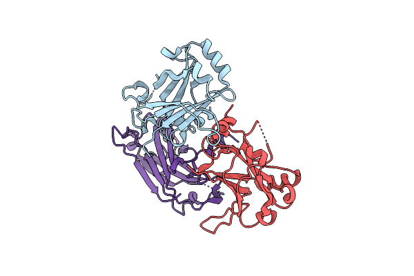
Organism: Vibrio cholerae serotype o1 (strain atcc 39315 / el tor inaba n16961)
Method: X-RAY DIFFRACTION Resolution:2.30 Å Release Date: 2025-07-30 Classification: TOXIN Ligands: SO4, ACT |
 |
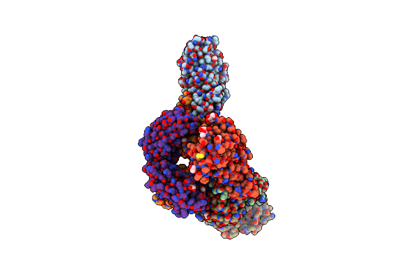
Organism: Acinetobacter baumannii
Method: X-RAY DIFFRACTION Resolution:2.67 Å Release Date: 2025-03-26 Classification: LYASE |
 |
Organism: Acinetobacter baumannii
Method: X-RAY DIFFRACTION Resolution:3.31 Å Release Date: 2024-12-04 Classification: HYDROLASE |
 |
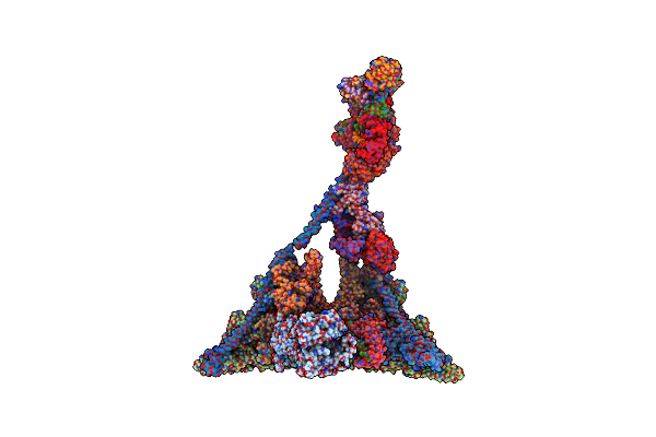
Organism: Homo sapiens
Method: ELECTRON MICROSCOPY Release Date: 2023-09-27 Classification: TRANSFERASE Ligands: ZN |
 |
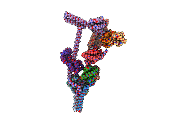
Organism: Homo sapiens
Method: ELECTRON MICROSCOPY Release Date: 2023-09-27 Classification: LIGASE Ligands: ZN |
 |
Organism: Rhodobacter capsulatus de442
Method: X-RAY DIFFRACTION Resolution:2.40 Å Release Date: 2023-09-13 Classification: IMMUNE SYSTEM |
 |
Organism: Notothenia coriiceps
Method: X-RAY DIFFRACTION Resolution:2.66 Å Release Date: 2023-07-12 Classification: IMMUNE SYSTEM |
 |
Organism: Spinacia oleracea
Method: ELECTRON MICROSCOPY Release Date: 2022-08-24 Classification: PLANT PROTEIN Ligands: ATP, ADP |
 |
Organism: Spinacia oleracea
Method: ELECTRON MICROSCOPY Release Date: 2022-05-25 Classification: PLANT PROTEIN |
 |
Crystal Structure Of Receptor Binding Domain Of Mers-Cov And Knih90-F1 Fab Complex
Organism: Homo sapiens, Middle east respiratory syndrome-related coronavirus
Method: X-RAY DIFFRACTION Resolution:2.05 Å Release Date: 2021-08-04 Classification: IMMUNE SYSTEM Ligands: EDO, NAG |
 |
Organism: Chlamydomonas reinhardtii
Method: ELECTRON MICROSCOPY Release Date: 2020-12-16 Classification: STRUCTURAL PROTEIN Ligands: PO4 |
 |
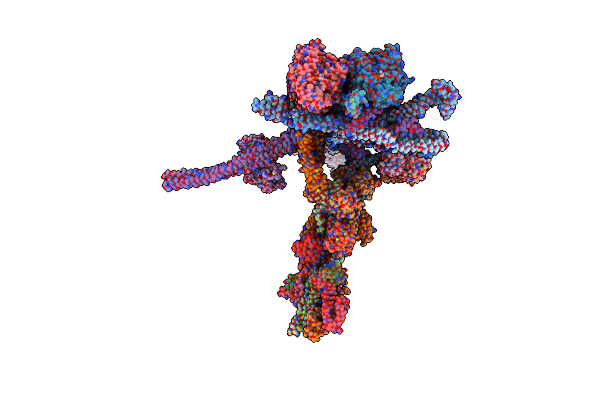
Stalk Of Radial Spoke 1 Attached With Doublet Microtubule From Chlamydomonas Reinhardtii
Organism: Chlamydomonas reinhardtii
Method: ELECTRON MICROSCOPY Release Date: 2020-12-16 Classification: STRUCTURAL PROTEIN |
 |
Organism: Chlamydomonas reinhardtii
Method: ELECTRON MICROSCOPY Release Date: 2020-12-16 Classification: STRUCTURAL PROTEIN Ligands: GDP, GTP, MG, ATP |
 |
Organism: Homo sapiens
Method: X-RAY DIFFRACTION Resolution:3.25 Å Release Date: 2019-11-20 Classification: TRANSFERASE Ligands: L1H |
 |
Organism: Homo sapiens
Method: X-RAY DIFFRACTION Resolution:2.73 Å Release Date: 2019-11-20 Classification: TRANSFERASE Ligands: L1K, DMS |
 |
Organism: Homo sapiens
Method: X-RAY DIFFRACTION Resolution:2.67 Å Release Date: 2019-11-20 Classification: TRANSFERASE Ligands: L3Z, DMS |
 |
Organism: Homo sapiens
Method: X-RAY DIFFRACTION Resolution:2.48 Å Release Date: 2019-09-11 Classification: STRUCTURAL PROTEIN Ligands: GOL, A0L |
 |
Organism: Homo sapiens
Method: X-RAY DIFFRACTION Resolution:2.41 Å Release Date: 2019-09-11 Classification: NUCLEAR PROTEIN Ligands: AE0 |
 |
Organism: Homo sapiens
Method: X-RAY DIFFRACTION Resolution:2.20 Å Release Date: 2018-09-26 Classification: TRANSFERASE Ligands: 9KO |