
Search Count: 21
All
Selected
 |
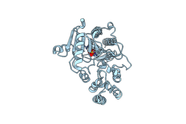
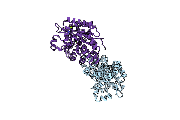
Urate Oxidase From Aspergillus Flavus With Its Inhibitor 9-Methyl Uric Acid By Continuous Serial Electron Diffraction (Serialed)
Organism: Aspergillus flavus
Method: ELECTRON CRYSTALLOGRAPHY Resolution:1.25 Å Release Date: 2026-04-29 Classification: OXIDOREDUCTASE Ligands: MUA |
 |
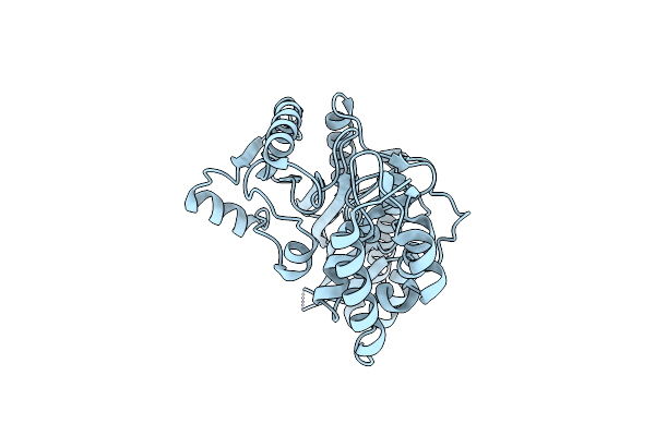
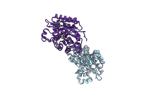
Urate Oxidase From Aspergillus Flavus With Its Substrate Uric Acid By Continuous Serial Electron Diffraction (Serialed)
Organism: Aspergillus flavus
Method: ELECTRON CRYSTALLOGRAPHY Resolution:1.75 Å Release Date: 2026-04-29 Classification: OXIDOREDUCTASE Ligands: OXY, URC |
 |
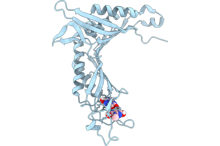
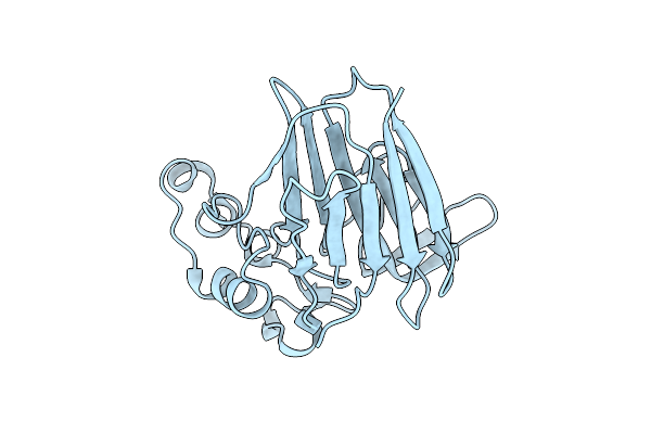
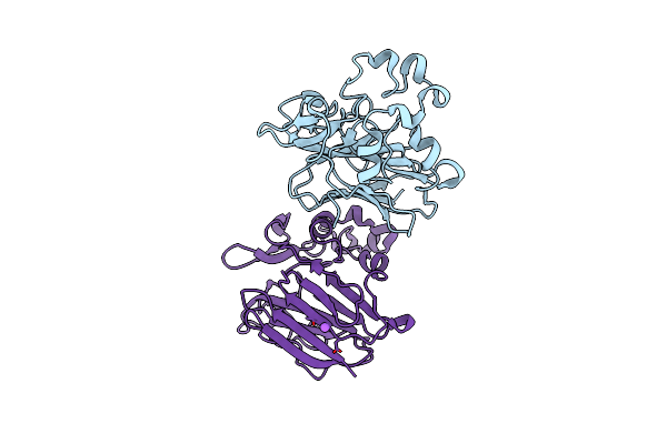
Structure Of Human Mth1 In Complex With 8Dg By Continuous Serial Electron Diffraction (Serialed)
Organism: Homo sapiens
Method: ELECTRON CRYSTALLOGRAPHY Resolution:1.66 Å Release Date: 2026-04-22 Classification: HYDROLASE Ligands: SO4, 8DG |
 |
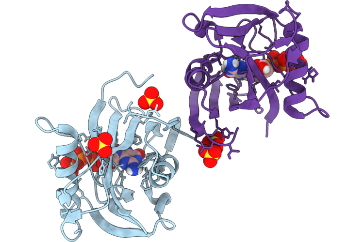
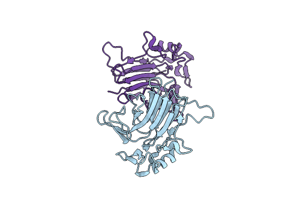
Structure Of Human Mth1 In Complex With 8Dg By Microed Using High Electron Fluence
Organism: Homo sapiens
Method: ELECTRON CRYSTALLOGRAPHY Resolution:2.32 Å Release Date: 2026-04-22 Classification: HYDROLASE Ligands: 8DG, SO4 |
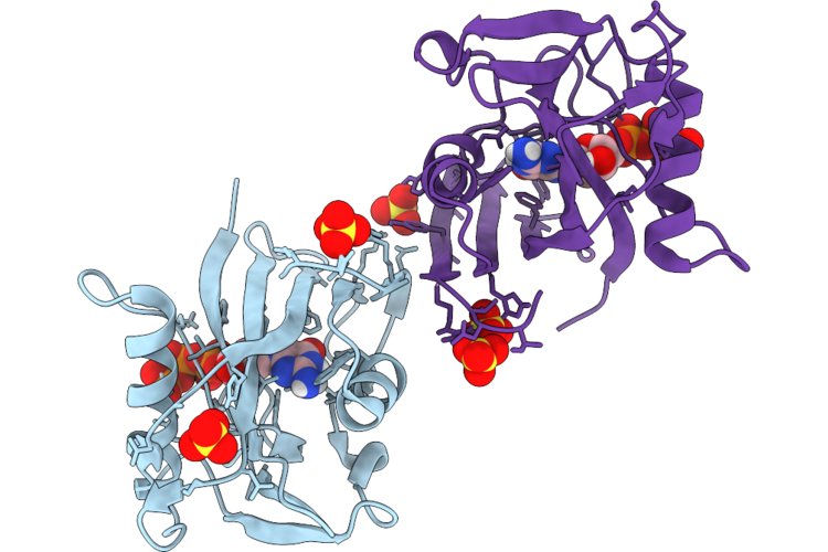 |
Structure Of Human Mth1 In Complex With 8Dg By Microed Using Low Electron Fluence
Organism: Homo sapiens
Method: ELECTRON CRYSTALLOGRAPHY Resolution:2.86 Å Release Date: 2026-04-22 Classification: HYDROLASE Ligands: 8DG, SO4 |
 |
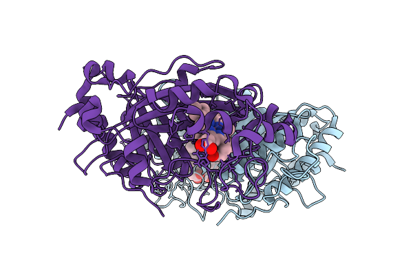
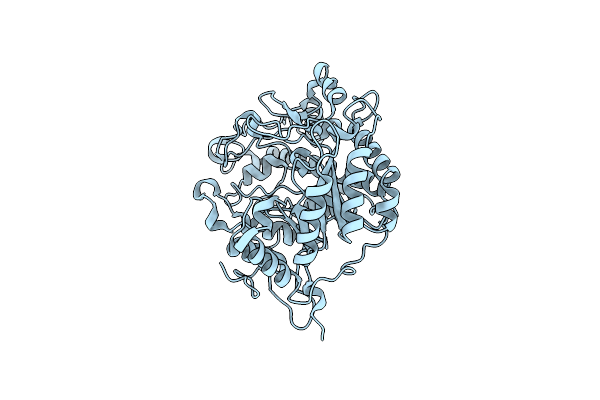
Organism: Gallus gallus
Method: ELECTRON CRYSTALLOGRAPHY Resolution:0.83 Å Release Date: 2026-04-22 Classification: HYDROLASE Ligands: ACT, CL |
 |
Dye Type Peroxidase Aa From Streptomyces Lividans With N3 Ligand By Serial Electron Diffraction (Serialed)
Organism: Streptomyces lividans
Method: ELECTRON CRYSTALLOGRAPHY Resolution:1.10 Å Release Date: 2025-07-16 Classification: OXIDOREDUCTASE Ligands: AZI, HEM |
 |
Dye Type Peroxidase Aa From Streptomyces Lividans By Microcrystal Electron Diffraction (Microed/3D Ed)
Organism: Streptomyces lividans
Method: ELECTRON CRYSTALLOGRAPHY Resolution:2.40 Å Release Date: 2025-07-16 Classification: OXIDOREDUCTASE Ligands: HEM |
 |
Dye Type Peroxidase Aa From Streptomyces Lividans By Serial Electron Diffraction (Serialed)
Organism: Streptomyces lividans
Method: ELECTRON CRYSTALLOGRAPHY Resolution:1.30 Å Release Date: 2025-07-16 Classification: OXIDOREDUCTASE Ligands: HEM |
 |
Organism: Dermatophagoides pteronyssinus
Method: X-RAY DIFFRACTION Resolution:1.90 Å Release Date: 2025-06-11 Classification: ALLERGEN |
 |
Organism: Dermatophagoides pteronyssinus
Method: X-RAY DIFFRACTION Resolution:1.80 Å Release Date: 2025-06-11 Classification: ALLERGEN Ligands: SO4 |
 |
Organism: Sarcoptes scabiei
Method: X-RAY DIFFRACTION Resolution:1.40 Å Release Date: 2025-06-11 Classification: ALLERGEN |
 |
Organism: Juniperus ashei
Method: X-RAY DIFFRACTION Resolution:1.40 Å Release Date: 2022-07-13 Classification: ALLERGEN Ligands: CL |
 |
Organism: Amycolatopsis rifamycinica
Method: X-RAY DIFFRACTION Resolution:1.87 Å Release Date: 2022-07-13 Classification: UNKNOWN FUNCTION |
 |
Organism: Puccinia graminis f. sp. tritici (strain crl 75-36-700-3 / race sccl)
Method: X-RAY DIFFRACTION Resolution:1.50 Å Release Date: 2022-07-13 Classification: UNKNOWN FUNCTION |
 |
Organism: Aspergillus oryzae
Method: X-RAY DIFFRACTION Resolution:1.21 Å Release Date: 2020-10-21 Classification: METAL BINDING PROTEIN Ligands: MG |
 |
Organism: Aspergillus oryzae
Method: X-RAY DIFFRACTION Resolution:1.53 Å Release Date: 2020-10-21 Classification: METAL BINDING PROTEIN Ligands: MG, CA |
 |
Organism: Triticum aestivum
Method: X-RAY DIFFRACTION Resolution:2.00 Å Release Date: 2018-12-19 Classification: HYDROLASE |
 |
Structure Of The Double Stranded Dna Binding Type Iv Secretion Protein Tran From Enterococcus
Organism: Enterococcus faecalis
Method: X-RAY DIFFRACTION Resolution:1.40 Å Release Date: 2014-07-30 Classification: DNA BINDING PROTEIN |
 |
Structure Of The Double Stranded Dna Binding Type Iv Secretion Protein Tran From Enterococcus
Organism: Enterococcus faecalis
Method: X-RAY DIFFRACTION Resolution:1.35 Å Release Date: 2014-07-30 Classification: DNA BINDING PROTEIN |