

Search Count: 19
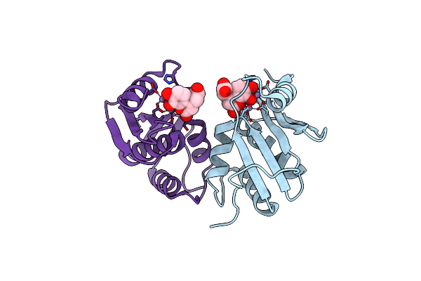 |
Structure Of A Dihydroxycoumarin Active-Site Inhibitor In Complex With The Rnase H Domain Of Hiv-1 Reverse Transcriptase
Organism: Human immunodeficiency virus type 1
Method: X-RAY DIFFRACTION Resolution:1.71 Å Release Date: 2014-06-04 Classification: HYDROLASE/HYDROLASE INHIBITOR Ligands: MN, F95 |
Organism: Human immunodeficiency virus type 1
Method: X-RAY DIFFRACTION
Release Date: 2014-06-04
Ligands: MN, F95
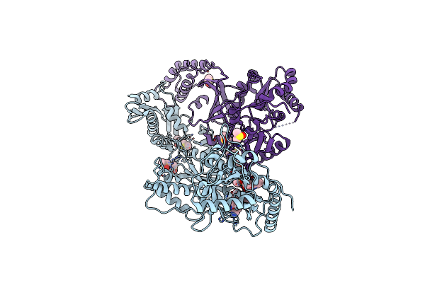 |
Hiv-1 Reverse Transcriptase With Bound Fragment At Nnrti Adjacent Site
Organism: Human immunodeficiency virus type 1
Method: X-RAY DIFFRACTION Resolution:1.85 Å Release Date: 2013-05-15 Classification: transferase/transferase inhibitor Ligands: 1QP, DMS, T27 |
Organism: Human immunodeficiency virus type 1
Method: X-RAY DIFFRACTION
Release Date: 2013-05-15
Ligands: 1QP, DMS, T27
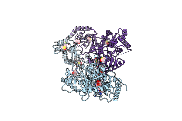 |
Hiv-1 Reverse Transcriptase With Bound Fragment At The Incoming Dntp Binding Site
Organism: Human immunodeficiency virus type 1
Method: X-RAY DIFFRACTION Resolution:1.80 Å Release Date: 2013-02-06 Classification: transferase/transferase inhibitor Ligands: T27, MG, DMS, 14N |
Organism: Human immunodeficiency virus type 1
Method: X-RAY DIFFRACTION
Release Date: 2013-02-06
Ligands: T27, MG, DMS, 14N
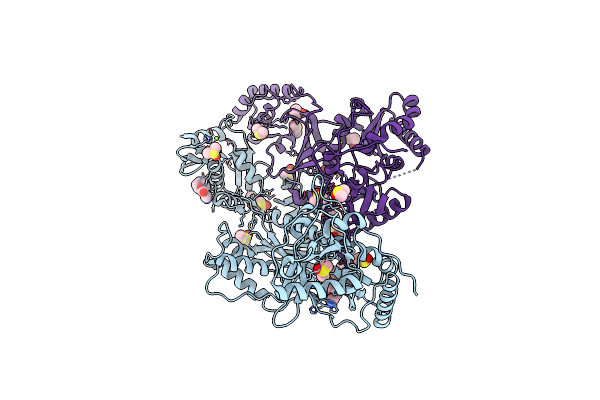 |
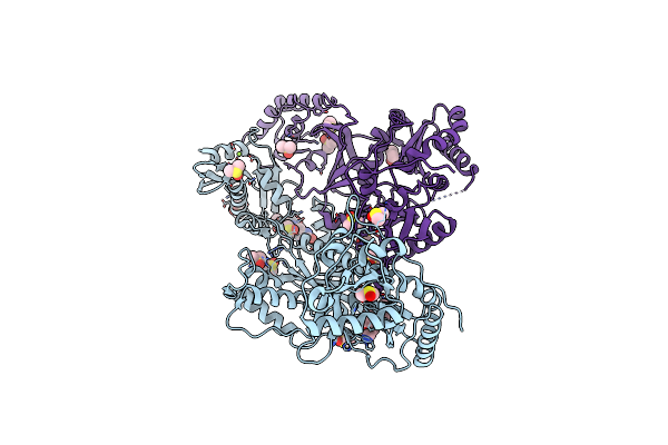
Hiv-1 Reverse Transcriptase With Bound Fragment At The Rnase H Primer Grip Site
Organism: Human immunodeficiency virus type 1
Method: X-RAY DIFFRACTION Resolution:1.95 Å Release Date: 2013-02-06 Classification: transferase/transferase inhibitor Ligands: T27, DMS, 1FF, MG |
Organism: Human immunodeficiency virus type 1
Method: X-RAY DIFFRACTION
Release Date: 2013-02-06
Ligands: T27, DMS, 1FF, MG
 |
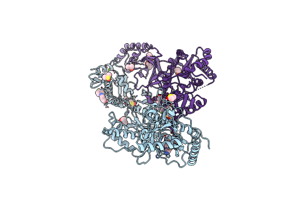
Hiv-1 Reverse Transcriptase With Bound Fragment At The 428 Site
Organism: Human immunodeficiency virus type 1
Method: X-RAY DIFFRACTION Resolution:2.10 Å Release Date: 2013-02-06 Classification: transferase/transferase inhibitor Ligands: T27, 1FE, MG, DMS |
Organism: Human immunodeficiency virus type 1
Method: X-RAY DIFFRACTION
Release Date: 2013-02-06
Ligands: T27, 1FE, MG, DMS
 |
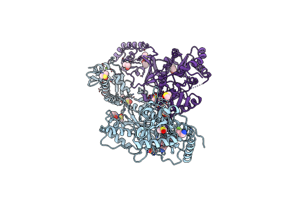
Detecting Allosteric Sites Of Hiv-1 Reverse Transcriptase By X-Ray Crystallographic Fragment Screening
Organism: Human immunodeficiency virus type 1
Method: X-RAY DIFFRACTION Resolution:2.05 Å Release Date: 2013-02-06 Classification: transferase/transferase inhibitor Ligands: T27, FMQ, DMS, MG |
Organism: Human immunodeficiency virus type 1
Method: X-RAY DIFFRACTION
Release Date: 2013-02-06
Ligands: T27, FMQ, DMS, MG
 |
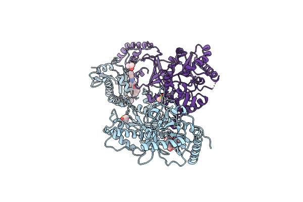
Hiv-1 Reverse Transcriptase With Bound Fragment At The Knuckles Site
Organism: Human immunodeficiency virus type 1
Method: X-RAY DIFFRACTION Resolution:2.10 Å Release Date: 2013-02-06 Classification: transferase/transferase inhibitor Ligands: T27, MG, DMS, 1FD |
Organism: Human immunodeficiency virus type 1
Method: X-RAY DIFFRACTION
Release Date: 2013-02-06
Ligands: T27, MG, DMS, 1FD
 |
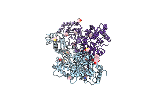
Hiv-1 Reverse Transcriptase With Bound Fragment At The 507 Site
Organism: Human immunodeficiency virus type 1
Method: X-RAY DIFFRACTION Resolution:2.50 Å Release Date: 2013-02-06 Classification: transferase/transferase inhibitor Ligands: 1FG, DMS |
Organism: Human immunodeficiency virus type 1
Method: X-RAY DIFFRACTION
Release Date: 2013-02-06
Ligands: 1FG, DMS
 |
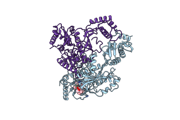
Hiv-1 Reverse Transcriptase With Bound Fragment Near Knuckles Site
Organism: Human immunodeficiency virus type 1, Human immunodeficiency virus type 1 bh10
Method: X-RAY DIFFRACTION Resolution:1.95 Å Release Date: 2013-02-06 Classification: transferase/transferase inhibitor Ligands: T27, DMS, J94 |
Organism: Human immunodeficiency virus type 1, Human immunodeficiency virus type 1 bh10
Method: X-RAY DIFFRACTION
Release Date: 2013-02-06
Ligands: T27, DMS, J94
 |
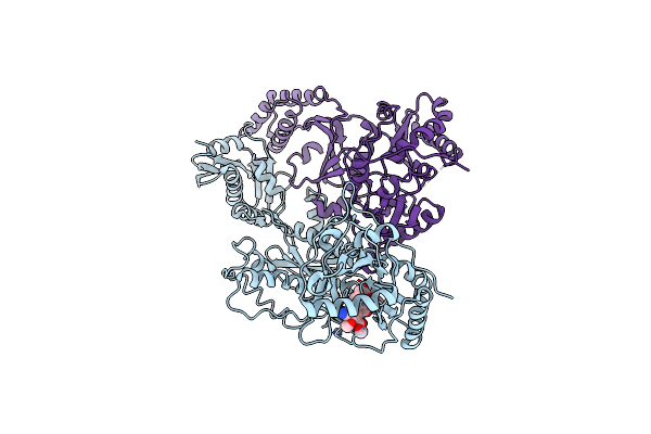
Crystal Structure Of Hiv-1 Reverse Transcriptase (Rt) In Complex With The Non-Nucleoside Rt Inhibitor (E)-S-Methyl 5-(1-(3,7-Dimethyl-2-Oxo-2,3-Dihydrobenzo[D]Oxazol-5-Yl)-5-(5-Methyl-1,3,4-Oxadiazol-2-Yl)Pent-1-Enyl)-2-Methoxy-3-Methylbenzothioate.
Organism: Human immunodeficiency virus type 1 bh10
Method: X-RAY DIFFRACTION Resolution:2.80 Å Release Date: 2010-04-07 Classification: transferase/hydrolase Ligands: UDR |
Organism: Human immunodeficiency virus type 1 bh10
Method: X-RAY DIFFRACTION
Release Date: 2010-04-07
Ligands: UDR
 |
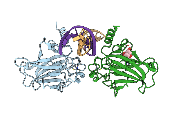
Crystal Structure Of The Hiv-1 Reverse Transcriptase (Rt) In Complex With The Alkenyldiarylmethane (Adam) Non-Nucleoside Rt Inhibitor Dimethyl 3,3'-(6-Methoxy-6-Oxohex-1-Ene-1,1-Diyl)Bis(5-Cyano-6-Methoxybenzoate).
Organism: Human immunodeficiency virus type 1 bh10
Method: X-RAY DIFFRACTION Resolution:2.55 Å Release Date: 2010-04-07 Classification: transferase/hydrolase Ligands: AC7 |
Organism: Human immunodeficiency virus type 1 bh10
Method: X-RAY DIFFRACTION
Release Date: 2010-04-07
Ligands: AC7
 |
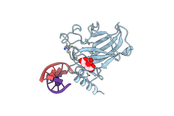
Crystal Structure Of A P53 Core Tetramer Bound To Dna
Organism: Mus musculus
Method: X-RAY DIFFRACTION Resolution:2.00 Å Release Date: 2008-12-16 Classification: TRANSCRIPTION/DNA Ligands: ZN, FLC |
Organism: Mus musculus
Method: X-RAY DIFFRACTION
Release Date: 2008-12-16
Ligands: ZN, FLC
 |
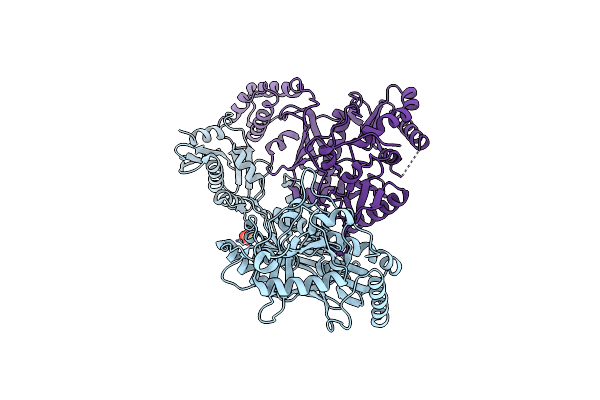
Crystal Structure Of A P53 Core Tetramer Bound To Dna
Organism: Mus musculus
Method: X-RAY DIFFRACTION Resolution:2.20 Å Release Date: 2008-12-16 Classification: TRANSCRIPTION/DNA Ligands: FLC, ZN |
Organism: Mus musculus
Method: X-RAY DIFFRACTION
Release Date: 2008-12-16
Ligands: FLC, ZN
 |
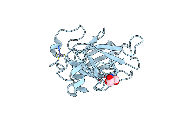
Crystal Structure Of An Engineered Form Of The Hiv-1 Reverse Transcriptase, Rt69A
Organism: Human immunodeficiency virus type 1 bh10, Human immunodeficiency virus type 1
Method: X-RAY DIFFRACTION Resolution:1.85 Å Release Date: 2008-10-07 Classification: TRANSFERASE Ligands: SO4 |
Organism: Human immunodeficiency virus type 1 bh10, Human immunodeficiency virus type 1
Method: X-RAY DIFFRACTION
Release Date: 2008-10-07
Ligands: SO4
 |
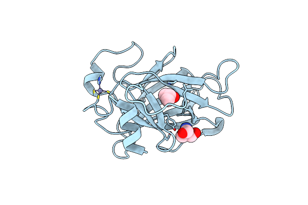
Crystal Structure Of The Mouse P53 Core Domain At 1.55 A
Organism: Mus musculus
Method: X-RAY DIFFRACTION Resolution:1.55 Å Release Date: 2006-12-05 Classification: TRANSCRIPTION, ANTITUMOR PROTEIN Ligands: ZN, TRS |
Organism: Mus musculus
Method: X-RAY DIFFRACTION
Release Date: 2006-12-05
Ligands: ZN, TRS
 |
Mouse P53 Core Domain Soaked With 2-Propanol
Organism: Mus musculus
Method: X-RAY DIFFRACTION Resolution:2.00 Å Release Date: 2006-12-05 Classification: TRANSCRIPTION, ANTITUMOR PROTEIN Ligands: ZN, TRS, IPA |
Organism: Mus musculus
Method: X-RAY DIFFRACTION
Release Date: 2006-12-05
Ligands: ZN, TRS, IPA
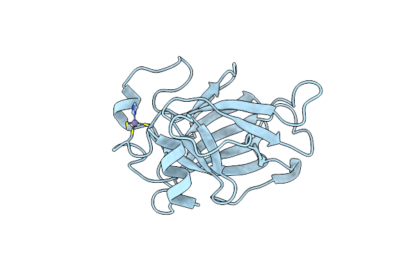 |
Crystal Structure Of The Mouse P53 Core Domain
Organism: Mus musculus
Method: X-RAY DIFFRACTION Resolution:2.02 Å Release Date: 2006-12-05 Classification: TRANSCRIPTION, ANTITUMOR PROTEIN Ligands: ZN |
Organism: Mus musculus
Method: X-RAY DIFFRACTION
Release Date: 2006-12-05
Ligands: ZN
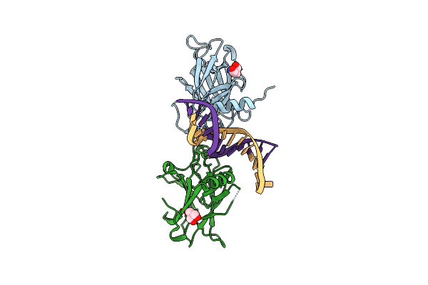 |
Crystal Structure Of A P53 Core Dimer Bound To Dna
Organism: Mus musculus
Method: X-RAY DIFFRACTION Resolution:2.30 Å Release Date: 2006-05-23 Classification: TRANSCRIPTION/DNA Ligands: ZN, TRS |
Organism: Mus musculus
Method: X-RAY DIFFRACTION
Release Date: 2006-05-23
Ligands: ZN, TRS
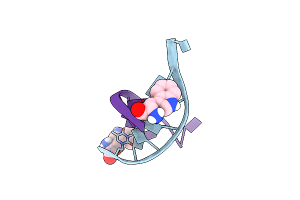 |
Solution Structure Of The Aminoacyl-Capped Oligodeoxyribonucleotide Duplex Trp-D(Tgcgcac)2
|
Method: SOLUTION NMR
Release Date: 1999-10-08
Ligands: TRP