

Search Count: 33
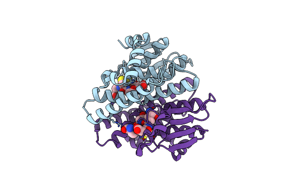 |
Crystal Structure Of A Glutathione S-Transferase Protein From Escherichia Coli Och 157:H7 Str. Sakai (Ecs3186, Target Efi-507414) With Bound Glutathione
Organism: Escherichia coli (strain k12)
Method: X-RAY DIFFRACTION Resolution:1.55 Å Release Date: 2016-02-10 Classification: TRANSFERASE Ligands: GSH |
Organism: Escherichia coli (strain k12)
Method: X-RAY DIFFRACTION
Release Date: 2016-02-10
Ligands: GSH
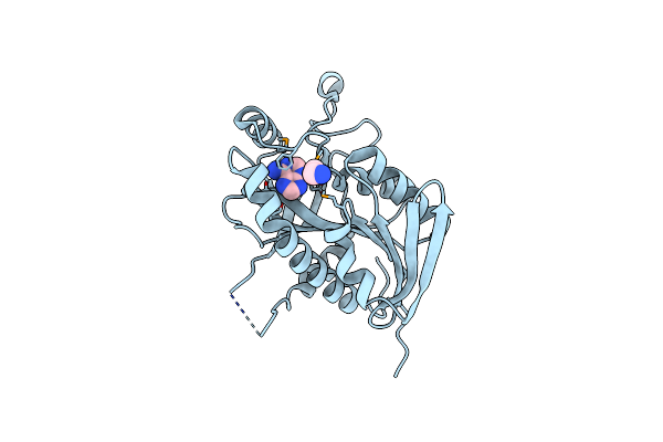 |
Crystal Structure Of A 5'-Methylthioadenosine/S-Adenosylhomocysteine (Mta/Sah) Nucleosidase (Mtan) From Colwellia Psychrerythraea 34H (Cps_4743, Target Psi-029300) In Complex With Adenine At 2.27 A Resolution
Organism: Colwellia psychrerythraea
Method: X-RAY DIFFRACTION Resolution:2.27 Å Release Date: 2015-11-04 Classification: HYDROLASE Ligands: GLY, ADE |
Organism: Colwellia psychrerythraea
Method: X-RAY DIFFRACTION
Release Date: 2015-11-04
Ligands: GLY, ADE
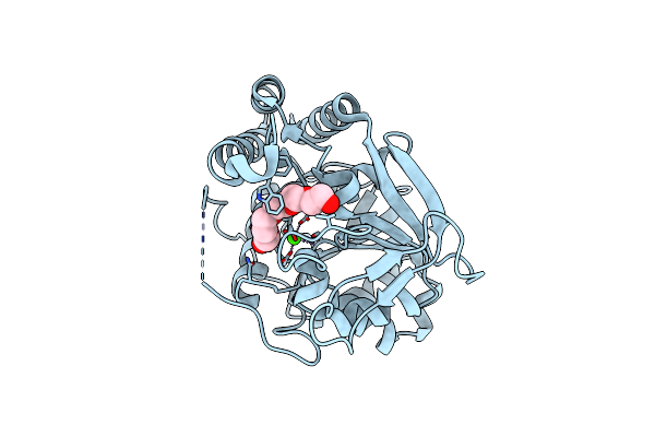 |
Crystal Structure Of A Putative Pyrimidine-Specific Ribonucleoside Hydrolase (Riha) Protein From Shewanella Loihica Pv-4 (Shew_0697, Target Psi-029635) With Divalent Cation And Peg 400 Bound At The Active Site
Organism: Shewanella loihica
Method: X-RAY DIFFRACTION Resolution:1.70 Å Release Date: 2014-11-12 Classification: HYDROLASE Ligands: CA, 1PE |
Organism: Shewanella loihica
Method: X-RAY DIFFRACTION
Release Date: 2014-11-12
Ligands: CA, 1PE
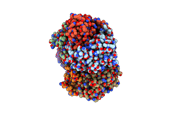 |
Crystal Structure Of A Putative Uridine Phosphorylase From Actinobacillus Succinogenes 130Z (Target Nysgrc-029667 )
Organism: Actinobacillus succinogenes
Method: X-RAY DIFFRACTION Resolution:2.00 Å Release Date: 2014-08-27 Classification: TRANSFERASE Ligands: GOL |
Organism: Actinobacillus succinogenes
Method: X-RAY DIFFRACTION
Release Date: 2014-08-27
Ligands: GOL
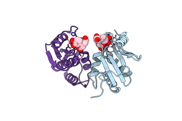 |
Structure Of A Dihydroxycoumarin Active-Site Inhibitor In Complex With The Rnase H Domain Of Hiv-1 Reverse Transcriptase
Organism: Human immunodeficiency virus type 1
Method: X-RAY DIFFRACTION Resolution:1.71 Å Release Date: 2014-06-04 Classification: HYDROLASE/HYDROLASE INHIBITOR Ligands: MN, F95 |
Organism: Human immunodeficiency virus type 1
Method: X-RAY DIFFRACTION
Release Date: 2014-06-04
Ligands: MN, F95
 |
Crystal Structure Of A Putative 5'-Methylthioadenosine/S-Adenosylhomocysteine Nucleosidase From Borrelia Burgdorferi B31 Bound To Adenine (Target Nysgrc-029268 )
Organism: Borrelia burgdorferi
Method: X-RAY DIFFRACTION Resolution:1.70 Å Release Date: 2013-08-14 Classification: HYDROLASE Ligands: ADE |
Organism: Borrelia burgdorferi
Method: X-RAY DIFFRACTION
Release Date: 2013-08-14
Ligands: ADE
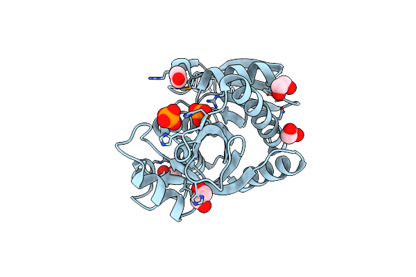 |
Crystal Structure Of A Putative Purine Nucleoside Phosphorylase From Vibrio Fischeri Es114 (Target Nysgrc-029521)
Organism: Vibrio fischeri
Method: X-RAY DIFFRACTION Resolution:1.48 Å Release Date: 2013-07-03 Classification: TRANSFERASE Ligands: PO4, EDO |
Organism: Vibrio fischeri
Method: X-RAY DIFFRACTION
Release Date: 2013-07-03
Ligands: PO4, EDO
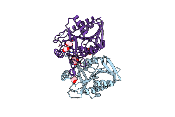 |
Crystal Structure Of A Putative Methylthioadenosine Nucleosidase From Weissella Paramesenteroides Atcc 33313 (Target Nysgrc-029342 )
Organism: Weissella paramesenteroides
Method: X-RAY DIFFRACTION Resolution:1.70 Å Release Date: 2013-05-29 Classification: HYDROLASE Ligands: EDO, GOL |
Organism: Weissella paramesenteroides
Method: X-RAY DIFFRACTION
Release Date: 2013-05-29
Ligands: EDO, GOL
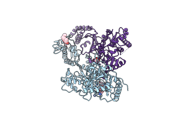 |
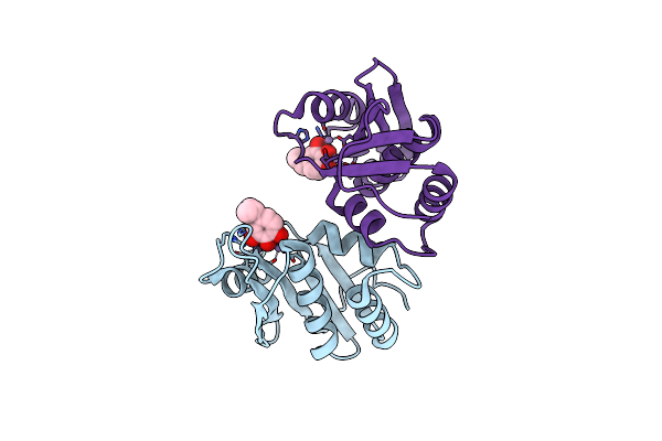
Hiv-1 Reverse Transcriptase In Complex With Manicol At The Rnase H Active Site And Tmc278 (Rilpivirine) At The Nnrti Binding Pocket
Organism: Human immunodeficiency virus type 1
Method: X-RAY DIFFRACTION Resolution:2.70 Å Release Date: 2011-12-21 Classification: TRANSFERASE,HYDROLASE/INHIBITOR Ligands: MNK, T27, MN, DMS, EDO |
Organism: Human immunodeficiency virus type 1
Method: X-RAY DIFFRACTION
Release Date: 2011-12-21
Ligands: MNK, T27, MN, DMS, EDO
 |
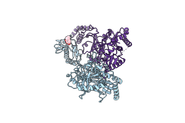
Hiv-1 Reverse Transcriptase Isolated Rnaseh Domain With The Inhibitor Beta-Thujaplicinol Bound At The Active Site
Organism: Human immunodeficiency virus type 1
Method: X-RAY DIFFRACTION Resolution:2.04 Å Release Date: 2010-02-09 Classification: HYDROLASE Ligands: MN, JTH |
Organism: Human immunodeficiency virus type 1
Method: X-RAY DIFFRACTION
Release Date: 2010-02-09
Ligands: MN, JTH
 |
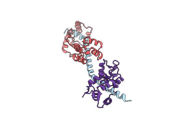
Hiv-1 Reverse Transcriptase With The Inhibitor Beta-Thujaplicinol Bound At The Rnase H Active Site
Organism: Human immunodeficiency virus type 1 bh10
Method: X-RAY DIFFRACTION Resolution:2.80 Å Release Date: 2010-01-26 Classification: TRANSFERASE Ligands: JTH, MN |
Organism: Human immunodeficiency virus type 1 bh10
Method: X-RAY DIFFRACTION
Release Date: 2010-01-26
Ligands: JTH, MN
 |
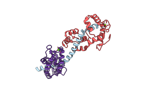
Calcium-Free Scallop Myosin Regulatory Domain With Elc-D19A Point Mutation
Organism: Argopecten irradians
Method: X-RAY DIFFRACTION Resolution:2.57 Å Release Date: 2009-12-01 Classification: CONTRACTILE PROTEIN Ligands: MG |
Organism: Argopecten irradians
Method: X-RAY DIFFRACTION
Release Date: 2009-12-01
Ligands: MG
 |
Calcium-Bound Scallop Myosin Regulatory Domain (Lever Arm) With Reconstituted Complete Light Chains
Organism: Argopecten irradians
Method: X-RAY DIFFRACTION Resolution:2.10 Å Release Date: 2009-12-01 Classification: CONTRACTILE PROTEIN Ligands: MG, CA |
Organism: Argopecten irradians
Method: X-RAY DIFFRACTION
Release Date: 2009-12-01
Ligands: MG, CA
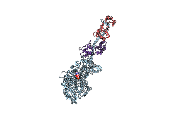 |
Crystal Structure Of Squid Mg.Adp Myosin S1
Organism: Loligo pealei, Todarodes pacificus
Method: X-RAY DIFFRACTION Resolution:3.10 Å Release Date: 2009-08-04 Classification: CONTRACTILE PROTEIN Ligands: MG, ADP |
Organism: Loligo pealei, Todarodes pacificus
Method: X-RAY DIFFRACTION
Release Date: 2009-08-04
Ligands: MG, ADP
 |
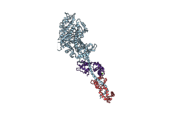
Crystal Structure Of Rigor-Like Squid Myosin S1
Organism: Loligo pealei, Todarodes pacificus
Method: X-RAY DIFFRACTION Resolution:2.60 Å Release Date: 2009-07-28 Classification: CONTRACTILE PROTEIN Ligands: MLI, CA |
Organism: Loligo pealei, Todarodes pacificus
Method: X-RAY DIFFRACTION
Release Date: 2009-07-28
Ligands: MLI, CA
 |
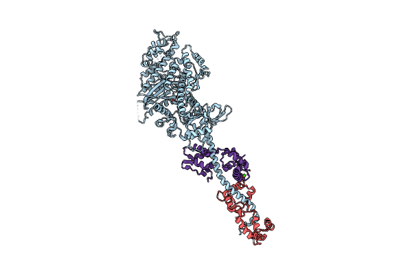
The Crystal Structure Of Rigor Like Squid Myosin S1 In The Absence Of Nucleotide
Organism: Loligo pealei, Todarodes pacificus
Method: X-RAY DIFFRACTION Resolution:3.40 Å Release Date: 2009-07-28 Classification: CONTRACTILE PROTEIN Ligands: CA |
Organism: Loligo pealei, Todarodes pacificus
Method: X-RAY DIFFRACTION
Release Date: 2009-07-28
Ligands: CA
 |
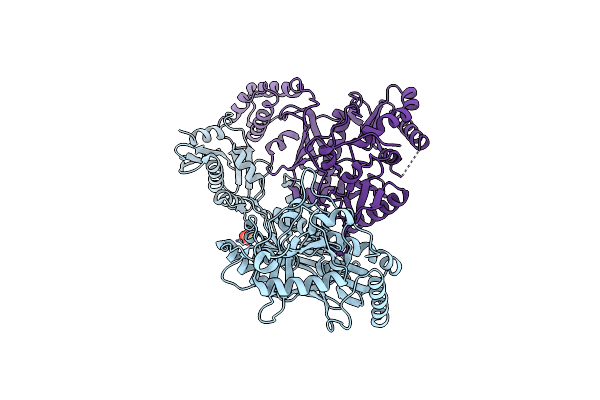
The Crystal Structure Of Squid Myosin S1 In The Presence Of So4 2-
Organism: Loligo pealei, Todarodes pacificus
Method: X-RAY DIFFRACTION Resolution:3.30 Å Release Date: 2009-07-28 Classification: CONTRACTILE PROTEIN Ligands: SO4, CA |
Organism: Loligo pealei, Todarodes pacificus
Method: X-RAY DIFFRACTION
Release Date: 2009-07-28
Ligands: SO4, CA
 |
Crystal Structure Of An Engineered Form Of The Hiv-1 Reverse Transcriptase, Rt69A
Organism: Human immunodeficiency virus type 1 bh10, Human immunodeficiency virus type 1
Method: X-RAY DIFFRACTION Resolution:1.85 Å Release Date: 2008-10-07 Classification: TRANSFERASE Ligands: SO4 |
Organism: Human immunodeficiency virus type 1 bh10, Human immunodeficiency virus type 1
Method: X-RAY DIFFRACTION
Release Date: 2008-10-07
Ligands: SO4
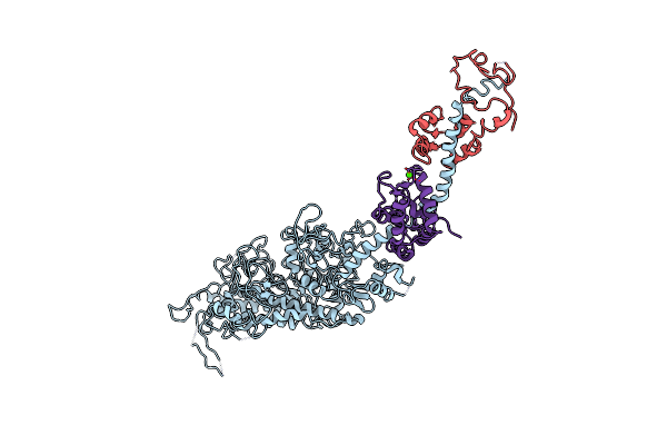 |
Placopecten Striated Muscle Myosin Ii
Organism: Placopecten magellanicus
Method: X-RAY DIFFRACTION Resolution:3.25 Å Release Date: 2008-02-26 Classification: CONTRACTILE PROTEIN Ligands: CA |
Organism: Placopecten magellanicus
Method: X-RAY DIFFRACTION
Release Date: 2008-02-26
Ligands: CA
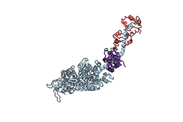 |
Rigor-Like Structures Of Muscle Myosins Reveal Key Mechanical Elements In The Transduction Pathways Of This Allosteric Motor
Organism: Placopecten magellanicus
Method: X-RAY DIFFRACTION Resolution:3.27 Å Release Date: 2007-05-29 Classification: CONTRACTILE PROTEIN Ligands: MG, CA |
Organism: Placopecten magellanicus
Method: X-RAY DIFFRACTION
Release Date: 2007-05-29
Ligands: MG, CA