

Search Count: 24
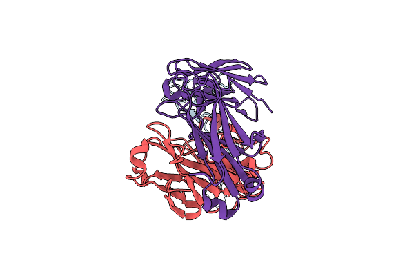 |
Fully Human Monoclonal Antibody Targeting The Cysteine-Rich Substrate-Interacting Region Of Adam17 On Cancer Cells.
Organism: Homo sapiens
Method: ELECTRON MICROSCOPY Release Date: 2024-11-20 Classification: ANTITUMOR PROTEIN/IMMUNE SYSTEM |
Organism: Homo sapiens
Method: ELECTRON MICROSCOPY
Release Date: 2024-11-20
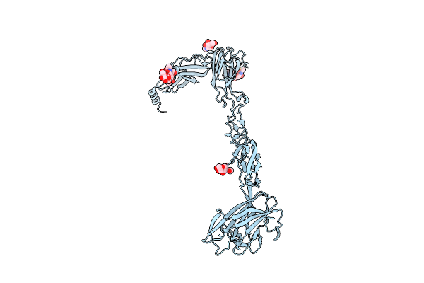 |
Crystal Structure Of The Ephb2 Extracellular Domain
Organism: Mus musculus
Method: X-RAY DIFFRACTION Resolution:3.15 Å Release Date: 2021-10-27 Classification: TRANSFERASE Ligands: NAG |
Organism: Mus musculus
Method: X-RAY DIFFRACTION
Release Date: 2021-10-27
Ligands: NAG
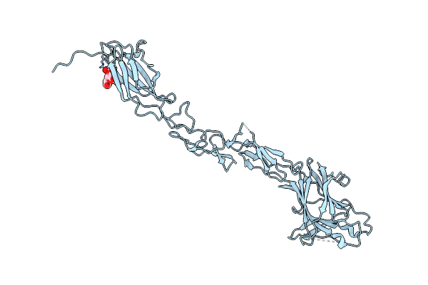 |
Ephb6 Receptor Ectodomain
Organism: Homo sapiens
Method: X-RAY DIFFRACTION Resolution:3.00 Å Release Date: 2021-04-07 Classification: TRANSFERASE |
Organism: Homo sapiens
Method: X-RAY DIFFRACTION
Release Date: 2021-04-07
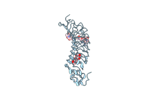 |
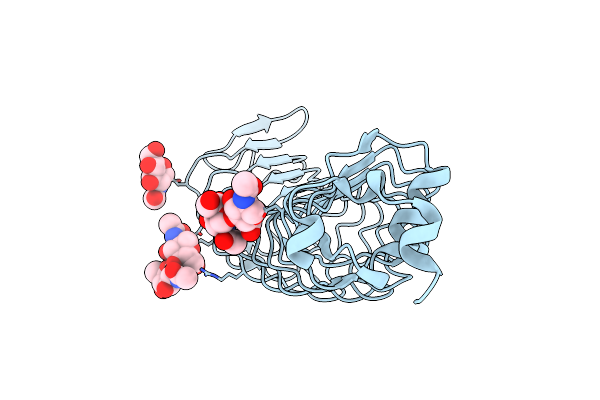
Crystal Structure Of The Nogo-Receptor-2 (27-330)
Organism: Rattus norvegicus
Method: X-RAY DIFFRACTION Resolution:2.10 Å Release Date: 2014-04-30 Classification: MEMBRANE PROTEIN Ligands: NAG |
Organism: Rattus norvegicus
Method: X-RAY DIFFRACTION
Release Date: 2014-04-30
Ligands: NAG
 |
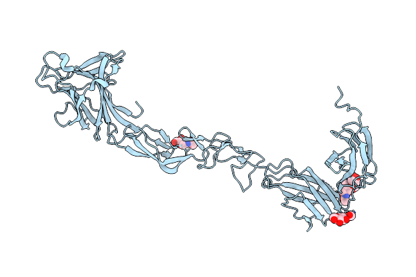
Crystal Structure Of Nogo-Receptor-2
Organism: Rattus norvegicus
Method: X-RAY DIFFRACTION Resolution:1.80 Å Release Date: 2014-04-09 Classification: MEMBRANE PROTEIN Ligands: NAG |
Organism: Rattus norvegicus
Method: X-RAY DIFFRACTION
Release Date: 2014-04-09
Ligands: NAG
 |
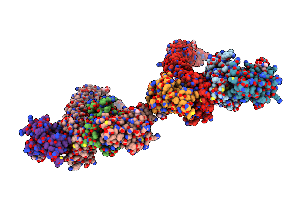
Crystal Structure Of Epha4 Ectodomain
Organism: Homo sapiens
Method: X-RAY DIFFRACTION Resolution:2.08 Å Release Date: 2013-10-30 Classification: TRANSFERASE Ligands: NAG |
Organism: Homo sapiens
Method: X-RAY DIFFRACTION
Release Date: 2013-10-30
Ligands: NAG
 |
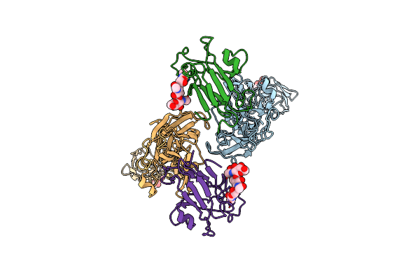
Epha4 Ectodomain Complex With Ephrin A5
Organism: Homo sapiens
Method: X-RAY DIFFRACTION Resolution:3.13 Å Release Date: 2013-10-30 Classification: TRANSFERASE Ligands: NAG |
Organism: Homo sapiens
Method: X-RAY DIFFRACTION
Release Date: 2013-10-30
Ligands: NAG
 |
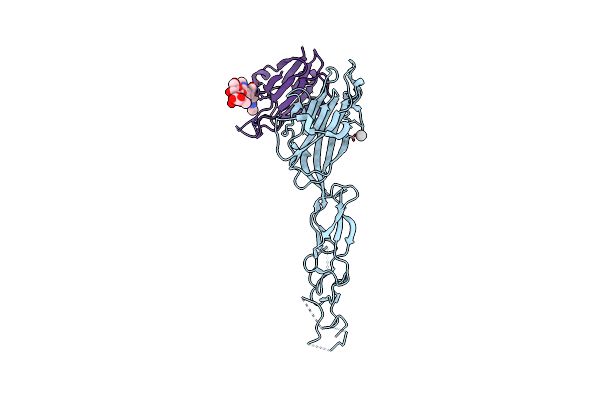
Crystal Structure Of Epha2 Ectodomain In Complex With Ephrin-A5
Organism: Homo sapiens
Method: X-RAY DIFFRACTION Resolution:3.51 Å Release Date: 2010-06-30 Classification: TRANSFERASE RECEPTOR/SIGNALLING PROTEIN Ligands: NAG |
Organism: Homo sapiens
Method: X-RAY DIFFRACTION
Release Date: 2010-06-30
Ligands: NAG
 |
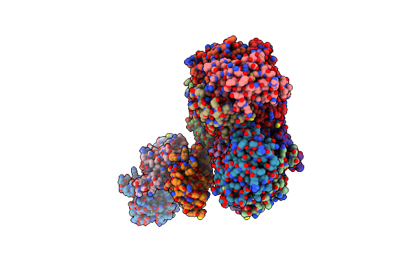
Crystal Structure Of The Human Ephrin A2 Lbd And Crd Domains In Complex With Ephrin A1
Organism: Homo sapiens
Method: X-RAY DIFFRACTION Resolution:2.81 Å Release Date: 2010-06-09 Classification: TRANSFERASE/SIGNALING PROTEIN Ligands: UNX |
Organism: Homo sapiens
Method: X-RAY DIFFRACTION
Release Date: 2010-06-09
Ligands: UNX
 |
Ligand Recognition By A-Class Eph Receptors: Crystal Structures Of The Epha2 Ligand-Binding Domain And The Epha2/Ephrin-A1 Complex
Organism: Homo sapiens
Method: X-RAY DIFFRACTION Resolution:2.00 Å Release Date: 2009-06-30 Classification: TRANSFERASE/SIGNALING PROTEIN |
Organism: Homo sapiens
Method: X-RAY DIFFRACTION
Release Date: 2009-06-30
 |
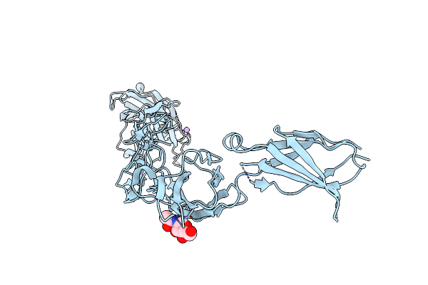
Ligand Recognition By A-Class Eph Receptors: Crystal Structures Of The Epha2 Ligand-Binding Domain And The Epha2/Ephrin-A1 Complex
Organism: Homo sapiens
Method: X-RAY DIFFRACTION Resolution:2.52 Å Release Date: 2009-06-30 Classification: TRANSFERASE |
Organism: Homo sapiens
Method: X-RAY DIFFRACTION
Release Date: 2009-06-30
 |
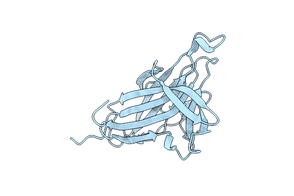
Crystal Structure Of The Human Ephrin A2 Ectodomain
Organism: Homo sapiens
Method: X-RAY DIFFRACTION Resolution:2.50 Å Release Date: 2009-01-27 Classification: TRANSFERASE, SIGNALING PROTEIN |
Organism: Homo sapiens
Method: X-RAY DIFFRACTION
Release Date: 2009-01-27
 |
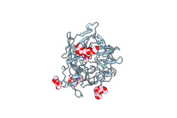
The Crystal Structure Of The Ligand-Binding Domain Of The Ephb2 Receptor At 2.0 A Resolution
Organism: Mus musculus
Method: X-RAY DIFFRACTION Resolution:2.00 Å Release Date: 2008-10-21 Classification: TRANSFERASE |
Organism: Mus musculus
Method: X-RAY DIFFRACTION
Release Date: 2008-10-21
 |
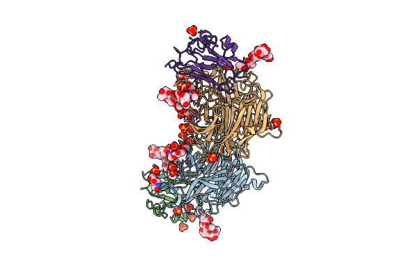
Crystal Structures Of The Nipah G Attachment Glycoprotein
Organism: Nipah virus
Method: X-RAY DIFFRACTION Resolution:2.31 Å Release Date: 2008-08-19 Classification: HYDROLASE Ligands: NAG, IOD |
Organism: Nipah virus
Method: X-RAY DIFFRACTION
Release Date: 2008-08-19
Ligands: NAG, IOD
 |
Crystal Structures Of Nipah Virus G Attachment Glycoprotein In Complex With Its Receptor Ephrin-B3
Organism: Nipah virus, Mus musculus
Method: X-RAY DIFFRACTION Resolution:3.01 Å Release Date: 2008-08-19 Classification: HYDROLASE/MEMBRANE PROTEIN Ligands: NAG, SO4 |
Organism: Nipah virus, Mus musculus
Method: X-RAY DIFFRACTION
Release Date: 2008-08-19
Ligands: NAG, SO4
 |
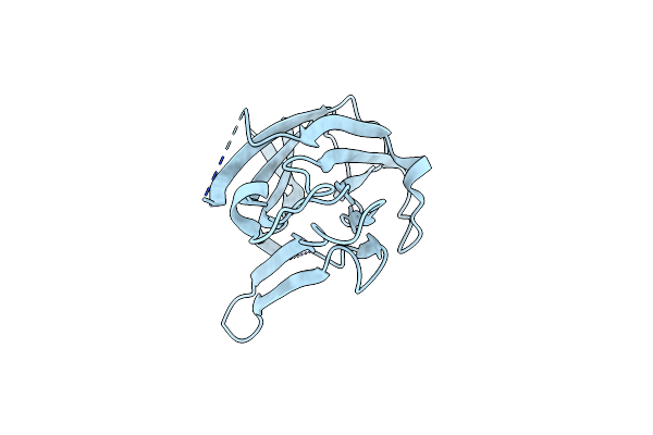
Crystal Structure Of The Human Ephrin A2- Ephrin A1 Complex
Organism: Homo sapiens
Method: X-RAY DIFFRACTION Resolution:2.65 Å Release Date: 2008-08-12 Classification: TRANSFERASE/SIGNALING PROTEIN |
Organism: Homo sapiens
Method: X-RAY DIFFRACTION
Release Date: 2008-08-12
 |
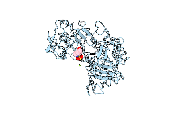
Crystal Structure Of The Ligand Binding Domain Of Human Ephrin A2 (Epha2) Receptor Protein Kinase
Organism: Homo sapiens
Method: X-RAY DIFFRACTION Resolution:1.95 Å Release Date: 2008-03-25 Classification: TRANSFERASE |
Organism: Homo sapiens
Method: X-RAY DIFFRACTION
Release Date: 2008-03-25
 |
Crystal Structure Of The Apo Form Of E. Coli Tryptophanase At 1.9 A Resolution
Organism: Escherichia coli
Method: X-RAY DIFFRACTION Resolution:1.90 Å Release Date: 2007-02-20 Classification: LYASE Ligands: CL, MG, EPE |
Organism: Escherichia coli
Method: X-RAY DIFFRACTION
Release Date: 2007-02-20
Ligands: CL, MG, EPE
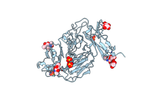 |
Tie2 Ligand-Binding Domain Crystal Structure
Organism: Homo sapiens
Method: X-RAY DIFFRACTION Resolution:2.90 Å Release Date: 2006-06-06 Classification: TRANSFERASE Ligands: NAG, NDG, SO4 |
Organism: Homo sapiens
Method: X-RAY DIFFRACTION
Release Date: 2006-06-06
Ligands: NAG, NDG, SO4
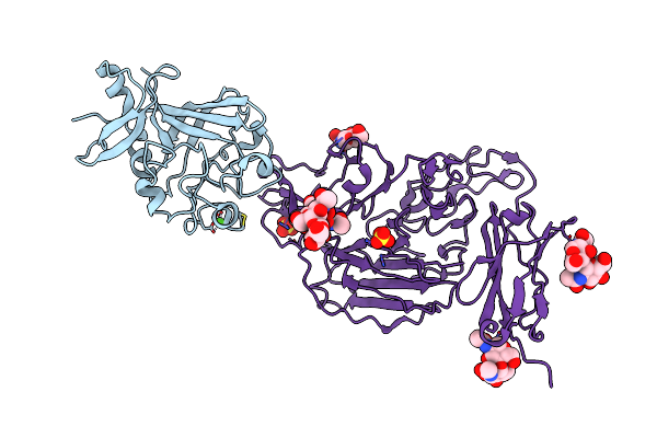 |
Angiopoietin-2/Tie2 Complex Crystal Structure
Organism: Homo sapiens
Method: X-RAY DIFFRACTION Resolution:3.70 Å Release Date: 2006-06-06 Classification: SIGNALING PROTEIN Ligands: CA, NAG, SO4 |
Organism: Homo sapiens
Method: X-RAY DIFFRACTION
Release Date: 2006-06-06
Ligands: CA, NAG, SO4