

Search Count: 20
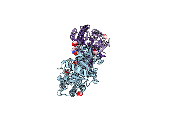 |
Protein Arginine Kinase Mcsb In The Parg-Bound State
Organism: Geobacillus stearothermophilus
Method: X-RAY DIFFRACTION Resolution:1.85 Å Release Date: 2019-02-27 Classification: SIGNALING PROTEIN Ligands: RPI, EDO |
Organism: Geobacillus stearothermophilus
Method: X-RAY DIFFRACTION
Release Date: 2019-02-27
Ligands: RPI, EDO
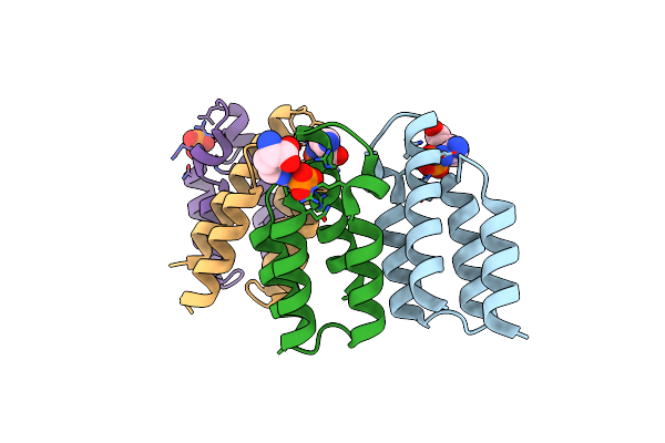 |
Ctsr C-Terminal Domain With Bound Phospho-Arginine
Organism: Bacillus subtilis
Method: X-RAY DIFFRACTION Resolution:2.49 Å Release Date: 2019-02-27 Classification: SIGNALING PROTEIN Ligands: RPI, PO4 |
Organism: Bacillus subtilis
Method: X-RAY DIFFRACTION
Release Date: 2019-02-27
Ligands: RPI, PO4
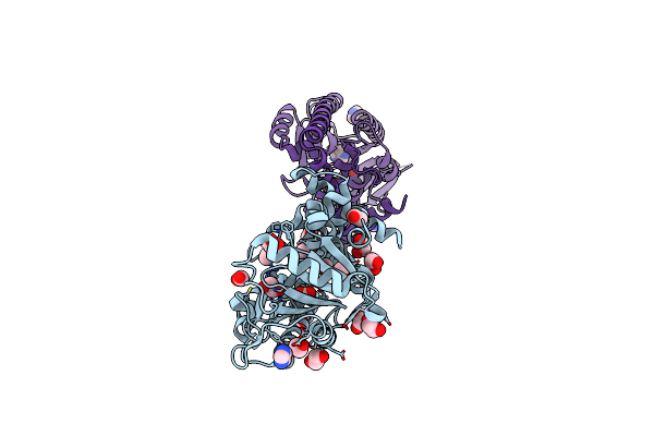 |
Protein Arginine Kinase Mcsb In The Apo State
Organism: Geobacillus stearothermophilus
Method: X-RAY DIFFRACTION Resolution:1.70 Å Release Date: 2019-02-06 Classification: SIGNALING PROTEIN Ligands: FMT, EDO, IMD |
Organism: Geobacillus stearothermophilus
Method: X-RAY DIFFRACTION
Release Date: 2019-02-06
Ligands: FMT, EDO, IMD
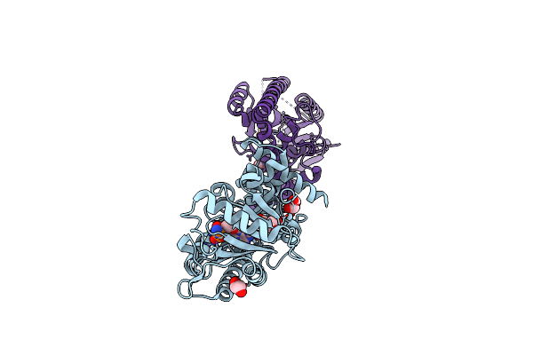 |
Protein Arginine Kinase Mcsb In The Amp-Pn-Bound State
Organism: Geobacillus stearothermophilus
Method: X-RAY DIFFRACTION Resolution:2.70 Å Release Date: 2019-02-06 Classification: SIGNALING PROTEIN Ligands: AN2, EDO |
Organism: Geobacillus stearothermophilus
Method: X-RAY DIFFRACTION
Release Date: 2019-02-06
Ligands: AN2, EDO
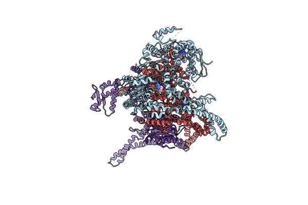 |
Crystal Structure Of Hsp104
Organism: Chaetomium thermophilum
Method: X-RAY DIFFRACTION Resolution:3.70 Å Release Date: 2016-12-07 Classification: CHAPERONE Ligands: ADP |
Organism: Chaetomium thermophilum
Method: X-RAY DIFFRACTION
Release Date: 2016-12-07
Ligands: ADP
 |
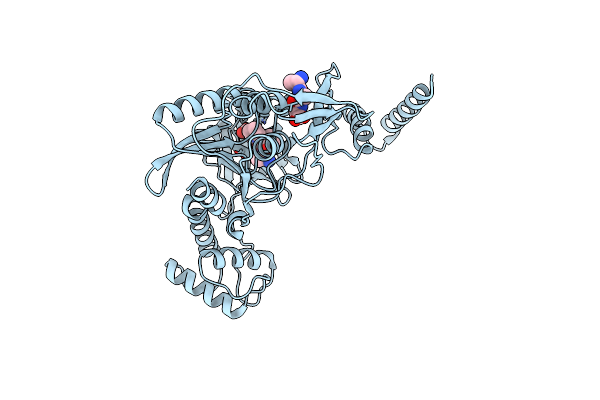
Clpc N-Terminal Domain With Bound Phospho-Arginine
Organism: Bacillus subtilis (strain 168)
Method: X-RAY DIFFRACTION Resolution:1.60 Å Release Date: 2016-10-12 Classification: HYDROLASE Ligands: SO4, RPI, ACT |
Organism: Bacillus subtilis (strain 168)
Method: X-RAY DIFFRACTION
Release Date: 2016-10-12
Ligands: SO4, RPI, ACT
 |
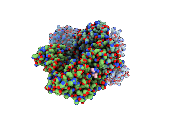
Crystal Structure Of The Protease Ctpb In An Active State
Organism: Bacillus subtilis subsp. subtilis str. 168, Escherichia coli
Method: X-RAY DIFFRACTION Resolution:1.90 Å Release Date: 2013-12-04 Classification: HYDROLASE |
Organism: Bacillus subtilis subsp. subtilis str. 168, Escherichia coli
Method: X-RAY DIFFRACTION
Release Date: 2013-12-04
 |
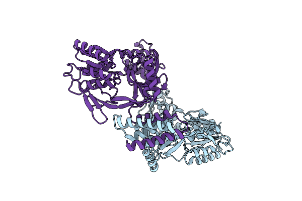
Crystal Structure Of The Protease Ctpb In An Active State
Organism: Bacillus subtilis subsp. subtilis str. 168, Escherichia coli
Method: X-RAY DIFFRACTION Resolution:2.70 Å Release Date: 2013-12-04 Classification: HYDROLASE/PEPTIDE |
Organism: Bacillus subtilis subsp. subtilis str. 168, Escherichia coli
Method: X-RAY DIFFRACTION
Release Date: 2013-12-04
 |
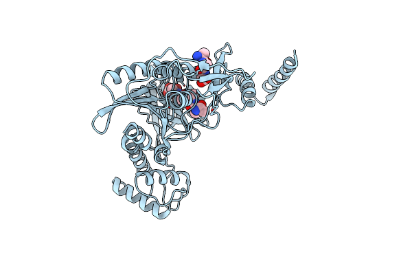
Crystal Structure Of The Protease Ctpb(S309A) Present In A Resting State
Organism: Bacillus subtilis subsp. subtilis str. 168
Method: X-RAY DIFFRACTION Resolution:1.80 Å Release Date: 2013-12-04 Classification: HYDROLASE |
Organism: Bacillus subtilis subsp. subtilis str. 168
Method: X-RAY DIFFRACTION
Release Date: 2013-12-04
 |
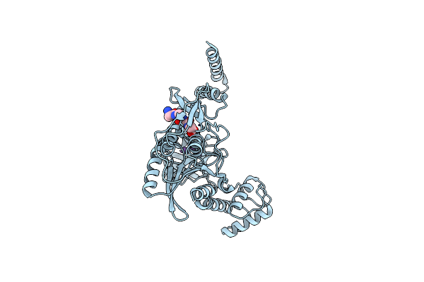
Crystal Structure Of The Ctpb R168A Mutant Present In An Active Conformation
Organism: Bacillus subtilis subsp. subtilis str. 168, Escherichia coli
Method: X-RAY DIFFRACTION Resolution:2.40 Å Release Date: 2013-12-04 Classification: HYDROLASE |
Organism: Bacillus subtilis subsp. subtilis str. 168, Escherichia coli
Method: X-RAY DIFFRACTION
Release Date: 2013-12-04
 |
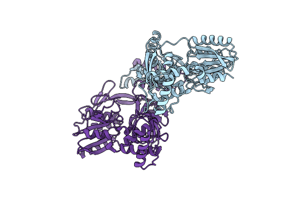
Crystal Structure Of Ctpb(S309A) In Complex With A Peptide Having A Val-Pro-Ala C-Terminus
Organism: Bacillus subtilis subsp. subtilis str. 168, Escherichia coli
Method: X-RAY DIFFRACTION Resolution:1.90 Å Release Date: 2013-12-04 Classification: HYDROLASE/PEPTIDE |
Organism: Bacillus subtilis subsp. subtilis str. 168, Escherichia coli
Method: X-RAY DIFFRACTION
Release Date: 2013-12-04
 |
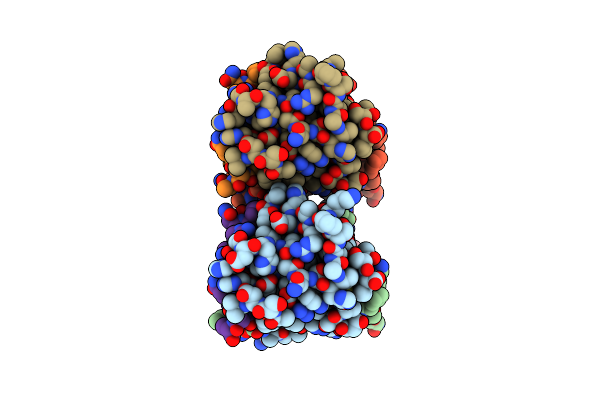
Crystal Structure Of The Ctpb(V118Y) Mutant
Organism: Bacillus subtilis subsp. subtilis str. 168
Method: X-RAY DIFFRACTION Resolution:1.95 Å Release Date: 2013-12-04 Classification: HYDROLASE |
Organism: Bacillus subtilis subsp. subtilis str. 168
Method: X-RAY DIFFRACTION
Release Date: 2013-12-04
 |
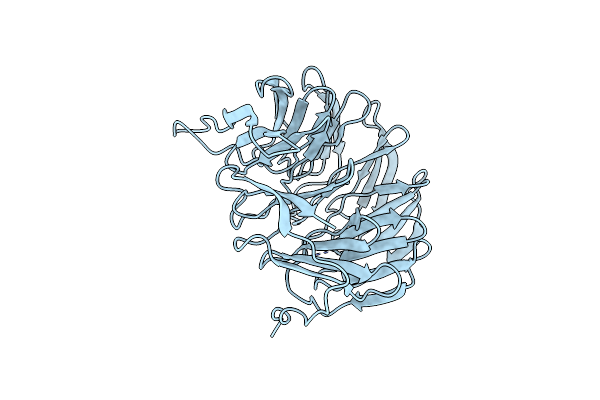
Crystal Structure Of The Spc24-Spc25/Cnn1 Binding Interface
Organism: Saccharomyces cerevisiae s288c
Method: X-RAY DIFFRACTION Resolution:2.01 Å Release Date: 2013-01-30 Classification: CELL CYCLE Ligands: GOL |
Organism: Saccharomyces cerevisiae s288c
Method: X-RAY DIFFRACTION
Release Date: 2013-01-30
Ligands: GOL
 |
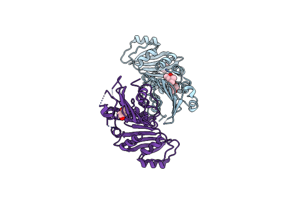
Crystal Structure Of The Lipoprotein Bamb
Organism: Escherichia coli
Method: X-RAY DIFFRACTION Resolution:1.80 Å Release Date: 2011-01-19 Classification: STRUCTURAL PROTEIN |
Organism: Escherichia coli
Method: X-RAY DIFFRACTION
Release Date: 2011-01-19
 |
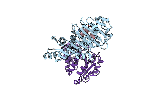
Structure Of Crystal Form I Of Tp0453
Organism: Treponema pallidum
Method: X-RAY DIFFRACTION Resolution:1.95 Å Release Date: 2010-10-27 Classification: MEMBRANE PROTEIN Ligands: MPD |
Organism: Treponema pallidum
Method: X-RAY DIFFRACTION
Release Date: 2010-10-27
Ligands: MPD
 |
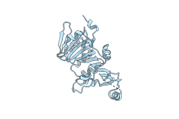
Structure Of Crystal Form I Of Tp0453
Organism: Treponema pallidum
Method: X-RAY DIFFRACTION Resolution:2.39 Å Release Date: 2010-10-27 Classification: MEMBRANE PROTEIN Ligands: MPD |
Organism: Treponema pallidum
Method: X-RAY DIFFRACTION
Release Date: 2010-10-27
Ligands: MPD
 |
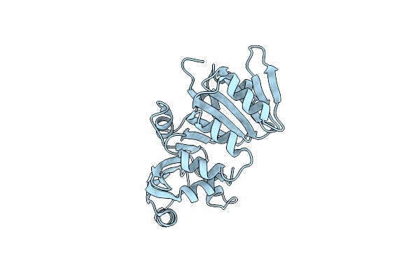
Structure Of Crystal Form Iv Of Tp0453
Organism: Treponema pallidum
Method: X-RAY DIFFRACTION Resolution:2.20 Å Release Date: 2010-10-27 Classification: MEMBRANE PROTEIN |
Organism: Treponema pallidum
Method: X-RAY DIFFRACTION
Release Date: 2010-10-27
 |
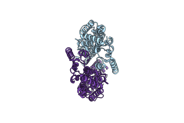
Structure Of Crystal Form Iii Of Tp0453
Organism: Treponema pallidum
Method: X-RAY DIFFRACTION Resolution:2.20 Å Release Date: 2010-10-27 Classification: MEMBRANE PROTEIN |
Organism: Treponema pallidum
Method: X-RAY DIFFRACTION
Release Date: 2010-10-27
 |
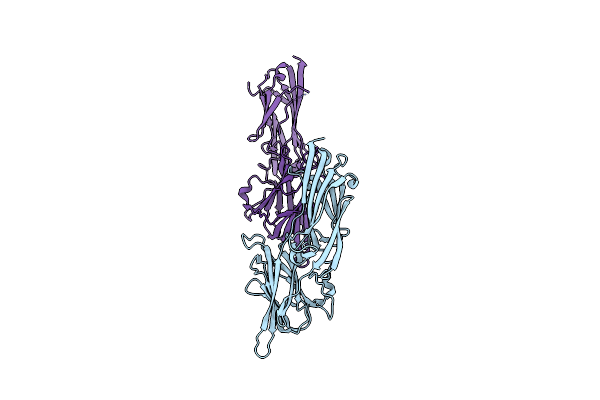
Crystal Structure Of The Globular Tail Of Myo4P
Organism: Saccharomyces cerevisiae
Method: X-RAY DIFFRACTION Resolution:2.30 Å Release Date: 2010-05-12 Classification: MOTOR PROTEIN |
Organism: Saccharomyces cerevisiae
Method: X-RAY DIFFRACTION
Release Date: 2010-05-12
 |
Crystal Structure Analysis Of Double Cysteine Mutant Of S.Epidermidis Adhesin Sdrg: Evidence For The Dock,Lock And Latch Ligand Binding Mechanism
Organism: Staphylococcus epidermidis
Method: X-RAY DIFFRACTION Resolution:2.80 Å Release Date: 2007-11-06 Classification: CELL ADHESION |
Organism: Staphylococcus epidermidis
Method: X-RAY DIFFRACTION
Release Date: 2007-11-06