

Search Count: 20
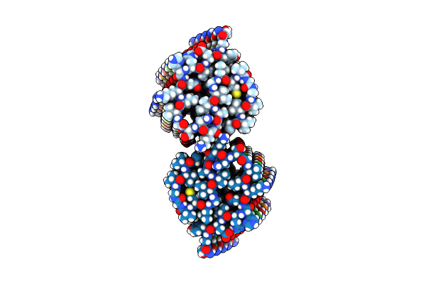 |
Cryo-Em Structure Of Amyloid-Beta Fibrils From Mouse Brain Carrying The Uppsala Abetaupp(1-42)Delta(19-24) Mutation
Organism: Mus musculus
Method: ELECTRON MICROSCOPY Release Date: 2025-06-11 Classification: PROTEIN FIBRIL |
Organism: Mus musculus
Method: ELECTRON MICROSCOPY
Release Date: 2025-06-11
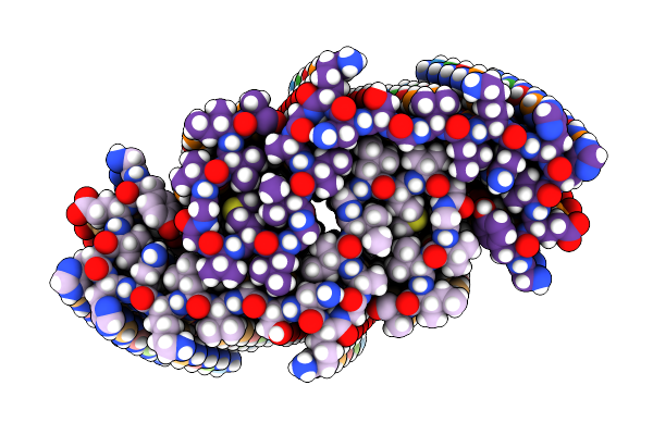 |
Cryo-Em Structure Of Amyloid-Beta Fibrils Carrying The Uppsala Abetaupp(1-42)Delta(19-24) Mutation - Polymorph 1
Organism: Homo sapiens
Method: ELECTRON MICROSCOPY Release Date: 2025-06-11 Classification: PROTEIN FIBRIL |
Organism: Homo sapiens
Method: ELECTRON MICROSCOPY
Release Date: 2025-06-11
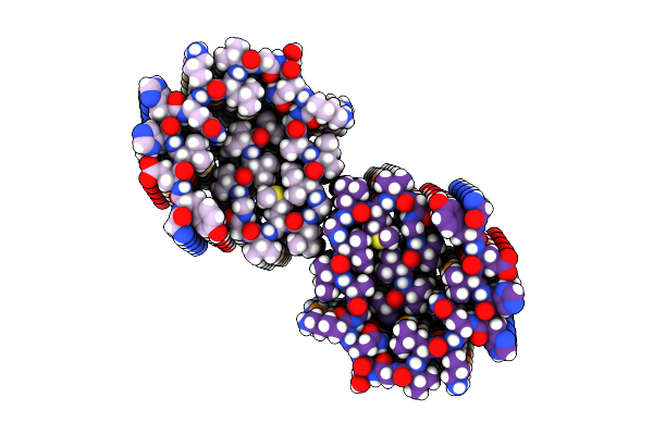 |
Cryo-Em Structure Of Amyloid-Beta Fibrils Carrying The Uppsala Abetaupp(1-42)Delta(19-24) Mutation - Polymorph 2
Organism: Homo sapiens
Method: ELECTRON MICROSCOPY Release Date: 2025-06-11 Classification: PROTEIN FIBRIL |
Organism: Homo sapiens
Method: ELECTRON MICROSCOPY
Release Date: 2025-06-11
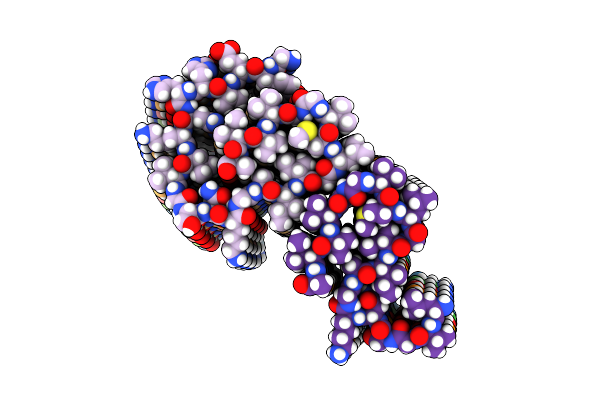 |
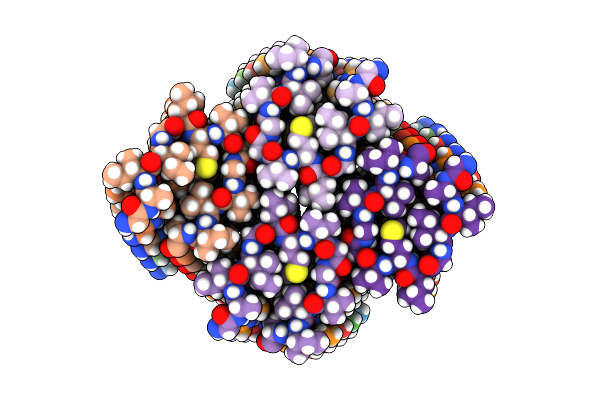
Cryo-Em Structure Of Amyloid-Beta Fibrils Carrying The Uppsala Abetaupp(1-42)Delta(19-24) Mutation - Polymorph 3
Organism: Homo sapiens
Method: ELECTRON MICROSCOPY Release Date: 2025-06-11 Classification: PROTEIN FIBRIL |
Organism: Homo sapiens
Method: ELECTRON MICROSCOPY
Release Date: 2025-06-11
 |
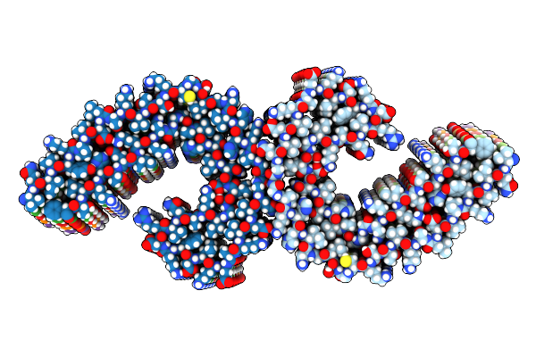
Cryo-Em Structure Of Amyloid-Beta Fibrils Carrying The Uppsala Abetaupp(1-42)Delta(19-24) Mutation - Polymorph 4
Organism: Homo sapiens
Method: ELECTRON MICROSCOPY Release Date: 2025-06-11 Classification: PROTEIN FIBRIL |
Organism: Homo sapiens
Method: ELECTRON MICROSCOPY
Release Date: 2025-06-11
 |
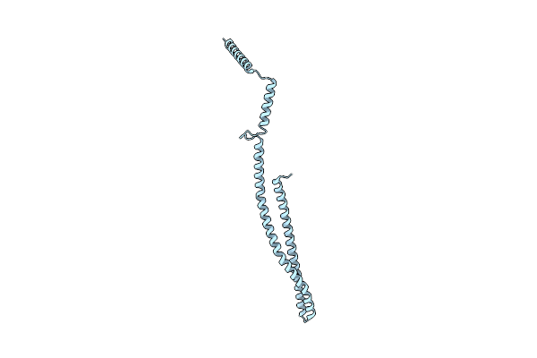
Cryo-Em Structure Of Tau Filaments From Individuals Carrying The Uppsala Abetaupp(1-42)Delta(19-24) Mutation
Organism: Homo sapiens
Method: ELECTRON MICROSCOPY Release Date: 2025-06-11 Classification: PROTEIN FIBRIL |
Organism: Homo sapiens
Method: ELECTRON MICROSCOPY
Release Date: 2025-06-11
 |
Helical Structure Of Pspa
Organism: Synechocystis sp. (strain pcc 6803 / kazusa)
Method: ELECTRON MICROSCOPY Release Date: 2021-08-04 Classification: LIPID BINDING PROTEIN |
Organism: Synechocystis sp. (strain pcc 6803 / kazusa)
Method: ELECTRON MICROSCOPY
Release Date: 2021-08-04
 |
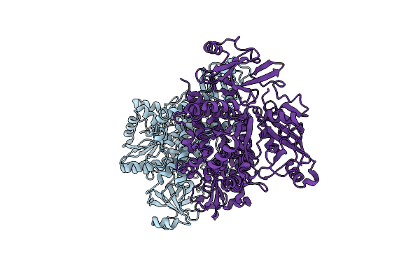
Crystal Structure Of Full-Length Cnfy (C866S) From Yersinia Pseudotuberculosis
Organism: Yersinia pseudotuberculosis
Method: X-RAY DIFFRACTION Resolution:2.70 Å Release Date: 2020-12-30 Classification: TOXIN Ligands: SO4, CL |
Organism: Yersinia pseudotuberculosis
Method: X-RAY DIFFRACTION
Release Date: 2020-12-30
Ligands: SO4, CL
 |
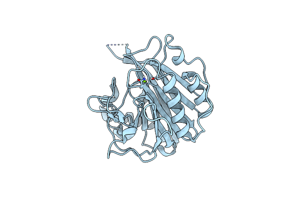
Crystal Structure Of Cnfy From Yersinia Pseudotuberculosis - N-Terminal Fragment Comprising Residues 1-704
Organism: Yersinia pseudotuberculosis
Method: X-RAY DIFFRACTION Resolution:3.28 Å Release Date: 2020-12-30 Classification: TOXIN |
Organism: Yersinia pseudotuberculosis
Method: X-RAY DIFFRACTION
Release Date: 2020-12-30
 |
Crystal Structure Of The C-Terminal Domain Of Cnfy From Yersinia Pseudotuberculosis
Organism: Yersinia pseudotuberculosis
Method: X-RAY DIFFRACTION Resolution:1.13 Å Release Date: 2020-12-30 Classification: TOXIN Ligands: MG |
Organism: Yersinia pseudotuberculosis
Method: X-RAY DIFFRACTION
Release Date: 2020-12-30
Ligands: MG
 |
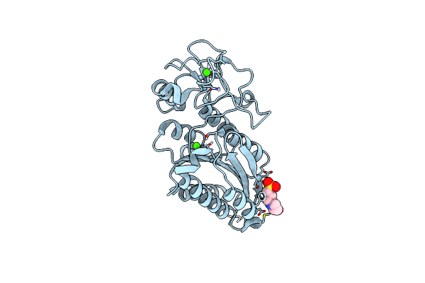
Crystal Structure Of Domains 4-5 Of Cnfy From Yersinia Pseudotuberculosis
Organism: Yersinia pseudotuberculosis
Method: X-RAY DIFFRACTION Resolution:1.80 Å Release Date: 2020-12-30 Classification: TOXIN Ligands: BU3, MG, NA, CL, SO4 |
Organism: Yersinia pseudotuberculosis
Method: X-RAY DIFFRACTION
Release Date: 2020-12-30
Ligands: BU3, MG, NA, CL, SO4
 |
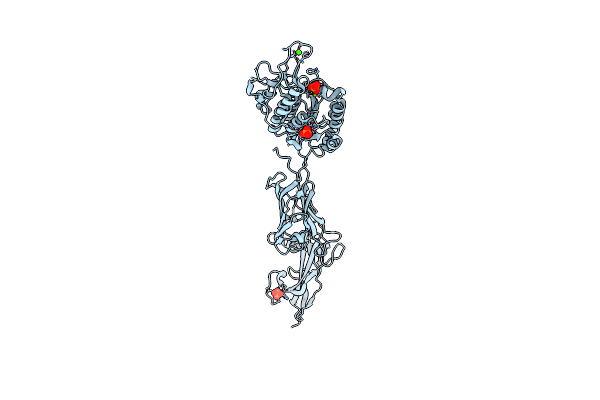
Crystal Structure Of The Vwf Domain Of The Type V Pili Tip Protein Mfa5 From Porphyromonas Gingivalis
Organism: Porphyromonas gingivalis atcc 33277
Method: X-RAY DIFFRACTION Resolution:1.85 Å Release Date: 2020-12-02 Classification: CELL ADHESION Ligands: MPO, CA |
Organism: Porphyromonas gingivalis atcc 33277
Method: X-RAY DIFFRACTION
Release Date: 2020-12-02
Ligands: MPO, CA
 |
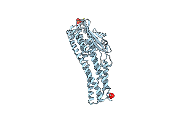
Crystal Structure Of Three N-Terminal Domains Of The Type V Pili Tip Protein Mfa5 From Porphyromonas Gingivalis
Organism: Porphyromonas gingivalis atcc 33277
Method: X-RAY DIFFRACTION Resolution:1.80 Å Release Date: 2020-12-02 Classification: CELL ADHESION Ligands: MG, PO4, CA |
Organism: Porphyromonas gingivalis atcc 33277
Method: X-RAY DIFFRACTION
Release Date: 2020-12-02
Ligands: MG, PO4, CA
 |
The Cytotoxin Makb From Vibrio Cholerae
Organism: Vibrio cholerae serotype o1 (strain atcc 39315 / el tor inaba n16961)
Method: X-RAY DIFFRACTION Resolution:2.10 Å Release Date: 2020-11-18 Classification: TOXIN Ligands: SO4 |
Organism: Vibrio cholerae serotype o1 (strain atcc 39315 / el tor inaba n16961)
Method: X-RAY DIFFRACTION
Release Date: 2020-11-18
Ligands: SO4
 |
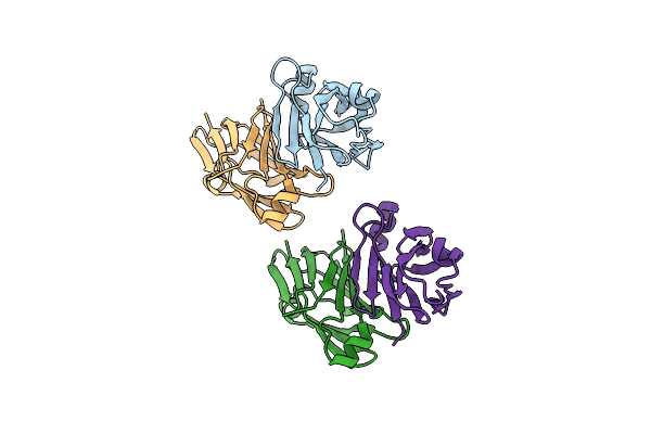
The Cytotoxin Make From Vibrio Cholerae
Organism: Vibrio cholerae
Method: X-RAY DIFFRACTION Resolution:1.98 Å Release Date: 2020-11-18 Classification: TOXIN Ligands: NI, SO4 |
Organism: Vibrio cholerae
Method: X-RAY DIFFRACTION
Release Date: 2020-11-18
Ligands: NI, SO4
 |
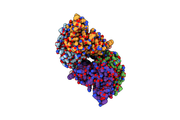
Makd From The Mak Operon Of Vibrio Cholerae
Organism: Vibrio cholerae o1 biovar el tor str. n16961
Method: X-RAY DIFFRACTION Resolution:2.02 Å Release Date: 2020-11-18 Classification: UNKNOWN FUNCTION |
Organism: Vibrio cholerae o1 biovar el tor str. n16961
Method: X-RAY DIFFRACTION
Release Date: 2020-11-18
 |
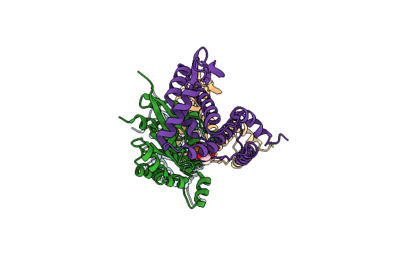
Shigella Ipgb2 In Complex With Human Rhoa, Gdp And Mg2+ (Complex A)
Organism: Homo sapiens, Shigella flexneri
Method: X-RAY DIFFRACTION Resolution:1.85 Å Release Date: 2010-03-31 Classification: Signaling Protein/RHOA-BINDING PROTEIN Ligands: GDP, MG |
Organism: Homo sapiens, Shigella flexneri
Method: X-RAY DIFFRACTION
Release Date: 2010-03-31
Ligands: GDP, MG
 |
Shigella Ipgb2 In Complex With Human Rhoa, Gdp And Mg2+ (Complex B)
Organism: Homo sapiens, Shigella flexneri
Method: X-RAY DIFFRACTION Resolution:2.28 Å Release Date: 2010-03-31 Classification: Signaling Protein/RHOA-BINDING PROTEIN Ligands: GDP, MG |
Organism: Homo sapiens, Shigella flexneri
Method: X-RAY DIFFRACTION
Release Date: 2010-03-31
Ligands: GDP, MG
 |
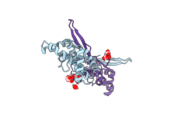
Shigella Ipgb2 In Complex With Human Rhoa And Gdp (Complex C)
Organism: Homo sapiens, Shigella flexneri
Method: X-RAY DIFFRACTION Resolution:1.68 Å Release Date: 2010-03-31 Classification: Signaling Protein/RHOA-BINDING PROTEIN Ligands: GDP, SO4 |
Organism: Homo sapiens, Shigella flexneri
Method: X-RAY DIFFRACTION
Release Date: 2010-03-31
Ligands: GDP, SO4
 |
Crystal Structure Of Ipgb2 From Shigella Flexneri
Organism: Shigella flexneri
Method: X-RAY DIFFRACTION Resolution:2.30 Å Release Date: 2010-03-31 Classification: RHOA-BINDING PROTEIN Ligands: FLC, FEO |
Organism: Shigella flexneri
Method: X-RAY DIFFRACTION
Release Date: 2010-03-31
Ligands: FLC, FEO