

Search Count: 25
 |
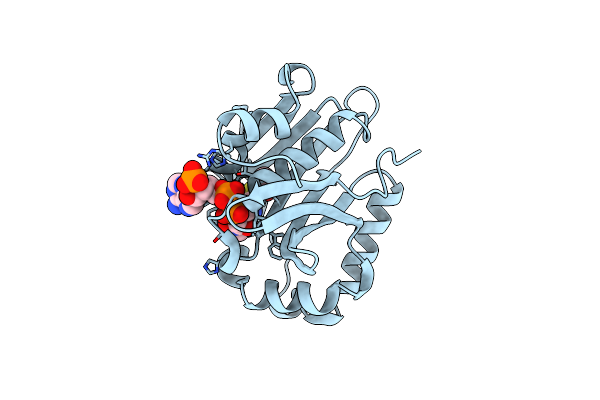
Crystal Structure Of Ilvd From Mycobacterium Tuberculosis
Organism: Mycobacterium tuberculosis
Method: X-RAY DIFFRACTION Resolution:1.88 Å Release Date: 2019-08-07 Classification: LYASE Ligands: MG, FES, PEG, NA |
Organism: Mycobacterium tuberculosis
Method: X-RAY DIFFRACTION
Release Date: 2019-08-07
Ligands: MG, FES, PEG, NA
 |
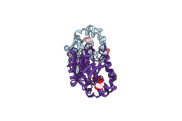
Mycobacterium Smegmatis Rimj With Accoa
Organism: Mycobacterium smegmatis
Method: X-RAY DIFFRACTION Resolution:1.90 Å Release Date: 2019-01-23 Classification: TRANSFERASE Ligands: ACO |
Organism: Mycobacterium smegmatis
Method: X-RAY DIFFRACTION
Release Date: 2019-01-23
Ligands: ACO
 |
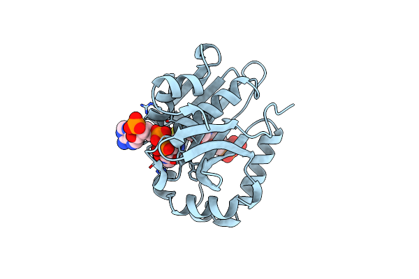
Mycobacterium Smegmatis Rimj (Apo Form)
Organism: Mycobacterium smegmatis
Method: X-RAY DIFFRACTION Resolution:1.40 Å Release Date: 2019-01-16 Classification: TRANSFERASE Ligands: GOL, CL |
Organism: Mycobacterium smegmatis
Method: X-RAY DIFFRACTION
Release Date: 2019-01-16
Ligands: GOL, CL
 |
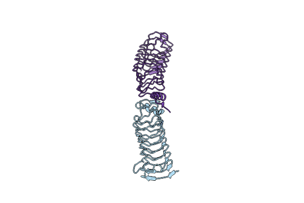
Mycobacterium Smegmatis Rimj In Complex With Coa-Disulfide
Organism: Mycobacterium smegmatis
Method: X-RAY DIFFRACTION Resolution:1.70 Å Release Date: 2019-01-16 Classification: TRANSFERASE Ligands: 5NG |
Organism: Mycobacterium smegmatis
Method: X-RAY DIFFRACTION
Release Date: 2019-01-16
Ligands: 5NG
 |
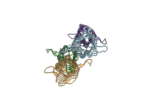
Structure Of Qnrb1 (R167E-Trypsin Treated), A Plasmid-Mediated Fluoroquinolone Resistance Protein
Organism: Klebsiella pneumoniae
Method: X-RAY DIFFRACTION Resolution:1.80 Å Release Date: 2010-10-27 Classification: CELL CYCLE |
Organism: Klebsiella pneumoniae
Method: X-RAY DIFFRACTION
Release Date: 2010-10-27
 |
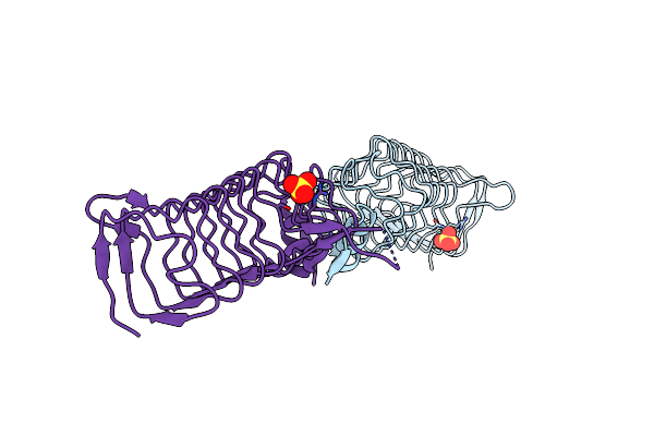
Structure Of Qnrb1 (Full Length), A Plasmid-Mediated Fluoroquinolone Resistance Protein
Organism: Klebsiella pneumoniae
Method: X-RAY DIFFRACTION Resolution:2.80 Å Release Date: 2010-10-20 Classification: CELL CYCLE |
Organism: Klebsiella pneumoniae
Method: X-RAY DIFFRACTION
Release Date: 2010-10-20
 |
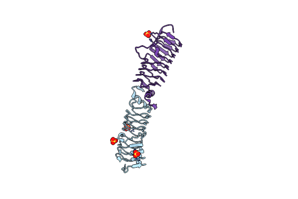
Structure Of Qnrb1 (M102R-Trypsin Treated), A Plasmid-Mediated Fluoroquinolone Resistance Protein
Organism: Klebsiella pneumoniae
Method: X-RAY DIFFRACTION Resolution:2.20 Å Release Date: 2010-10-20 Classification: CELL CYCLE Ligands: SO4 |
Organism: Klebsiella pneumoniae
Method: X-RAY DIFFRACTION
Release Date: 2010-10-20
Ligands: SO4
 |
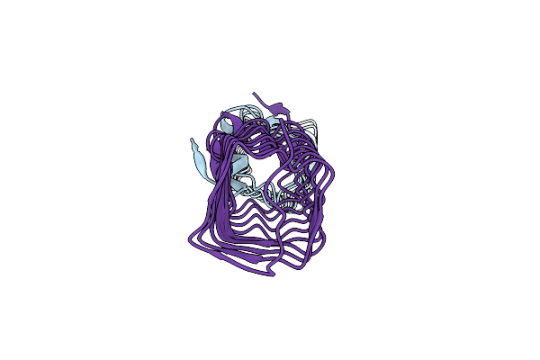
Structure Of The Pentapeptide Repeat Protein Albg, A Resistance Factor For The Topoisomerase Poison Albicidin.
Organism: Xanthomonas albilineans
Method: X-RAY DIFFRACTION Resolution:2.00 Å Release Date: 2010-10-13 Classification: CELL CYCLE Ligands: SO4 |
Organism: Xanthomonas albilineans
Method: X-RAY DIFFRACTION
Release Date: 2010-10-13
Ligands: SO4
 |
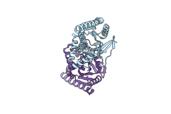
Structure Of The Pentapeptide Repeat Protein Albg, A Resistance Factor For The Topoisomerase Poison Albicidin.
Organism: Xanthomonas albilineans
Method: X-RAY DIFFRACTION Resolution:2.39 Å Release Date: 2010-10-13 Classification: CELL CYCLE |
Organism: Xanthomonas albilineans
Method: X-RAY DIFFRACTION
Release Date: 2010-10-13
 |
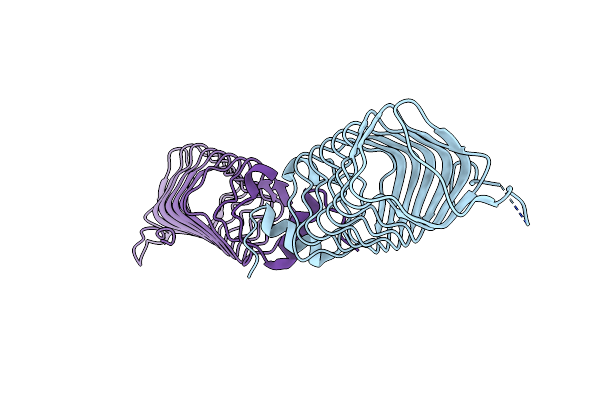
Structure Of The Mycobacterium Tuberculosis Protein, Rv2275, Demonstrates That Cyclodipeptide Synthetases Are Related To Type I Trna-Synthetases.
Organism: Mycobacterium tuberculosis
Method: X-RAY DIFFRACTION Resolution:2.02 Å Release Date: 2010-09-22 Classification: LIGASE |
Organism: Mycobacterium tuberculosis
Method: X-RAY DIFFRACTION
Release Date: 2010-09-22
 |
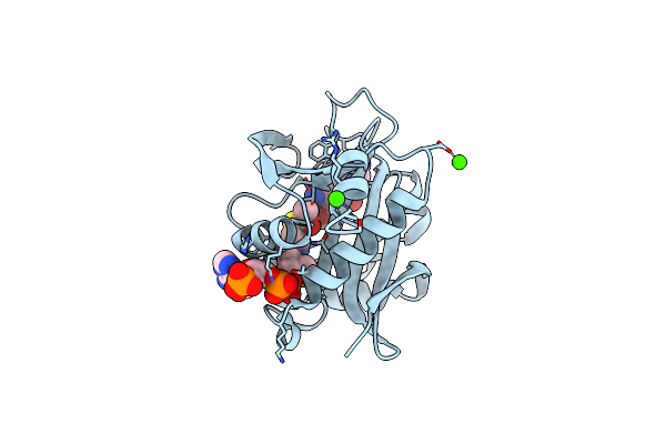
Structure Of The Pentapeptide Repeat Protein Efsqnr, A Dna Gyrase Inhibitor. Free Amines Modified By Cyclic Pentylation With Glutaraldehyde.
Organism: Enterococcus faecalis
Method: X-RAY DIFFRACTION Resolution:1.60 Å Release Date: 2009-05-05 Classification: INHIBITOR Ligands: CL |
Organism: Enterococcus faecalis
Method: X-RAY DIFFRACTION
Release Date: 2009-05-05
Ligands: CL
 |
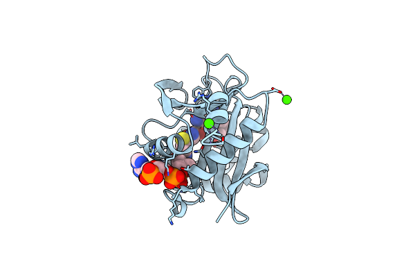
Structure Of Aac(6')-Ib In Complex With Kanamycin C And Acetylcoa.
Organism: Escherichia coli
Method: X-RAY DIFFRACTION Resolution:2.20 Å Release Date: 2008-09-02 Classification: TRANSFERASE Ligands: ACO, KNC, CA |
Organism: Escherichia coli
Method: X-RAY DIFFRACTION
Release Date: 2008-09-02
Ligands: ACO, KNC, CA
 |
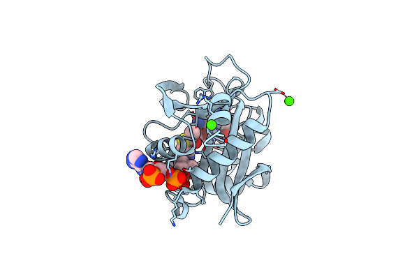
Structure Of Aac(6')-Ib In Complex With Ribostamycin And Coenzyme A.
Organism: Escherichia coli
Method: X-RAY DIFFRACTION Resolution:1.70 Å Release Date: 2008-09-02 Classification: TRANSFERASE Ligands: COA, RIO, CA |
Organism: Escherichia coli
Method: X-RAY DIFFRACTION
Release Date: 2008-09-02
Ligands: COA, RIO, CA
 |
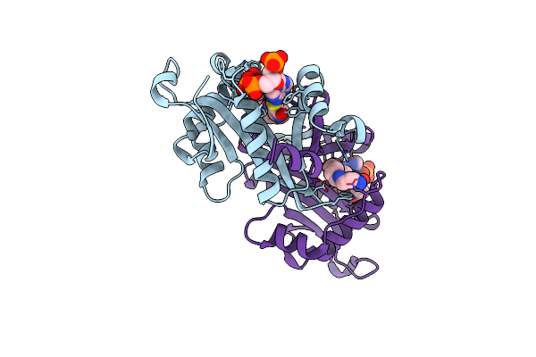
Structure Of Aac(6')-Ib In Complex With Parmomycin And Acetylcoa.
Organism: Escherichia coli
Method: X-RAY DIFFRACTION Resolution:1.80 Å Release Date: 2008-09-02 Classification: TRANSFERASE Ligands: ACO, PAR, CA |
Organism: Escherichia coli
Method: X-RAY DIFFRACTION
Release Date: 2008-09-02
Ligands: ACO, PAR, CA
 |
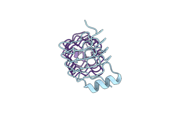
Crystal Structure Of Human Spermine,Spermidine Acetyltransferase In Complex With A Bisubstrate Analog (N1-Acetylspermine-S-Coa).
Organism: Homo sapiens
Method: X-RAY DIFFRACTION Resolution:2.30 Å Release Date: 2007-06-05 Classification: TRANSFERASE Ligands: NHQ |
Organism: Homo sapiens
Method: X-RAY DIFFRACTION
Release Date: 2007-06-05
Ligands: NHQ
 |
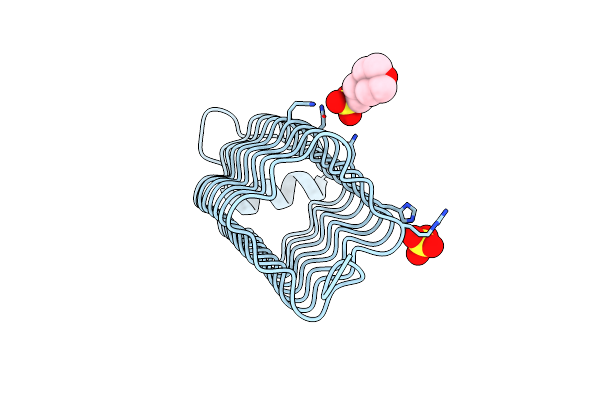
Structure Of Np275, A Pentapeptide Repeat Protein From Nostoc Punctiforme
Organism: Nostoc punctiforme
Method: X-RAY DIFFRACTION Resolution:2.10 Å Release Date: 2006-12-07 Classification: TOXIN Ligands: CL |
Organism: Nostoc punctiforme
Method: X-RAY DIFFRACTION
Release Date: 2006-12-07
Ligands: CL
 |
Structure Of The Fusion Of Np275 And Np276, Pentapeptide Repeat Proteins From Nostoc Punctiforme
Organism: Nostoc punctiforme
Method: X-RAY DIFFRACTION Resolution:1.50 Å Release Date: 2006-12-07 Classification: TOXIN Ligands: MES, SO4 |
Organism: Nostoc punctiforme
Method: X-RAY DIFFRACTION
Release Date: 2006-12-07
Ligands: MES, SO4
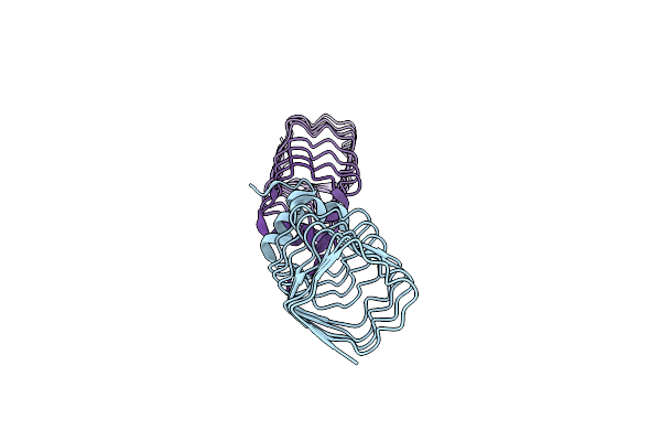 |
The Structure Of Mfpa (Rv3361C, C2 Crystal Form). The Pentapeptide Repeat Protein From Mycobacterium Tuberculosis Folds As A Right- Handed Quadrilateral Beta-Helix.
Organism: Mycobacterium tuberculosis
Method: X-RAY DIFFRACTION Resolution:2.20 Å Release Date: 2005-06-07 Classification: PENTAPEPTIDE REPEAT PROTEIN |
Organism: Mycobacterium tuberculosis
Method: X-RAY DIFFRACTION
Release Date: 2005-06-07
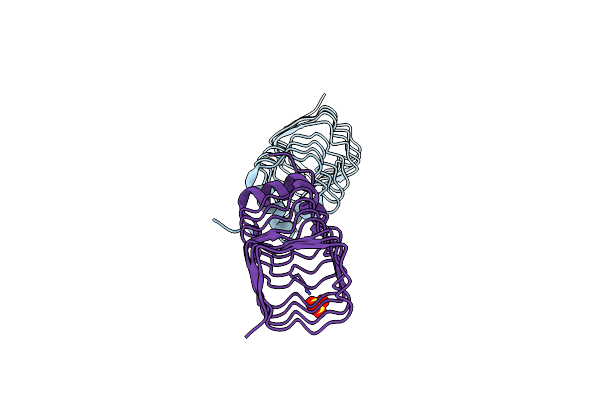 |
The Structure Of Mfpa (Rv3361C, P21 Crystal Form). The Pentapeptide Repeat Protein From Mycobacterium Tuberculosis Folds As A Right- Handed Quadrilateral Beta-Helix.
Organism: Mycobacterium tuberculosis
Method: X-RAY DIFFRACTION Resolution:2.00 Å Release Date: 2005-06-07 Classification: PENTAPEPTIDE REPEAT PROTEIN Ligands: SO4 |
Organism: Mycobacterium tuberculosis
Method: X-RAY DIFFRACTION
Release Date: 2005-06-07
Ligands: SO4
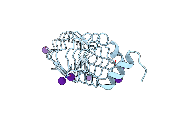 |
The Structure Of Mfpa (Rv3361C, C2221 Crystal Form). The Pentapeptide Repeat Protein From Mycobacterium Tuberculosis Folds As A Right- Handed Quadrilateral Beta-Helix.
Organism: Mycobacterium tuberculosis
Method: X-RAY DIFFRACTION Resolution:2.20 Å Release Date: 2005-06-07 Classification: PENTAPEPTIDE REPEAT PROTEIN Ligands: CS |
Organism: Mycobacterium tuberculosis
Method: X-RAY DIFFRACTION
Release Date: 2005-06-07
Ligands: CS