

Search Count: 78
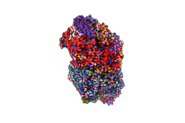 |
Xfel Structure Of Cyanobacterial Photosystem Ii Under Dark Conditions
|
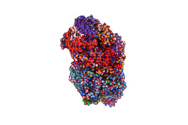 |
Xfel Structure Of Cyanobacterial Photosystem Ii Following One Flash (1F) With A 20-Nanosecond Delay
|
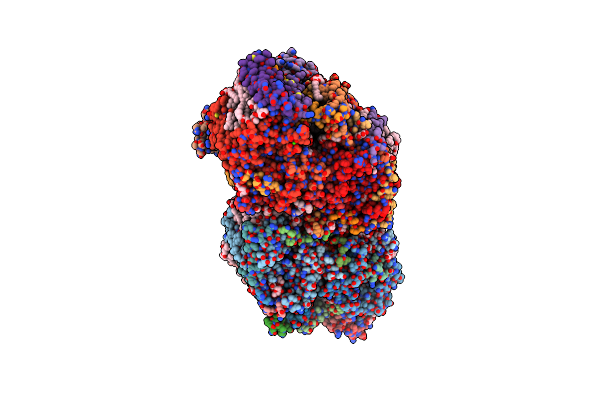 |
Xfel Structure Of Cyanobacterial Photosystem Ii Following One Flash (1F) With A 200-Nanosecond Delay
|
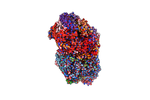 |
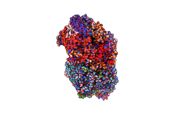
Xfel Structure Of Cyanobacterial Photosystem Ii Following One Flash (1F) With A 1-Microsecond Delay
|
 |
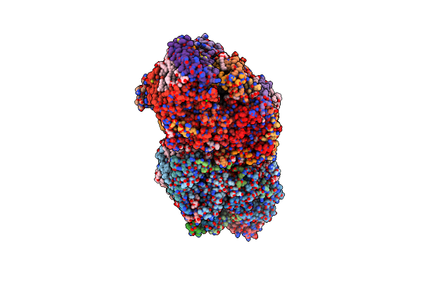
Xfel Structure Of Cyanobacterial Photosystem Ii Following One Flash (1F) With A 30-Microsecond Delay
|
 |
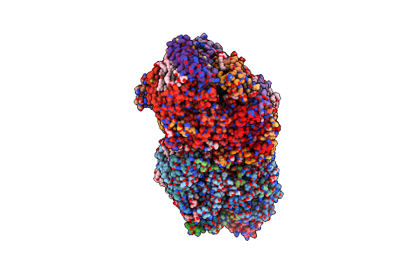
Xfel Structure Of Cyanobacterial Photosystem Ii Following One Flash (1F) With A 200-Microsecond Delay
|
 |
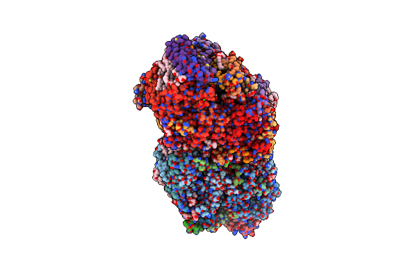
Xfel Structure Of Cyanobacterial Photosystem Ii Following One Flash (1F) With A 5-Millisecond Delay
|
 |
Xfel Structure Of Cyanobacterial Photosystem Ii Following One Flash (1F) With A 5-Millisecond Delay (Single Conformation)
|
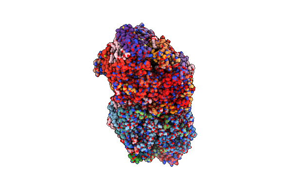 |
Xfel Structure Of Cyanobacterial Photosystem Ii Following Two Flashes (2F) With A 20-Nanosecond Delay
|
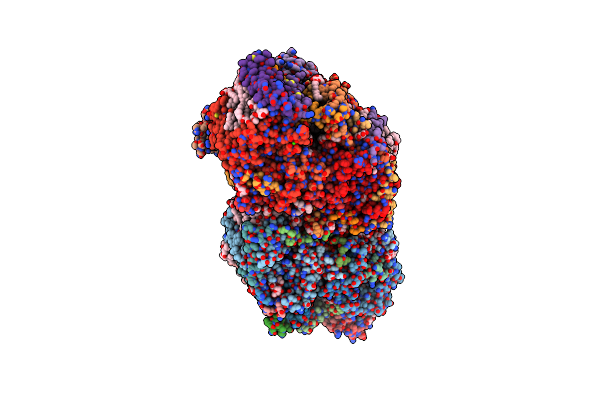 |
Xfel Structure Of Cyanobacterial Photosystem Ii Following Two Flashes (2F) With A 200-Nanosecond Delay
|
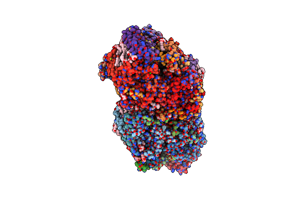 |
Xfel Structure Of Cyanobacterial Photosystem Ii Following Two Flashes (2F) With A 1-Microsecond Delay
|
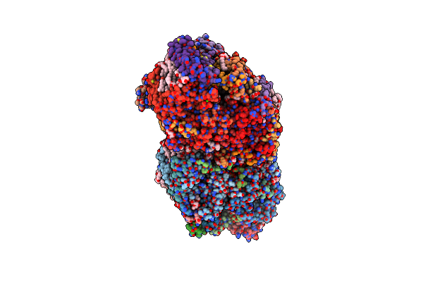 |
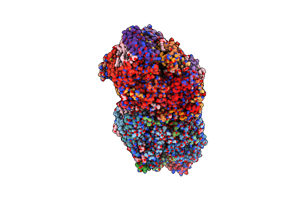
Xfel Structure Of Cyanobacterial Photosystem Ii Following Two Flashes (2F) With A 30-Microsecond Delay
|
 |
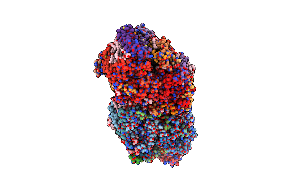
Xfel Structure Of Cyanobacterial Photosystem Ii Following Two Flashes (2F) With A 200-Microsecond Delay
|
 |
Xfel Structure Of Cyanobacterial Photosystem Ii Following Two Flashes (2F) With A 5-Millisecond Delay
|
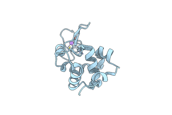 |
Crystal Structure Of Hen Egg-White Lysozyme
Organism: Gallus gallus
Method: X-RAY DIFFRACTION Resolution:2.01 Å Release Date: 2017-12-20 Classification: HYDROLASE |
Organism: Gallus gallus
Method: X-RAY DIFFRACTION
Release Date: 2017-12-20
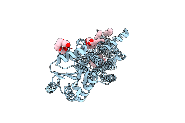 |
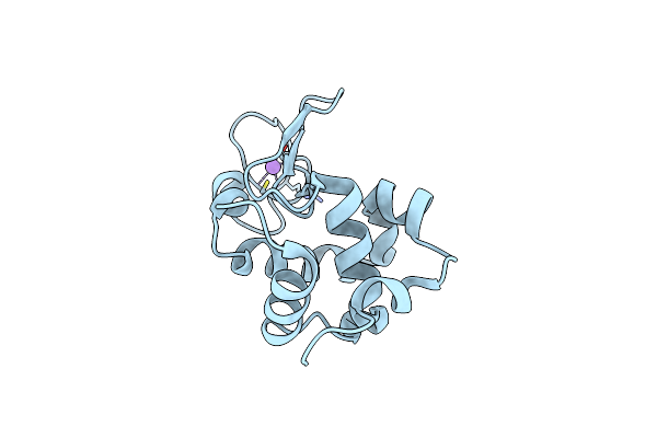
Crystal Structures Of Human Orexin 2 Receptor Bound To The Selective Antagonist Empa Determined By Serial Femtosecond Crystallography At Sacla
Organism: Homo sapiens, Pyrococcus abyssi (strain ge5 / orsay)
Method: X-RAY DIFFRACTION Resolution:2.30 Å Release Date: 2017-12-13 Classification: SIGNALING PROTEIN Ligands: 7MA, OLA, 1PE |
Organism: Homo sapiens, Pyrococcus abyssi (strain ge5 / orsay)
Method: X-RAY DIFFRACTION
Release Date: 2017-12-13
Ligands: 7MA, OLA, 1PE
 |
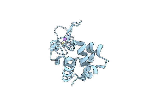
Crystal Structure Of Hen Egg-White Lysozyme
Organism: Gallus gallus
Method: X-RAY DIFFRACTION Resolution:1.80 Å Release Date: 2017-12-06 Classification: HYDROLASE |
Organism: Gallus gallus
Method: X-RAY DIFFRACTION
Release Date: 2017-12-06
 |
Crystal Structure Of Hen Egg-White Lysozyme
Organism: Gallus gallus
Method: X-RAY DIFFRACTION Resolution:1.45 Å Release Date: 2017-12-06 Classification: HYDROLASE |
Organism: Gallus gallus
Method: X-RAY DIFFRACTION
Release Date: 2017-12-06
 |
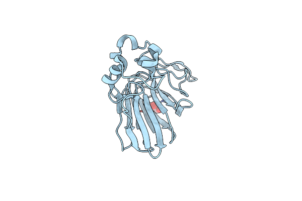
Crystal Structure Of Human Orexin 2 Receptor Bound To The Selective Antagonist Empa Determined By The Synchrotron Light Source At Spring-8.
Organism: Homo sapiens, Pyrococcus abyssi ge5
Method: X-RAY DIFFRACTION Resolution:1.96 Å Release Date: 2017-11-29 Classification: SIGNALING PROTEIN Ligands: 7MA, OLA, 1PE |
Organism: Homo sapiens, Pyrococcus abyssi ge5
Method: X-RAY DIFFRACTION
Release Date: 2017-11-29
Ligands: 7MA, OLA, 1PE
 |
Thaumatin Structure Determined By Sacla At 1.55 Angstrom
Organism: Thaumatococcus daniellii
Method: X-RAY DIFFRACTION Resolution:1.55 Å Release Date: 2017-11-29 Classification: PLANT PROTEIN Ligands: TLA |
Organism: Thaumatococcus daniellii
Method: X-RAY DIFFRACTION
Release Date: 2017-11-29
Ligands: TLA