

Search Count: 18
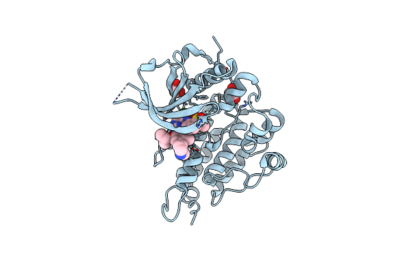 |
Crystal Structure Of Anaplastic Lymphoma Kinase Complexed With Ldk378
Organism: Homo sapiens
Method: X-RAY DIFFRACTION Resolution:2.01 Å Release Date: 2014-04-09 Classification: TRANSFERASE/TRANSFERASE INHIBITOR Ligands: GOL, 4MK |
Organism: Homo sapiens
Method: X-RAY DIFFRACTION
Release Date: 2014-04-09
Ligands: GOL, 4MK
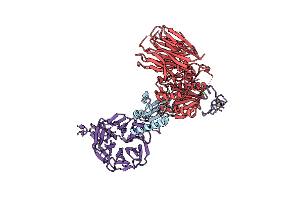 |
The X-Ray Crystal Structure Of Pcsk9 In Complex With The Ldl Receptor
Organism: Homo sapiens
Method: X-RAY DIFFRACTION Resolution:7.01 Å Release Date: 2011-03-16 Classification: PROTEIN BINDING Ligands: CA |
Organism: Homo sapiens
Method: X-RAY DIFFRACTION
Release Date: 2011-03-16
Ligands: CA
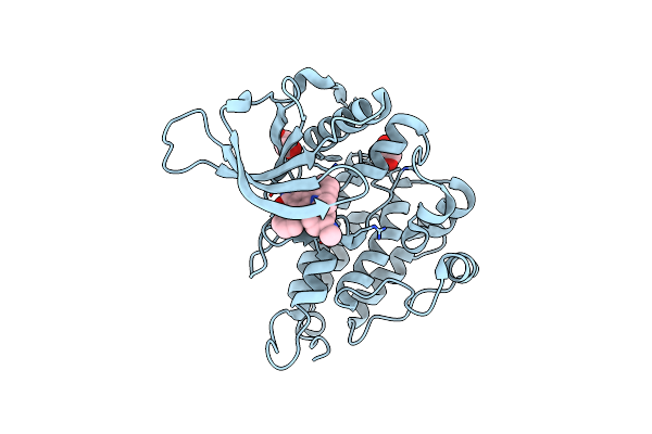 |
Crystal Structure Of The Anaplastic Lymphoma Kinase Catalytic Domain
Organism: Homo sapiens
Method: X-RAY DIFFRACTION Resolution:1.95 Å Release Date: 2010-08-04 Classification: TRANSFERASE Ligands: STU, GOL |
Organism: Homo sapiens
Method: X-RAY DIFFRACTION
Release Date: 2010-08-04
Ligands: STU, GOL
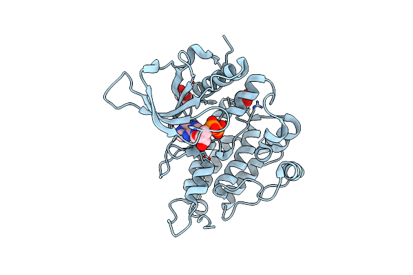 |
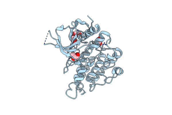
Crystal Structure Of The Anaplastic Lymphoma Kinase Catalytic Domain
Organism: Homo sapiens
Method: X-RAY DIFFRACTION Resolution:2.10 Å Release Date: 2010-08-04 Classification: TRANSFERASE Ligands: GOL, ADP |
Organism: Homo sapiens
Method: X-RAY DIFFRACTION
Release Date: 2010-08-04
Ligands: GOL, ADP
 |
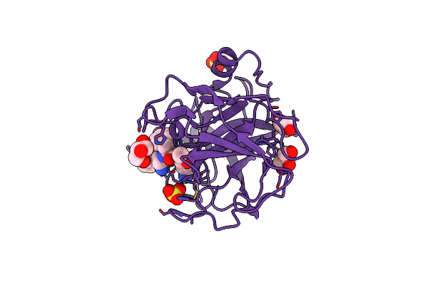
Crystal Structure Of The Anaplastic Lymphoma Kinase Catalytic Domain
Organism: Homo sapiens
Method: X-RAY DIFFRACTION Resolution:1.80 Å Release Date: 2010-07-28 Classification: TRANSFERASE Ligands: GOL |
Organism: Homo sapiens
Method: X-RAY DIFFRACTION
Release Date: 2010-07-28
Ligands: GOL
 |
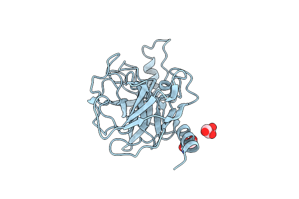
The X-Ray Structure Of Human Prostasin In Complex With Dffr-Chloromethyl Ketone Inhibitor
Organism: Homo sapiens, synthetic construct
Method: X-RAY DIFFRACTION Resolution:1.70 Å Release Date: 2009-06-16 Classification: HYDROLASE Ligands: SO4, GOL |
Organism: Homo sapiens, synthetic construct
Method: X-RAY DIFFRACTION
Release Date: 2009-06-16
Ligands: SO4, GOL
 |
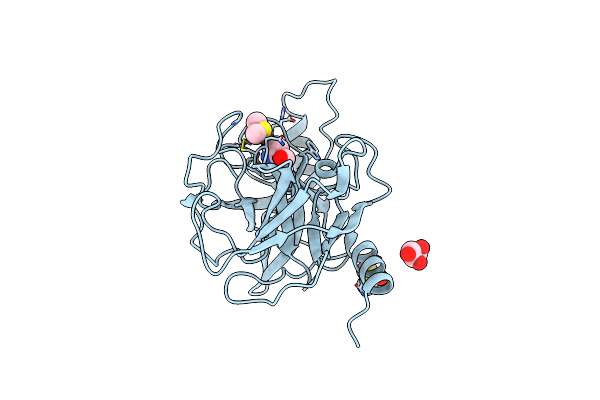
The Crystal Structure Of Apo Prostasin At 1.7 Angstroms Resolution
Organism: Homo sapiens
Method: X-RAY DIFFRACTION Resolution:1.70 Å Release Date: 2009-05-05 Classification: HYDROLASE Ligands: GOL |
Organism: Homo sapiens
Method: X-RAY DIFFRACTION
Release Date: 2009-05-05
Ligands: GOL
 |
The Crystal Structure Of Prostasin Complexed With Camostat At 1.6 Angstroms Resolution
Organism: Homo sapiens
Method: X-RAY DIFFRACTION Resolution:1.60 Å Release Date: 2009-05-05 Classification: HYDROLASE Ligands: GOL, 1JZ, DMS |
Organism: Homo sapiens
Method: X-RAY DIFFRACTION
Release Date: 2009-05-05
Ligands: GOL, 1JZ, DMS
 |
Structure Of Prostasin In Complex With Aprotinin
Organism: Homo sapiens, Bos taurus
Method: X-RAY DIFFRACTION Resolution:2.80 Å Release Date: 2009-05-05 Classification: HYDROLASE/INHIBITOR |
Organism: Homo sapiens, Bos taurus
Method: X-RAY DIFFRACTION
Release Date: 2009-05-05
 |
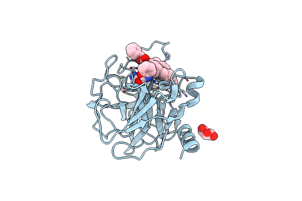
Structure Of Prostasin At 1.3 Angstroms Resolution In Complex With A Calcium Ion.
Organism: Homo sapiens
Method: X-RAY DIFFRACTION Resolution:1.30 Å Release Date: 2009-04-21 Classification: HYDROLASE Ligands: CA, GOL |
Organism: Homo sapiens
Method: X-RAY DIFFRACTION
Release Date: 2009-04-21
Ligands: CA, GOL
 |
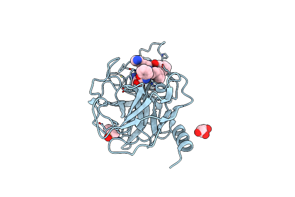
The X-Ray Structure Of Human Prostasin In Complex With A Covalent Benzoxazole Inhibitor
Organism: Homo sapiens
Method: X-RAY DIFFRACTION Resolution:1.70 Å Release Date: 2008-09-09 Classification: HYDROLASE Ligands: B3C, GOL |
Organism: Homo sapiens
Method: X-RAY DIFFRACTION
Release Date: 2008-09-09
Ligands: B3C, GOL
 |
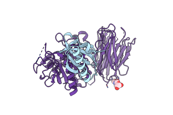
X-Ray Structure Of Human Prostasin In Complex With Benzoxazole Warhead Peptidomimic, Lysine In P3
Organism: Homo sapiens
Method: X-RAY DIFFRACTION Resolution:1.60 Å Release Date: 2008-09-09 Classification: HYDROLASE Ligands: GOL, B4C, DMS |
Organism: Homo sapiens
Method: X-RAY DIFFRACTION
Release Date: 2008-09-09
Ligands: GOL, B4C, DMS
 |
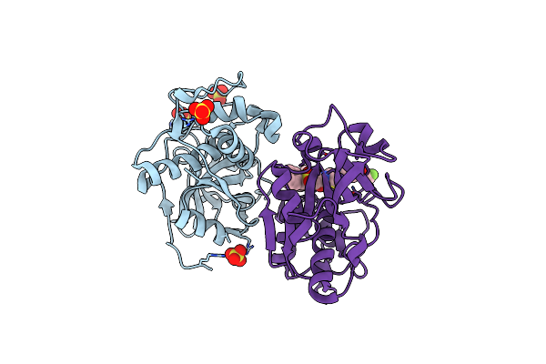
The Crystal Structure Of Pcsk9 At 1.9 Angstroms Resolution Reveals Structural Homology To Resistin Within The C-Terminal Domain
Organism: Homo sapiens
Method: X-RAY DIFFRACTION Resolution:1.90 Å Release Date: 2007-09-18 Classification: HYDROLASE Ligands: NAG, CA |
Organism: Homo sapiens
Method: X-RAY DIFFRACTION
Release Date: 2007-09-18
Ligands: NAG, CA
 |
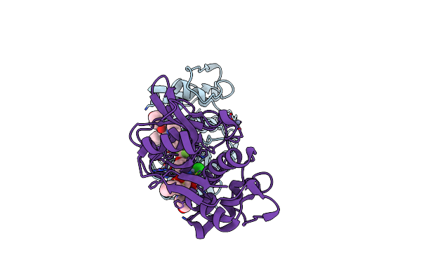
Cathepsin S In Complex With Non Covalent Arylaminoethyl Amide.
Organism: Homo sapiens
Method: X-RAY DIFFRACTION Resolution:1.55 Å Release Date: 2007-05-08 Classification: HYDROLASE Ligands: SO4, GNQ |
Organism: Homo sapiens
Method: X-RAY DIFFRACTION
Release Date: 2007-05-08
Ligands: SO4, GNQ
 |
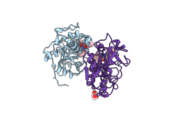
Crystal Structure Of Cathepsin S In Complex With A Zinc Mediated Non-Covalent Arylaminoethyl Amide
Organism: Homo sapiens
Method: X-RAY DIFFRACTION Resolution:1.80 Å Release Date: 2006-08-15 Classification: HYDROLASE Ligands: ZN, CL, GNQ |
Organism: Homo sapiens
Method: X-RAY DIFFRACTION
Release Date: 2006-08-15
Ligands: ZN, CL, GNQ
 |
Cathepsin S In Complex With Non-Covalent 2-(Benzoxazol-2-Ylamino)-Acetamide
Organism: Homo sapiens
Method: X-RAY DIFFRACTION Resolution:1.90 Å Release Date: 2006-04-04 Classification: HYDROLASE Ligands: GNF, GOL |
Organism: Homo sapiens
Method: X-RAY DIFFRACTION
Release Date: 2006-04-04
Ligands: GNF, GOL
 |
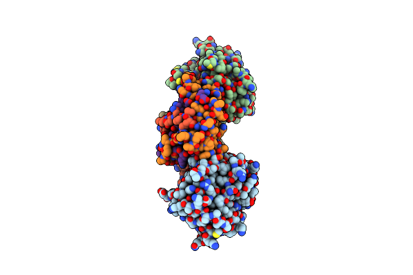
The Oligomeric Structure Of Human Granzyme A Reveals The Molecular Determinants Of Substrate Specificity
Organism: Homo sapiens
Method: X-RAY DIFFRACTION Resolution:2.40 Å Release Date: 2003-07-01 Classification: HYDROLASE/HYDROLASE INHIBITOR Ligands: 0G6, SO4 |
Organism: Homo sapiens
Method: X-RAY DIFFRACTION
Release Date: 2003-07-01
Ligands: 0G6, SO4
 |
Rat Granzyme B [N66Q] Complexed To Ecotin [81-84 Iepd]
Organism: Rattus norvegicus, Escherichia coli
Method: X-RAY DIFFRACTION Resolution:2.20 Å Release Date: 2000-09-13 Classification: HYDROLASE/HYDROLASE INHIBITOR |
Organism: Rattus norvegicus, Escherichia coli
Method: X-RAY DIFFRACTION
Release Date: 2000-09-13