

Search Count: 20
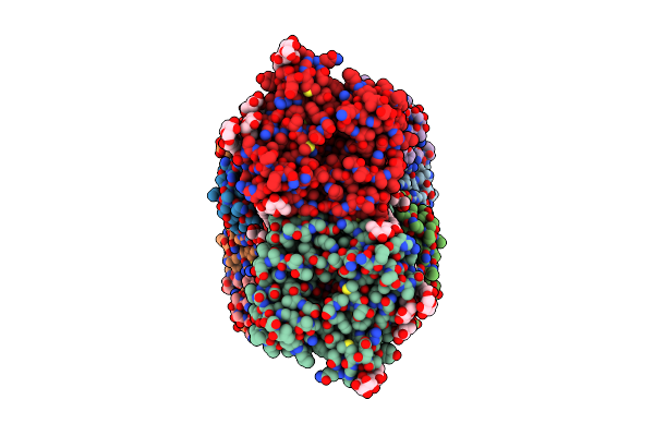 |
Ktrab Complex - Ktra8 Ring With A Ktrb Dimer On Each Side
Organism: Vibrio alginolyticus
Method: ELECTRON MICROSCOPY Resolution:2.82 Å Release Date: 2023-05-10 Classification: MEMBRANE PROTEIN Ligands: MG, ADP, LMT, K |
Organism: Vibrio alginolyticus
Method: ELECTRON MICROSCOPY
Release Date: 2023-05-10
Ligands: MG, ADP, LMT, K
 |
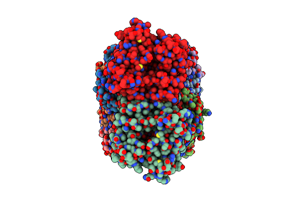
Native Ktrab Complex
Organism: Vibrio alginolyticus
Method: ELECTRON MICROSCOPY Resolution:3.24 Å Release Date: 2023-05-10 Classification: MEMBRANE PROTEIN Ligands: ADP, MG, LMT, K |
Organism: Vibrio alginolyticus
Method: ELECTRON MICROSCOPY
Release Date: 2023-05-10
Ligands: ADP, MG, LMT, K
 |
Ktrab Complex With N-Terminal Deletion Of Ktrb 1-19
Organism: Vibrio alginolyticus
Method: ELECTRON MICROSCOPY Resolution:3.56 Å Release Date: 2023-05-10 Classification: MEMBRANE PROTEIN Ligands: MG, ADP, K |
Organism: Vibrio alginolyticus
Method: ELECTRON MICROSCOPY
Release Date: 2023-05-10
Ligands: MG, ADP, K
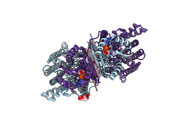 |
Kima From B. Subtilis With Nucleotide Second-Messenger C-Di-Amp Bound
Organism: Bacillus subtilis
Method: ELECTRON MICROSCOPY Release Date: 2023-01-18 Classification: MEMBRANE PROTEIN Ligands: 2BA, LMT, K |
Organism: Bacillus subtilis
Method: ELECTRON MICROSCOPY
Release Date: 2023-01-18
Ligands: 2BA, LMT, K
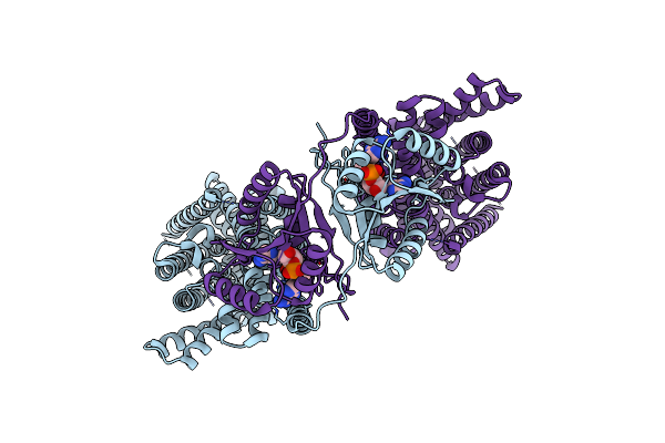 |
Upright Kima Dimer With Bound C-Di-Amp From B. Subtilis
Organism: Bacillus subtilis
Method: ELECTRON MICROSCOPY Release Date: 2023-01-18 Classification: MEMBRANE PROTEIN Ligands: 2BA |
Organism: Bacillus subtilis
Method: ELECTRON MICROSCOPY
Release Date: 2023-01-18
Ligands: 2BA
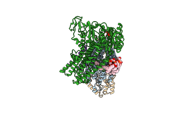 |
Cryo-Em Map Of The Wt Kdpfabc Complex In The E1-P Tight Conformation, Stabilised With The Inhibitor Orthovanadate
Organism: Escherichia coli
Method: ELECTRON MICROSCOPY Release Date: 2022-11-16 Classification: MEMBRANE PROTEIN Ligands: K, CDL, VO4 |
Organism: Escherichia coli
Method: ELECTRON MICROSCOPY
Release Date: 2022-11-16
Ligands: K, CDL, VO4
 |
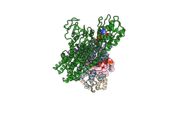
Cryo-Em Map Of The Wt Kdpfabc Complex In The E1-P Tight Conformation, Under Turnover Conditions
Organism: Escherichia coli
Method: ELECTRON MICROSCOPY Release Date: 2022-11-16 Classification: MEMBRANE PROTEIN Ligands: K, CDL |
Organism: Escherichia coli
Method: ELECTRON MICROSCOPY
Release Date: 2022-11-16
Ligands: K, CDL
 |
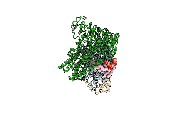
Cryo-Em Map Of The Wt Kdpfabc Complex In The E1_Atpearly Conformation, Under Turnover Conditions
Organism: Escherichia coli
Method: ELECTRON MICROSCOPY Release Date: 2022-11-16 Classification: MEMBRANE PROTEIN Ligands: K, CDL, ATP |
Organism: Escherichia coli
Method: ELECTRON MICROSCOPY
Release Date: 2022-11-16
Ligands: K, CDL, ATP
 |
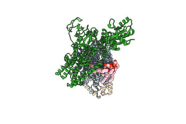
Cryo-Em Structure Of The Kdpfabc Complex In A Nucleotide-Free E1 Conformation Loaded With K+
Organism: Escherichia coli
Method: ELECTRON MICROSCOPY Release Date: 2022-11-16 Classification: MEMBRANE PROTEIN Ligands: K, CDL |
Organism: Escherichia coli
Method: ELECTRON MICROSCOPY
Release Date: 2022-11-16
Ligands: K, CDL
 |
Cryo-Em Structure Of The Kdpfabc Complex In A Nucleotide-Free E1 Conformation Loaded With K+
Organism: Escherichia coli
Method: ELECTRON MICROSCOPY Release Date: 2022-11-16 Classification: MEMBRANE PROTEIN Ligands: K, CDL |
Organism: Escherichia coli
Method: ELECTRON MICROSCOPY
Release Date: 2022-11-16
Ligands: K, CDL
 |
Cryo-Em Structure Of The Kdpfabc Complex In A Nucleotide-Free E1 Conformation Loaded With K+
Organism: Escherichia coli
Method: ELECTRON MICROSCOPY Release Date: 2022-11-16 Classification: MEMBRANE PROTEIN Ligands: K, CDL |
Organism: Escherichia coli
Method: ELECTRON MICROSCOPY
Release Date: 2022-11-16
Ligands: K, CDL
 |
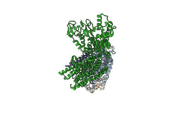
Cryo-Em Map Of The Wt Kdpfabc Complex In The E1-P_Adp Conformation, Under Turnover Conditions
Organism: Escherichia coli
Method: ELECTRON MICROSCOPY Release Date: 2022-11-16 Classification: MEMBRANE PROTEIN Ligands: K, CDL, ADP |
Organism: Escherichia coli
Method: ELECTRON MICROSCOPY
Release Date: 2022-11-16
Ligands: K, CDL, ADP
 |
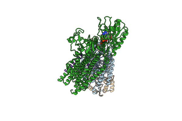
Cryo-Em Map Of The Unphosphorylated Kdpfabc Complex In The E2-P Conformation, Under Turnover Conditions
Organism: Escherichia coli k-12
Method: ELECTRON MICROSCOPY Release Date: 2022-11-16 Classification: MEMBRANE PROTEIN Ligands: K |
Organism: Escherichia coli k-12
Method: ELECTRON MICROSCOPY
Release Date: 2022-11-16
Ligands: K
 |
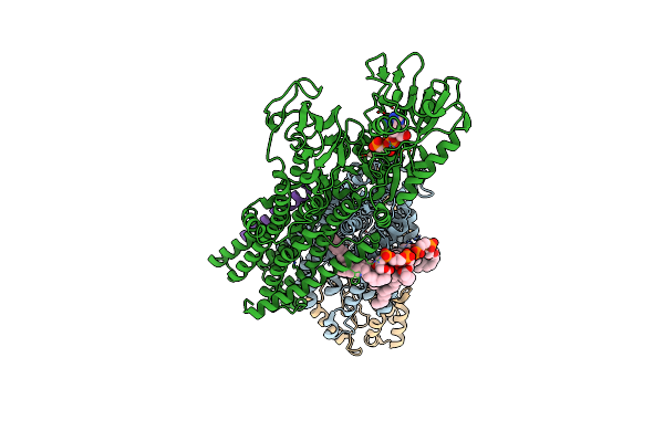
Cryo-Em Map Of The Unphosphorylated Kdpfabc Complex In The E1-P_Adp Conformation, Under Turnover Conditions
Organism: Escherichia coli
Method: ELECTRON MICROSCOPY Release Date: 2022-11-16 Classification: MEMBRANE PROTEIN Ligands: K, ADP, MG |
Organism: Escherichia coli
Method: ELECTRON MICROSCOPY
Release Date: 2022-11-16
Ligands: K, ADP, MG
 |
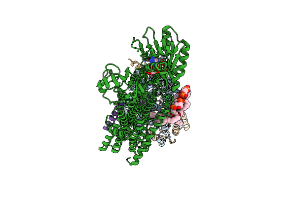
Cryo-Em Structure Of The Kdpfabc Complex In An E1-Atp Conformation Loaded With K+
Organism: Escherichia coli
Method: ELECTRON MICROSCOPY Release Date: 2021-07-28 Classification: MEMBRANE PROTEIN Ligands: K, CDL, ACP |
Organism: Escherichia coli
Method: ELECTRON MICROSCOPY
Release Date: 2021-07-28
Ligands: K, CDL, ACP
 |
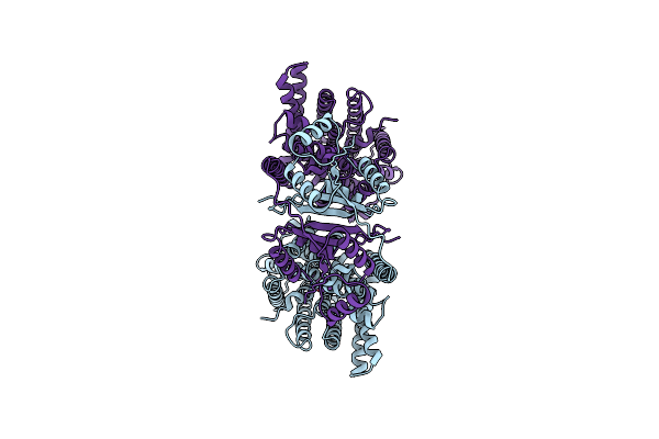
Rb-Loaded Cryo-Em Structure Of The E1-Atp Kdpfabc Complex.
Organism: Escherichia coli
Method: ELECTRON MICROSCOPY Release Date: 2021-07-28 Classification: MEMBRANE PROTEIN Ligands: RB, CDL, ACP |
Organism: Escherichia coli
Method: ELECTRON MICROSCOPY
Release Date: 2021-07-28
Ligands: RB, CDL, ACP
 |
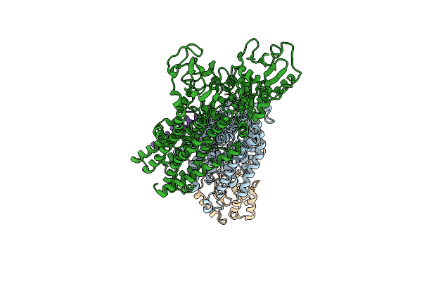
Kima From Bacillus Subtilis In Inward-Facing, Occluded State
Organism: Bacillus subtilis
Method: ELECTRON MICROSCOPY Release Date: 2020-02-12 Classification: TRANSPORT PROTEIN Ligands: K |
Organism: Bacillus subtilis
Method: ELECTRON MICROSCOPY
Release Date: 2020-02-12
Ligands: K
 |
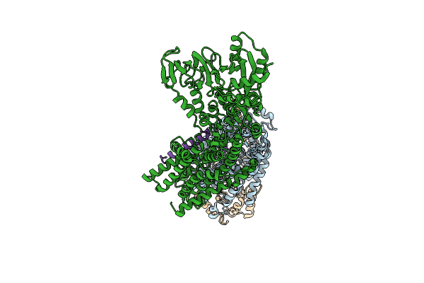
Cryo-Em Structure Of The Kdpfabc Complex In An E1 Outward-Facing State (State 1)
Organism: Escherichia coli (strain k12)
Method: ELECTRON MICROSCOPY Release Date: 2018-12-05 Classification: MEMBRANE PROTEIN Ligands: K |
Organism: Escherichia coli (strain k12)
Method: ELECTRON MICROSCOPY
Release Date: 2018-12-05
Ligands: K
 |
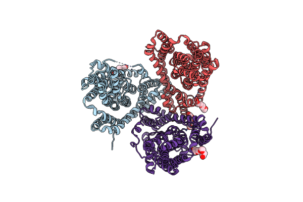
Cryo-Em Structure Of The Kdpfabc Complex In An E2 Inward-Facing State (State 2)
Organism: Escherichia coli (strain k12)
Method: ELECTRON MICROSCOPY Release Date: 2018-12-05 Classification: MEMBRANE PROTEIN Ligands: K |
Organism: Escherichia coli (strain k12)
Method: ELECTRON MICROSCOPY
Release Date: 2018-12-05
Ligands: K
 |
Crystal Structure Of A Substrate-Free Glutamate Transporter Homologue From Thermococcus Kodakarensis
Organism: Thermococcus kodakarensis
Method: X-RAY DIFFRACTION Resolution:3.00 Å Release Date: 2013-09-11 Classification: TRANSPORT PROTEIN, membrane protein Ligands: PG4 |
Organism: Thermococcus kodakarensis
Method: X-RAY DIFFRACTION
Release Date: 2013-09-11
Ligands: PG4