
Search Count: 48
All
Selected
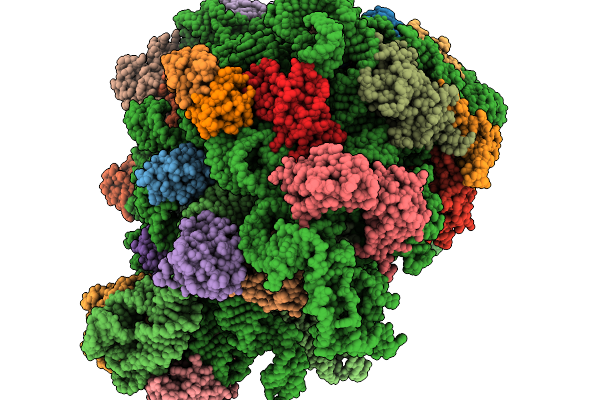 |
Structure Of E.Coli Ribosome With Filamin Mutant Y719E Nascent Chain At Linker Length Of 47 Amino Acids, With Trna
Organism: Dictyostelium discoideum, Escherichia coli
Method: ELECTRON MICROSCOPY Release Date: 2026-02-04 Classification: RIBOSOME |
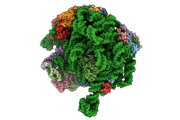 |
Structure Of E.Coli Ribosome With Nascent Chain At Linker Length Of 31 Amino Acids, With Mrna, P-Site And A-Site Trnas
Organism: Dictyostelium discoideum, Escherichia coli
Method: ELECTRON MICROSCOPY Release Date: 2026-01-28 Classification: RIBOSOME |
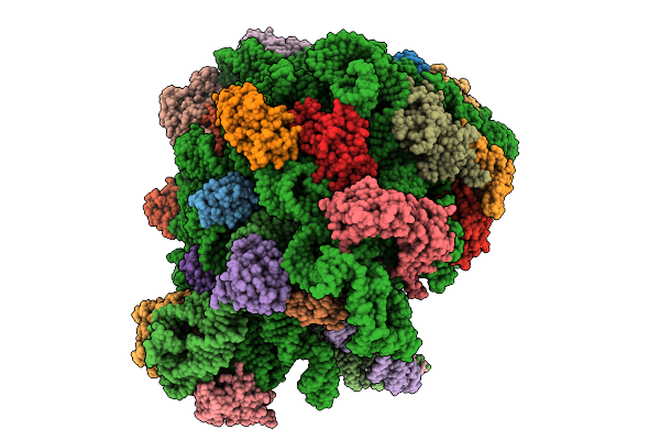 |
Structure Of Wt E.Coli Ribosome With Complexed Filament Nascent Chain At Length 34, With Mrna, P-Site And A-Site Trnas, And Mrna
Organism: Dictyostelium discoideum, Escherichia coli
Method: ELECTRON MICROSCOPY Release Date: 2026-01-21 Classification: RIBOSOME |
 |
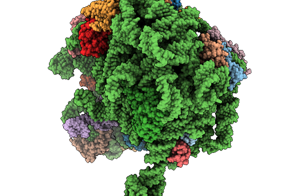
Structure Of Wt E.Coli Ribosome With Complexed Filament Nascent Chain At Length 47, With P-Site Trna
Organism: Dictyostelium discoideum, Escherichia coli
Method: ELECTRON MICROSCOPY Release Date: 2026-01-14 Classification: RIBOSOME |
 |
Structure Of Wt E.Coli Ribosome With Complexed Filament Nascent Chain At Length 31, With P-Site Trnas
Organism: Dictyostelium discoideum, Escherichia coli
Method: ELECTRON MICROSCOPY Release Date: 2026-01-14 Classification: RIBOSOME |
 |
Organism: Homo sapiens
Method: X-RAY DIFFRACTION Resolution:2.11 Å Release Date: 2025-08-13 Classification: STRUCTURAL PROTEIN/INHIBITOR Ligands: A1BW7 |
 |
Organism: Homo sapiens
Method: X-RAY DIFFRACTION Resolution:1.58 Å Release Date: 2025-08-13 Classification: STRUCTURAL PROTEIN/INHIBITOR Ligands: A1BW8 |
 |
Organism: Homo sapiens
Method: X-RAY DIFFRACTION Resolution:1.58 Å Release Date: 2025-08-13 Classification: STRUCTURAL PROTEIN/INHIBITOR Ligands: A1BW9 |
 |
Organism: Severe acute respiratory syndrome coronavirus 2
Method: X-RAY DIFFRACTION Resolution:2.39 Å Release Date: 2025-02-05 Classification: VIRAL PROTEIN Ligands: MG, SAH, EDO, ZN, M7G |
 |
Organism: Mus musculus
Method: ELECTRON MICROSCOPY Release Date: 2024-05-01 Classification: MEMBRANE PROTEIN Ligands: NAG |
 |
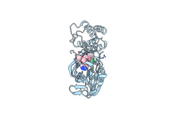
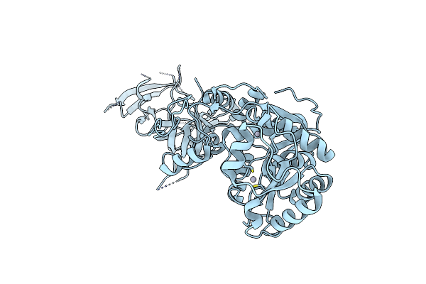
Structure Of Bovine Pka Bound To (R)-N-(4-(1H-Pyrrolo[2,3-B]Pyridin-4-Yl)Phenyl)-2-Amino-4-Methylpentanamide
Organism: Bos taurus
Method: X-RAY DIFFRACTION Resolution:1.70 Å Release Date: 2024-02-21 Classification: CYTOSOLIC PROTEIN Ligands: ZWG |
 |
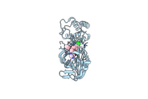
Sars-Cov-2 3Clpro In Complex With N-(4-(1H-Pyrazol-4-Yl)Phenyl)-N-(3-Chlorobenzyl)-2-(Pyridin-3-Yl)Acetamide
Organism: Severe acute respiratory syndrome coronavirus 2
Method: X-RAY DIFFRACTION Resolution:2.20 Å Release Date: 2023-01-11 Classification: VIRAL PROTEIN,HYDROLASE/INHIBITOR Ligands: I2D |
 |
Sars-Cov-2 3Clpro In Complex With N-(4-(1H-Imidazol-4-Yl)Phenyl)-N-(3-Chloro-5-Fluorobenzyl)-2-(Isoquinolin-4-Yl)Acetamide
Organism: Severe acute respiratory syndrome coronavirus 2
Method: X-RAY DIFFRACTION Resolution:2.40 Å Release Date: 2023-01-11 Classification: VIRAL PROTEIN,HYDROLASE/INHIBITOR Ligands: I2N |
 |
Organism: Mycolicibacterium smegmatis mc2 155
Method: X-RAY DIFFRACTION Resolution:1.87 Å Release Date: 2022-12-21 Classification: HYDROLASE Ligands: K, ACT, SO4, GOL, MG, PEG |
 |
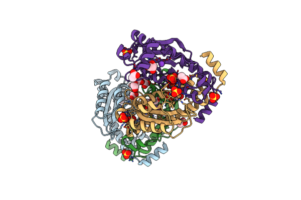
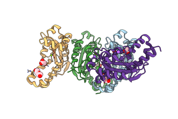
Crystal Structure Of The Chicken Isoleucyl-Trna Synthetase 1 (Iars1) Une-I Complexed With Glutamyl-Trna Synthetase 1 (Ears1)
Organism: Gallus gallus
Method: X-RAY DIFFRACTION Resolution:2.40 Å Release Date: 2022-11-23 Classification: TRANSLATION |
 |
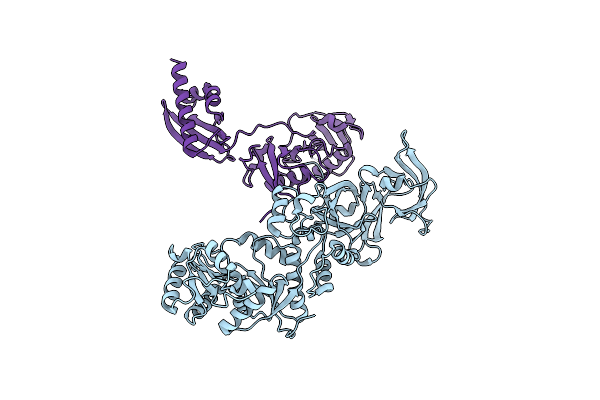
Organism: Gallus gallus
Method: X-RAY DIFFRACTION Resolution:2.50 Å Release Date: 2022-11-23 Classification: TRANSLATION Ligands: HG |
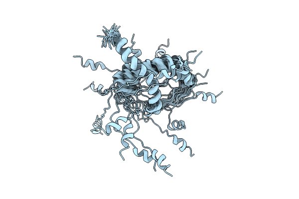 |
Organism: Human immunodeficiency virus 1
Method: SOLUTION NMR Release Date: 2022-10-26 Classification: ANTIVIRAL PROTEIN |
 |
Organism: Escherichia coli k-12
Method: X-RAY DIFFRACTION Resolution:2.30 Å Release Date: 2022-09-28 Classification: HYDROLASE Ligands: ACT, MG, NA, PEG |
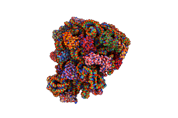 |
70S E. Coli Ribosome With An Extended Ul23 Loop From Candidatus Marinimicrobia And A Stalled Filamin Domain 5 Nascent Chain
Organism: Dictyostelium discoideum, Escherichia coli
Method: ELECTRON MICROSCOPY Release Date: 2022-08-10 Classification: RIBOSOME |
 |
70S E. Coli Ribosome With An Extended Ul23 Loop From Candidatus Marinimicrobia
Organism: Escherichia coli
Method: ELECTRON MICROSCOPY Release Date: 2022-08-10 Classification: RIBOSOME |