

Search Count: 10
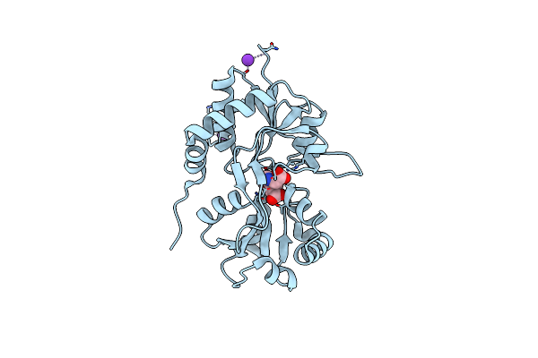 |
Crystal Structure Of The Kainate Receptor Gluk3 Ligand Binding Domain In Complex With Kainate
Organism: Rattus norvegicus
Method: X-RAY DIFFRACTION Resolution:2.35 Å Release Date: 2012-05-02 Classification: MEMBRANE PROTEIN Ligands: KAI, CL, K |
Organism: Rattus norvegicus
Method: X-RAY DIFFRACTION
Release Date: 2012-05-02
Ligands: KAI, CL, K
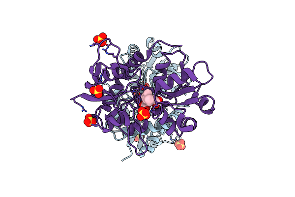 |
Crystal Structure Of The Kainate Receptor Gluk1 Ligand-Binding Domain In Complex With Kainate In The Absence Of Glycerol
Organism: Rattus norvegicus
Method: X-RAY DIFFRACTION Resolution:2.00 Å Release Date: 2012-04-25 Classification: MEMBRANE PROTEIN Ligands: KAI, SO4, CL |
Organism: Rattus norvegicus
Method: X-RAY DIFFRACTION
Release Date: 2012-04-25
Ligands: KAI, SO4, CL
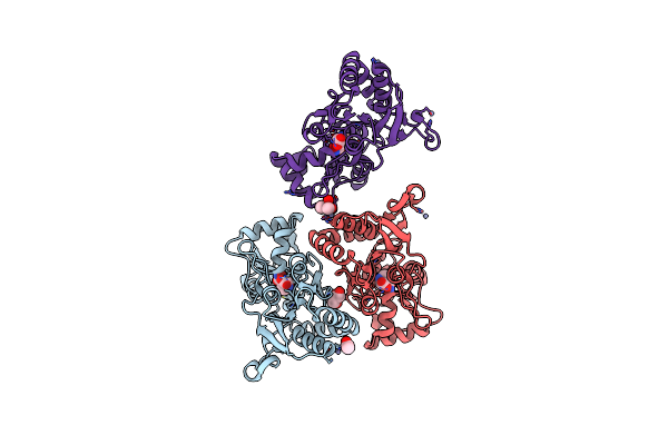 |
X-Ray Structure Of The Ligand-Binding Core Of Glua2 In Complex With (S)-7-Hpca At 2.5 A Resolution
Organism: Rattus norvegicus
Method: X-RAY DIFFRACTION Resolution:2.48 Å Release Date: 2010-12-29 Classification: MEMBRANE PROTEIN Ligands: ZN, HA7, GOL, ACY, CAC |
Organism: Rattus norvegicus
Method: X-RAY DIFFRACTION
Release Date: 2010-12-29
Ligands: ZN, HA7, GOL, ACY, CAC
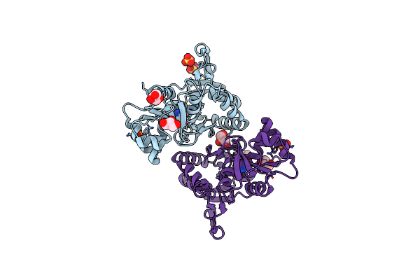 |
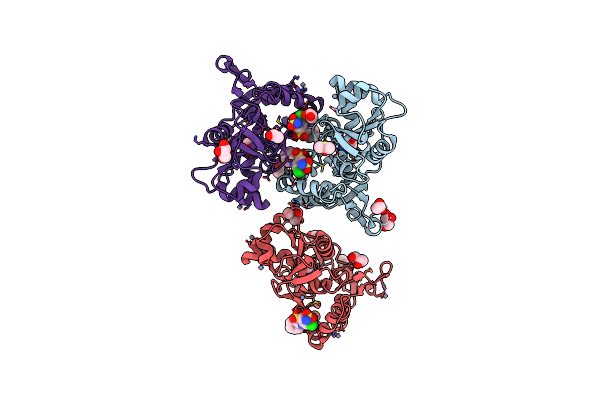
X-Ray Structure Of The Ligand-Binding Core Of Glua2 In Complex With (R)-5-Hpca At 2.1 A Resolution
Organism: Rattus norvegicus
Method: X-RAY DIFFRACTION Resolution:2.10 Å Release Date: 2010-12-29 Classification: MEMBRANE PROTEIN Ligands: HA5, SO4, GOL, CL |
Organism: Rattus norvegicus
Method: X-RAY DIFFRACTION
Release Date: 2010-12-29
Ligands: HA5, SO4, GOL, CL
 |
Crystal Structure Of The Iglur2 Ligand-Binding Core (S1S2J-N754S) In Complex With Glutamate And Cyclothiazide At 2.25 A Resolution
Organism: Rattus norvegicus
Method: X-RAY DIFFRACTION Resolution:2.25 Å Release Date: 2009-07-28 Classification: MEMBRANE PROTEIN Ligands: GLU, CYZ, ZN, GOL, ACT, DMS, CAC |
Organism: Rattus norvegicus
Method: X-RAY DIFFRACTION
Release Date: 2009-07-28
Ligands: GLU, CYZ, ZN, GOL, ACT, DMS, CAC
 |
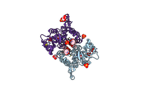
Crystal Structure Of The Iglur2 Ligand-Binding Core (S1S2J-N754S) In Complex With Glutamate And Ns1493 At 1.85 A Resolution
Organism: Rattus norvegicus
Method: X-RAY DIFFRACTION Resolution:1.85 Å Release Date: 2009-07-28 Classification: MEMBRANE PROTEIN Ligands: GLU, NS3, SO4, GOL, FLC |
Organism: Rattus norvegicus
Method: X-RAY DIFFRACTION
Release Date: 2009-07-28
Ligands: GLU, NS3, SO4, GOL, FLC
 |
Crystal Structure Of The Iglur2 Ligand-Binding Core (S1S2J-N754S) In Complex With Glutamate And Ns5206 At 2.10 A Resolution
Organism: Rattus norvegicus
Method: X-RAY DIFFRACTION Resolution:2.10 Å Release Date: 2009-07-28 Classification: MEMBRANE PROTEIN Ligands: GLU, NS6, GOL, SO4, DMS |
Organism: Rattus norvegicus
Method: X-RAY DIFFRACTION
Release Date: 2009-07-28
Ligands: GLU, NS6, GOL, SO4, DMS
 |
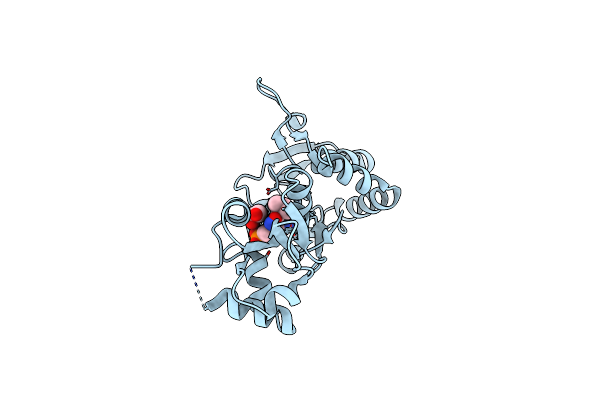
Crystal Structure Of The Iglur2 Ligand-Binding Core (S1S2J-N754S) In Complex With Glutamate And Ns5217 At 1.50 A Resolution
Organism: Rattus norvegicus
Method: X-RAY DIFFRACTION Resolution:1.49 Å Release Date: 2009-07-28 Classification: MEMBRANE PROTEIN Ligands: GLU, NS7, SO4, GOL, DMS |
Organism: Rattus norvegicus
Method: X-RAY DIFFRACTION
Release Date: 2009-07-28
Ligands: GLU, NS7, SO4, GOL, DMS
 |
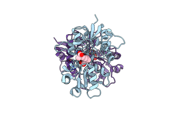
Crystal Structure Of The Ligand-Binding Core Of Iglur5 In Complex With The Antagonist (S)-Atpo At 1.85 A Resolution
Organism: Rattus norvegicus
Method: X-RAY DIFFRACTION Resolution:1.85 Å Release Date: 2007-07-03 Classification: MEMBRANE PROTEIN Ligands: AT1, GOL |
Organism: Rattus norvegicus
Method: X-RAY DIFFRACTION
Release Date: 2007-07-03
Ligands: AT1, GOL
 |
Crystal Structure Of The Ligand-Binding Core Of Iglur5 In Complex With The Partial Agonist Domoic Acid At 2.5 A Resolution
Organism: Rattus norvegicus
Method: X-RAY DIFFRACTION Resolution:2.50 Å Release Date: 2007-07-03 Classification: MEMBRANE PROTEIN Ligands: DOQ |
Organism: Rattus norvegicus
Method: X-RAY DIFFRACTION
Release Date: 2007-07-03
Ligands: DOQ