

Search Count: 29
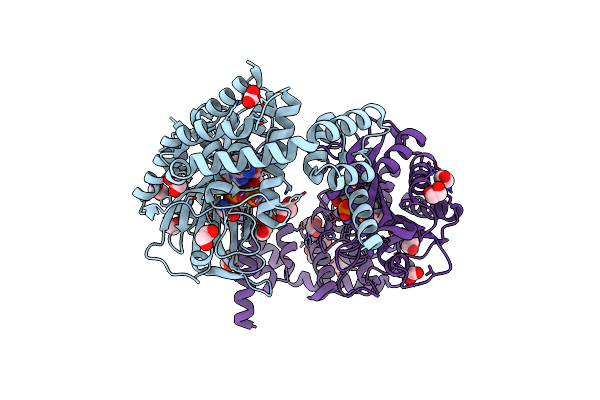 |
Crystal Structure Of Alanyl-Trna Synthetase In Complex With Atp And L-Alanine
Organism: Methylomonas sp. dh-1
Method: X-RAY DIFFRACTION Resolution:1.90 Å Release Date: 2025-06-18 Classification: LIGASE Ligands: ATP, ALA, EDO, FMT, MLT, CIT |
Organism: Methylomonas sp. dh-1
Method: X-RAY DIFFRACTION
Release Date: 2025-06-18
Ligands: ATP, ALA, EDO, FMT, MLT, CIT
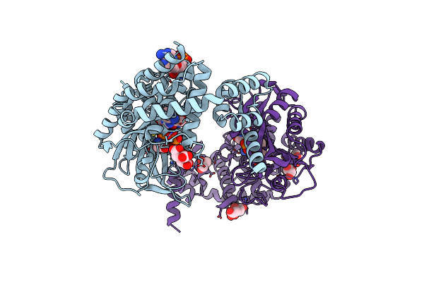 |
Crystal Structure Of Alanyl-Trna Synthetase L219M Mutant In Complex With Atp And L-Alanine
Organism: Methylomonas sp. dh-1
Method: X-RAY DIFFRACTION Resolution:2.18 Å Release Date: 2025-06-18 Classification: LIGASE Ligands: ATP, ALA, CIT, TRS, GOL |
Organism: Methylomonas sp. dh-1
Method: X-RAY DIFFRACTION
Release Date: 2025-06-18
Ligands: ATP, ALA, CIT, TRS, GOL
 |
Pks Domains-Fused Ampc Ec2
Organism: Bacillus amyloliquefaciens, Escherichia coli
Method: X-RAY DIFFRACTION Resolution:2.77 Å Release Date: 2024-06-05 Classification: PROTEIN BINDING |
Organism: Bacillus amyloliquefaciens, Escherichia coli
Method: X-RAY DIFFRACTION
Release Date: 2024-06-05
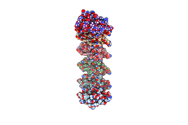 |
X-Ray Crystal Structure Of Bacillus Subtilis Comea
Organism: Bacillus subtilis subsp. subtilis
Method: X-RAY DIFFRACTION Resolution:3.20 Å Release Date: 2022-12-28 Classification: DNA BINDING PROTEIN |
Organism: Bacillus subtilis subsp. subtilis
Method: X-RAY DIFFRACTION
Release Date: 2022-12-28
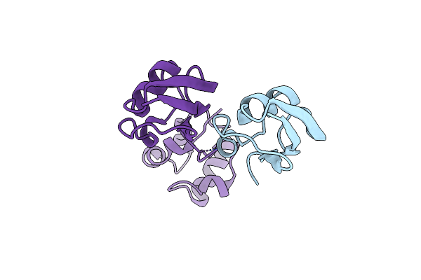 |
X-Ray Crystal Structure Of Geobacillus Stearothermophilus Comea
Organism: Geobacillus stearothermophilus atcc 7953
Method: X-RAY DIFFRACTION Resolution:3.05 Å Release Date: 2022-12-28 Classification: DNA BINDING PROTEIN |
Organism: Geobacillus stearothermophilus atcc 7953
Method: X-RAY DIFFRACTION
Release Date: 2022-12-28
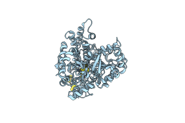 |
Chcodh2 A559W Mutant In Anaerobic Condition
Organism: Carboxydothermus hydrogenoformans
Method: X-RAY DIFFRACTION Resolution:1.74 Å Release Date: 2022-10-19 Classification: OXIDOREDUCTASE Ligands: SF4, FES, NFS |
Organism: Carboxydothermus hydrogenoformans
Method: X-RAY DIFFRACTION
Release Date: 2022-10-19
Ligands: SF4, FES, NFS
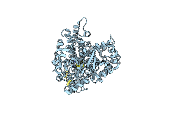 |
Tunnel-Redesigned O2-Tolerant Co Dehydrogenase For Removal Of Co In Real Flue Gas (Chcodh2 A559H Mutant In Anaerobic Condition)
Organism: Carboxydothermus hydrogenoformans
Method: X-RAY DIFFRACTION Resolution:1.80 Å Release Date: 2022-10-19 Classification: OXIDOREDUCTASE Ligands: SF4, FES, NFS |
Organism: Carboxydothermus hydrogenoformans
Method: X-RAY DIFFRACTION
Release Date: 2022-10-19
Ligands: SF4, FES, NFS
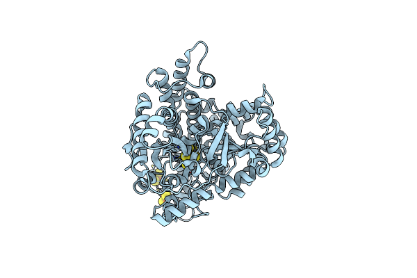 |
Tunnel-Redesigned O2-Tolerant Co Dehydrogenase For Removal Of Co In Real Flue Gas (Chcodh2 A559S Mutant In Anaerobic Condition)
Organism: Carboxydothermus hydrogenoformans
Method: X-RAY DIFFRACTION Resolution:1.84 Å Release Date: 2022-10-19 Classification: OXIDOREDUCTASE Ligands: SF4, FES, NFS |
Organism: Carboxydothermus hydrogenoformans
Method: X-RAY DIFFRACTION
Release Date: 2022-10-19
Ligands: SF4, FES, NFS
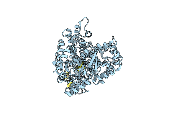 |
Tunnel-Redesigned O2-Tolerant Co Dehydrogenase For Removal Of Co In Real Flue Gas (Aerobic Chcodh2 A559W Mutant)
Organism: Carboxydothermus hydrogenoformans z-2901
Method: X-RAY DIFFRACTION Resolution:2.26 Å Release Date: 2022-10-05 Classification: OXIDOREDUCTASE Ligands: SF4, FES, NFS |
Organism: Carboxydothermus hydrogenoformans z-2901
Method: X-RAY DIFFRACTION
Release Date: 2022-10-05
Ligands: SF4, FES, NFS
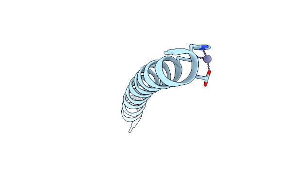 |
Crystal Structure Of A De Novo Three-Stranded Coiled Coil Peptide Containing A Dual Tris-Thiolate Binding Site For Heavy Metal Complexes
Organism: Synthetic construct
Method: X-RAY DIFFRACTION Resolution:2.08 Å Release Date: 2022-06-01 Classification: BIOSYNTHETIC PROTEIN Ligands: ZN |
Organism: Synthetic construct
Method: X-RAY DIFFRACTION
Release Date: 2022-06-01
Ligands: ZN
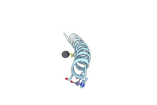 |
Crystal Structure Of A De Novo Three-Stranded Coiled Coil Peptide Containing Trigonal Pyrmidal Pb(Ii) Complexes In The Dual Tris-Thiolate Site
Organism: Synthetic construct
Method: X-RAY DIFFRACTION Resolution:1.29 Å Release Date: 2022-06-01 Classification: BIOSYNTHETIC PROTEIN Ligands: ZN, PB, CL |
Organism: Synthetic construct
Method: X-RAY DIFFRACTION
Release Date: 2022-06-01
Ligands: ZN, PB, CL
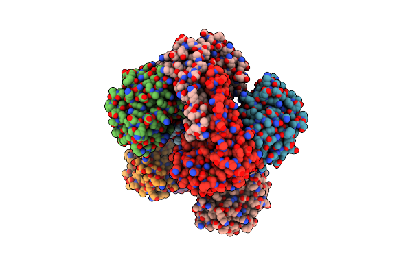 |
Crystal Structure Analysis Of The G73A Mutant Of Superoxide Dismutase From Trichoderma Reesei
Organism: Hypocrea jecorina (strain qm6a)
Method: X-RAY DIFFRACTION Resolution:2.80 Å Release Date: 2022-02-16 Classification: OXIDOREDUCTASE Ligands: MN |
Organism: Hypocrea jecorina (strain qm6a)
Method: X-RAY DIFFRACTION
Release Date: 2022-02-16
Ligands: MN
 |
Crystal Structure Of N-Truncated Human B12 Chaperone Cbld(C262S)-Thiolato-Cob(Iii)Alamin Complex (108-296)
Organism: Homo sapiens
Method: X-RAY DIFFRACTION Resolution:2.50 Å Release Date: 2020-09-16 Classification: OXIDOREDUCTASE Ligands: B12 |
Organism: Homo sapiens
Method: X-RAY DIFFRACTION
Release Date: 2020-09-16
Ligands: B12
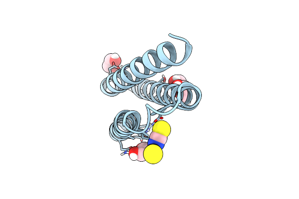 |
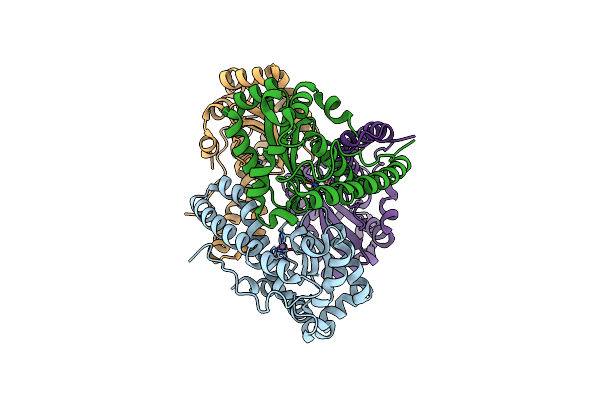
Elongated Version Of A De Novo Designed Three Helix Bundle Structure (Gra3D)
Organism: Synthetic construct
Method: X-RAY DIFFRACTION Resolution:1.34 Å Release Date: 2018-10-03 Classification: DE NOVO PROTEIN Ligands: ZN, SCN, CL, EDO |
Organism: Synthetic construct
Method: X-RAY DIFFRACTION
Release Date: 2018-10-03
Ligands: ZN, SCN, CL, EDO
 |
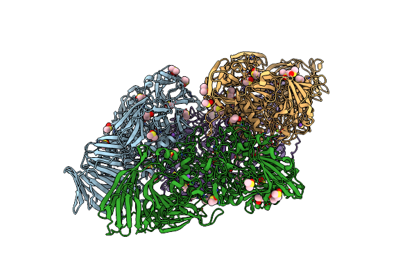
Crystal Structure Of Mn Superoxide Dismutase Mutant M27V From Trichoderma Reesei
Organism: Hypocrea jecorina (strain qm6a)
Method: X-RAY DIFFRACTION Resolution:1.62 Å Release Date: 2017-10-11 Classification: OXIDOREDUCTASE Ligands: MN |
Organism: Hypocrea jecorina (strain qm6a)
Method: X-RAY DIFFRACTION
Release Date: 2017-10-11
Ligands: MN
 |
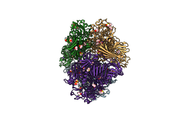
E. Coli (Lacz) Beta-Galactosidase (N460D)
Organism: Escherichia coli
Method: X-RAY DIFFRACTION Resolution:2.80 Å Release Date: 2012-04-11 Classification: HYDROLASE Ligands: MG, NA, DMS |
Organism: Escherichia coli
Method: X-RAY DIFFRACTION
Release Date: 2012-04-11
Ligands: MG, NA, DMS
 |
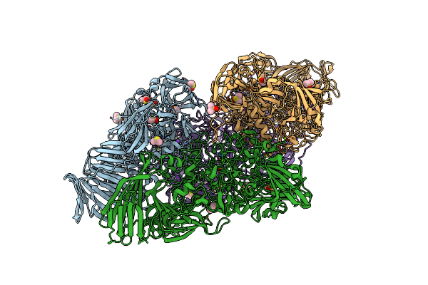
E. Coli (Lacz) Beta-Galactosidase (N460D) In Complex With Iptg
Organism: Escherichia coli
Method: X-RAY DIFFRACTION Resolution:2.00 Å Release Date: 2012-04-11 Classification: HYDROLASE Ligands: MG, NA, DMS, IPT |
Organism: Escherichia coli
Method: X-RAY DIFFRACTION
Release Date: 2012-04-11
Ligands: MG, NA, DMS, IPT
 |
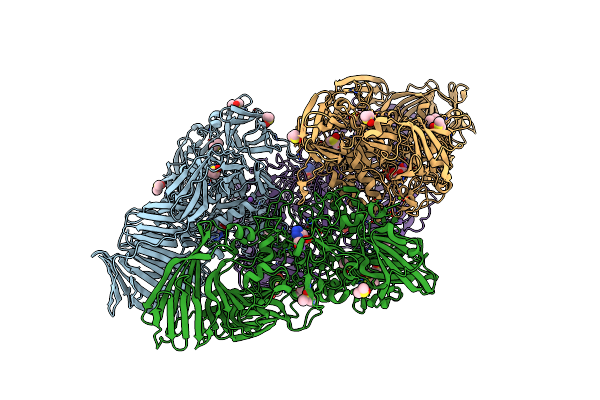
E. Coli (Lacz) Beta-Galactosidase (N460S)
Organism: Escherichia coli
Method: X-RAY DIFFRACTION Resolution:2.70 Å Release Date: 2012-04-11 Classification: HYDROLASE Ligands: MG, NA, DMS |
Organism: Escherichia coli
Method: X-RAY DIFFRACTION
Release Date: 2012-04-11
Ligands: MG, NA, DMS
 |
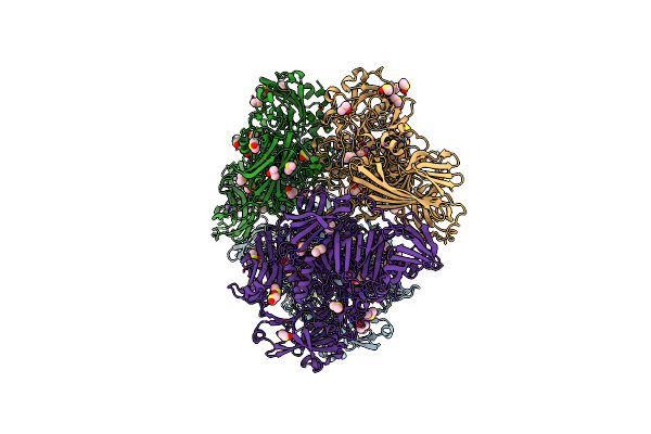
E. Coli (Lacz) Beta-Galactosidase (N460S) In Complex With Galactotetrazole
Organism: Escherichia coli
Method: X-RAY DIFFRACTION Resolution:2.87 Å Release Date: 2012-04-11 Classification: HYDROLASE Ligands: GTZ, MG, NA, DMS |
Organism: Escherichia coli
Method: X-RAY DIFFRACTION
Release Date: 2012-04-11
Ligands: GTZ, MG, NA, DMS
 |
E. Coli (Lacz) Beta-Galactosidase (N460S) In Complex With Iptg
Organism: Escherichia coli
Method: X-RAY DIFFRACTION Resolution:2.05 Å Release Date: 2012-04-11 Classification: HYDROLASE Ligands: MG, NA, IPT, DMS |
Organism: Escherichia coli
Method: X-RAY DIFFRACTION
Release Date: 2012-04-11
Ligands: MG, NA, IPT, DMS