

Search Count: 15
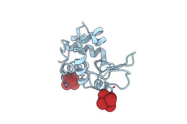 |
K2Ptbr6 Binding To Lysozyme
Organism: Gallus gallus
Method: X-RAY DIFFRACTION Resolution:1.80 Å Release Date: 2010-12-08 Classification: HYDROLASE Ligands: 6BP |
Organism: Gallus gallus
Method: X-RAY DIFFRACTION
Release Date: 2010-12-08
Ligands: 6BP
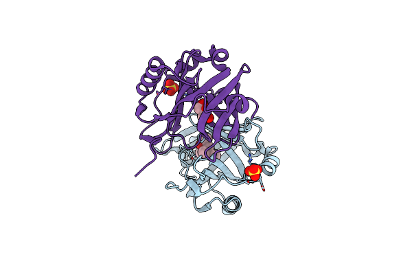 |
The Structure And Refinement Of Apocrustacyanin C2 To 1.6A Resolution And The Search For Differences Between This Protein And The Homologous Apoproteins A1 And C1.
Organism: Homarus gammarus
Method: X-RAY DIFFRACTION Resolution:1.60 Å Release Date: 2004-04-27 Classification: PROTEIN BINDING Ligands: SO4, MPD, GOL |
Organism: Homarus gammarus
Method: X-RAY DIFFRACTION
Release Date: 2004-04-27
Ligands: SO4, MPD, GOL
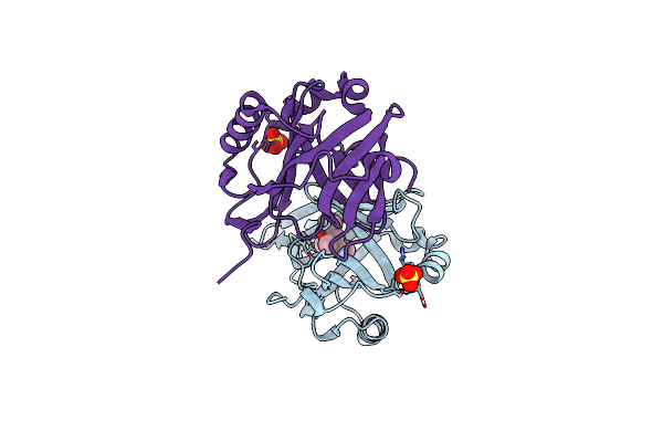 |
The Structure And Refinement Of Apocrustacyanin C2 To 1.3A Resolution And The Search For Differences Between This Protein And The Homologous Apoproteins A1 And C1
Organism: Homarus gammarus
Method: X-RAY DIFFRACTION Resolution:1.30 Å Release Date: 2004-03-02 Classification: PROTEIN BINDING Ligands: SO4, MPD |
Organism: Homarus gammarus
Method: X-RAY DIFFRACTION
Release Date: 2004-03-02
Ligands: SO4, MPD
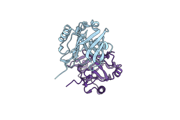 |
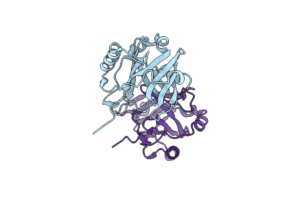
Apocrustacyanin C1 Crystals Grown In Space And Earth Using Vapour Diffusion Geometry
Organism: Homarus gammarus
Method: X-RAY DIFFRACTION Resolution:1.85 Å Release Date: 2003-07-03 Classification: TRANSPORT PROTEIN |
Organism: Homarus gammarus
Method: X-RAY DIFFRACTION
Release Date: 2003-07-03
 |
Apocrustacyanin C1 Crystals Grown In Space And Earth Using Vapour Diffusion Geometry
Organism: Homarus gammarus
Method: X-RAY DIFFRACTION Resolution:2.00 Å Release Date: 2003-07-03 Classification: TRANSPORT PROTEIN |
Organism: Homarus gammarus
Method: X-RAY DIFFRACTION
Release Date: 2003-07-03
 |
Time-Resolved And Static-Ensemble Structural Chemistry Of Hydroxymethylbilane Synthase
Organism: Escherichia coli
Method: X-RAY DIFFRACTION Resolution:1.66 Å Release Date: 2003-01-16 Classification: TRANSFERASE Ligands: DPM |
Organism: Escherichia coli
Method: X-RAY DIFFRACTION
Release Date: 2003-01-16
Ligands: DPM
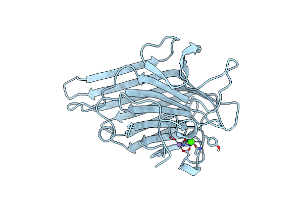 |
Direct Determination Of The Positions Of Deuterium Atoms Of Bound Water In Concanavalin A By Neutron Laue Crystallography
Organism: Canavalia ensiformis
Method: NEUTRON DIFFRACTION Resolution:2.40 Å Release Date: 2000-05-08 Classification: SUGAR BINDING PROTEIN Ligands: MN, CA |
Organism: Canavalia ensiformis
Method: NEUTRON DIFFRACTION
Release Date: 2000-05-08
Ligands: MN, CA
 |
X-Ray Refinement Of D2O Soaked Crystal Of Concanavalin A
Organism: Canavalia ensiformis
Method: X-RAY DIFFRACTION Resolution:1.80 Å Release Date: 2000-04-30 Classification: LECTIN Ligands: MN, CA |
Organism: Canavalia ensiformis
Method: X-RAY DIFFRACTION
Release Date: 2000-04-30
Ligands: MN, CA
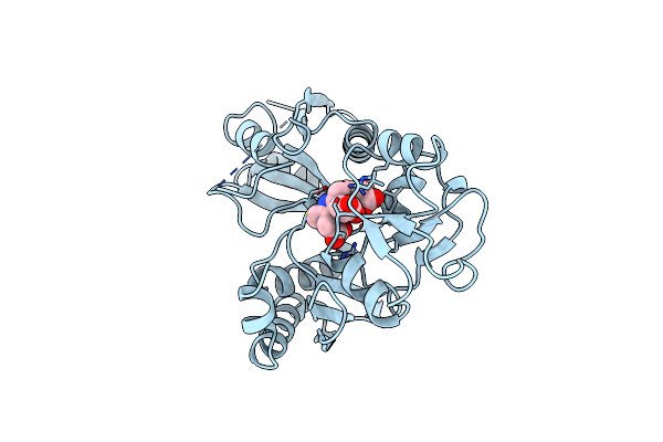 |
Reduced Form Hydroxymethylbilane Synthase (K59Q Mutant) Crystal Structure After 2 Hours In A Flow Cell Determined By Time-Resolved Laue Diffraction
Organism: Escherichia coli
Method: X-RAY DIFFRACTION Resolution:2.30 Å Release Date: 1999-03-02 Classification: TRANSFERASE Ligands: DPM |
Organism: Escherichia coli
Method: X-RAY DIFFRACTION
Release Date: 1999-03-02
Ligands: DPM
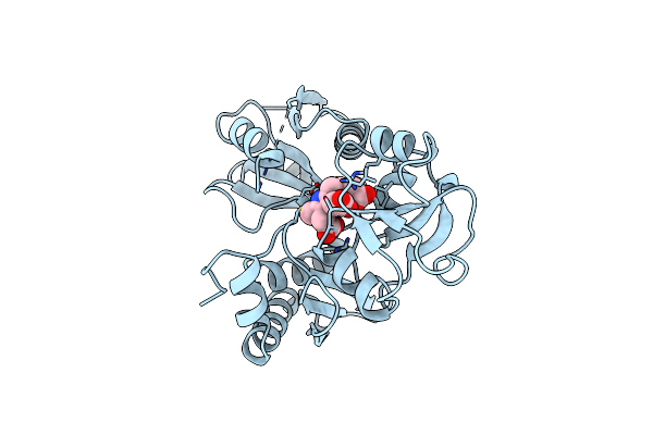 |
Hydroxymethylbilane Synthase
Organism: Escherichia coli
Method: X-RAY DIFFRACTION Resolution:2.30 Å Release Date: 1999-02-02 Classification: TRANSFERASE Ligands: DPM |
Organism: Escherichia coli
Method: X-RAY DIFFRACTION
Release Date: 1999-02-02
Ligands: DPM
 |
Low Temperature Orthorhombic Lysozyme
Organism: Gallus gallus
Method: X-RAY DIFFRACTION Resolution:2.10 Å Release Date: 1997-11-12 Classification: HYDROLASE |
Organism: Gallus gallus
Method: X-RAY DIFFRACTION
Release Date: 1997-11-12
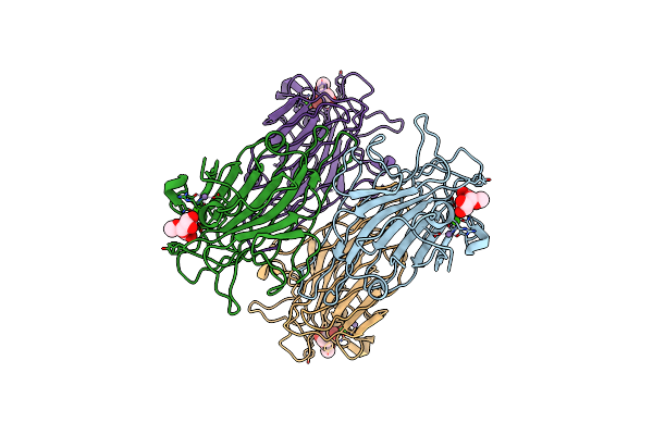 |
Refined Structure Of Concanavalin A Complexed With Alpha-Methyl-D-Mannopyranoside At 2.0 Angstroms Resolution And Comparison With The Saccharide-Free Structure
Organism: Canavalia ensiformis
Method: X-RAY DIFFRACTION Resolution:2.00 Å Release Date: 1994-05-31 Classification: LECTIN(AGGLUTININ) Ligands: MMA, MN, CA, CL |
Organism: Canavalia ensiformis
Method: X-RAY DIFFRACTION
Release Date: 1994-05-31
Ligands: MMA, MN, CA, CL
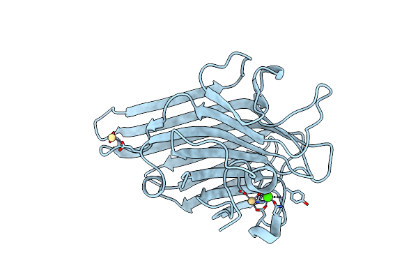 |
The Refined Structure Of Cadmium Substituted Concanavalin A At 2.0 Angstroms Resolution
Organism: Canavalia ensiformis
Method: X-RAY DIFFRACTION Resolution:2.00 Å Release Date: 1994-01-31 Classification: LECTIN(AGGLUTININ) Ligands: CD, CA |
Organism: Canavalia ensiformis
Method: X-RAY DIFFRACTION
Release Date: 1994-01-31
Ligands: CD, CA
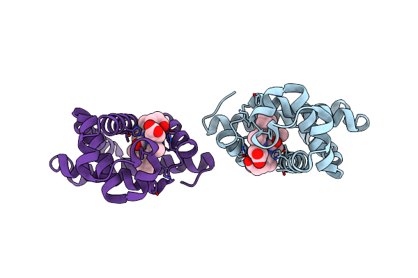 |
Distal Pocket Polarity In Ligand Binding To Myoglobin: Deoxy And Carbonmonoxy Forms Of A Threonine68 (E11) Mutant Investigated By X-Ray Crystallography And Infrared Spectroscopy
Organism: Sus scrofa
Method: X-RAY DIFFRACTION Resolution:2.90 Å Release Date: 1994-01-31 Classification: OXYGEN STORAGE Ligands: HEM, CMO |
Organism: Sus scrofa
Method: X-RAY DIFFRACTION
Release Date: 1994-01-31
Ligands: HEM, CMO
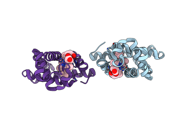 |
Distal Pocket Polarity In Ligand Binding To Myoglobin: Deoxy And Carbonmonoxy Forms Of A Threonine68 (E11) Mutant Investigated By X-Ray Crystallography And Infrared Spectroscopy
Organism: Sus scrofa
Method: X-RAY DIFFRACTION Resolution:2.10 Å Release Date: 1994-01-31 Classification: OXYGEN STORAGE Ligands: HEM |
Organism: Sus scrofa
Method: X-RAY DIFFRACTION
Release Date: 1994-01-31
Ligands: HEM