
Search Count: 1,469
 |
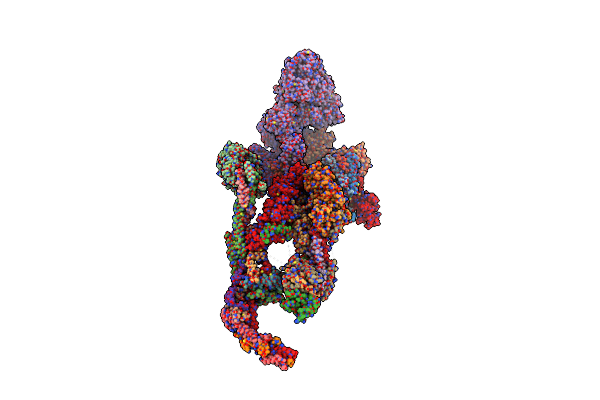
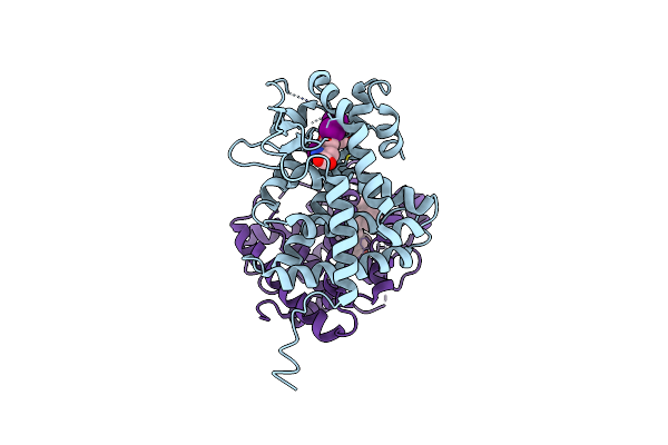
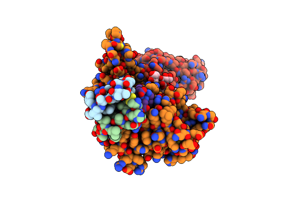
Structure Of Human Transcriptional Mediator Complex
Organism: Homo sapiens
Method: ELECTRON MICROSCOPY Release Date: 2024-10-09 Classification: TRANSCRIPTION Ligands: ZN |
 |
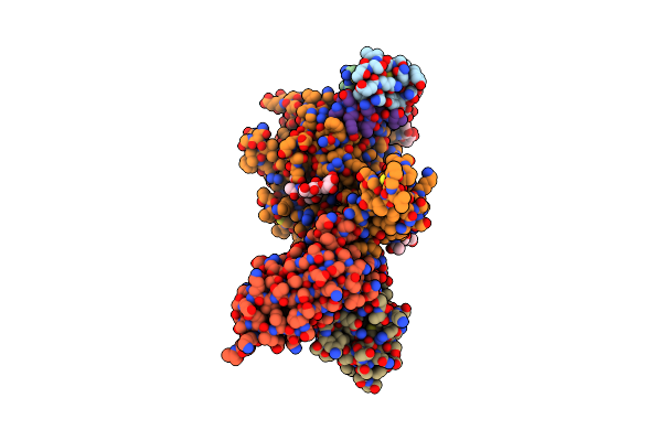
The Idrc Bound Human Core Mediator Complex
Organism: Homo sapiens
Method: ELECTRON MICROSCOPY Release Date: 2024-10-09 Classification: TRANSCRIPTION Ligands: ZN |
 |
Cryoem Structure Of Tr-Trap
Organism: Homo sapiens
Method: ELECTRON MICROSCOPY Release Date: 2024-07-03 Classification: STRUCTURAL PROTEIN |
 |
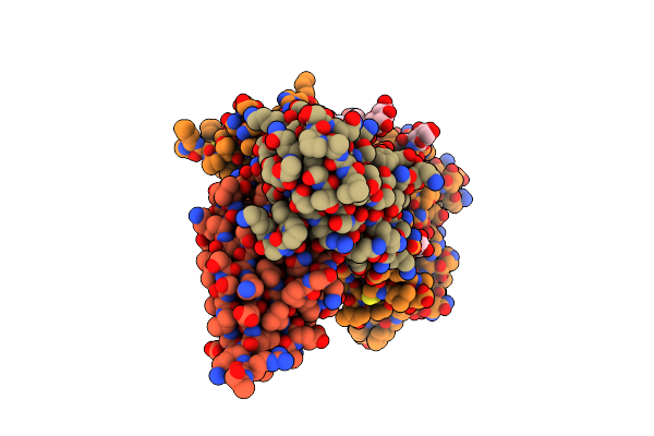
Atomic Model Of The Mammalian Mouse Mediator Complex With Ckm Module
Organism: Mus musculus
Method: ELECTRON MICROSCOPY Release Date: 2024-07-03 Classification: GENE REGULATION |
 |
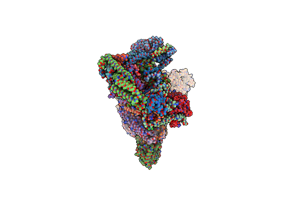
Structure Of The Human Mediator-Bound Transcription Pre-Initiation Complex
Method: ELECTRON MICROSCOPY
Release Date: 2021-03-24 Classification: TRANSCRIPTION/DNA Ligands: MG, ZN, SF4 |
 |
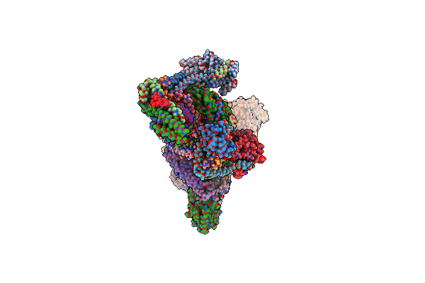
Atomic Model Of The Mammalian Mediator Complex With Med26 Subunit
Organism: Mus musculus
Method: ELECTRON MICROSCOPY Release Date: 2024-06-12 Classification: GENE REGULATION |
 |
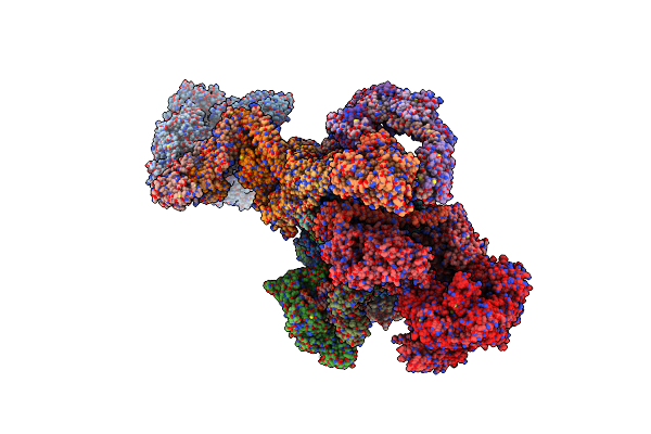
Atomic Model Of The Mammalian Mediator Complex
Organism: Mus musculus
Method: ELECTRON MICROSCOPY Release Date: 2021-03-10 Classification: GENE REGULATION |
 |
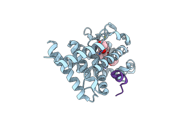
Crystal Structure Of The Ligand Binding Domains Of The Thyroid Receptor:Retinoid X Receptor Complexed With 3,3',5 Triiodo-L-Thyronine And 9-Cis Retinoic Acid
Organism: Gallus gallus, Homo sapiens
Method: X-RAY DIFFRACTION Resolution:2.95 Å Release Date: 2012-04-18 Classification: HORMONE RECEPTOR/HORMONE RECEPTOR Ligands: T3, 9CR |
 |
Crystal Structure Of Human Follicle Stimulating Hormone Complexed With Its Receptor
Organism: Homo sapiens
Method: X-RAY DIFFRACTION Resolution:2.92 Å Release Date: 2005-01-25 Classification: Hormone/Growth Factor Ligands: NAG, SO4 |
 |
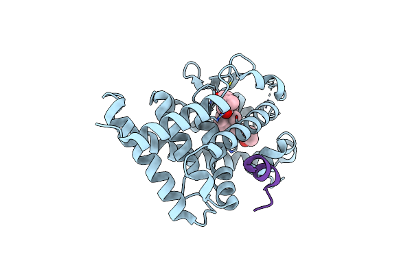
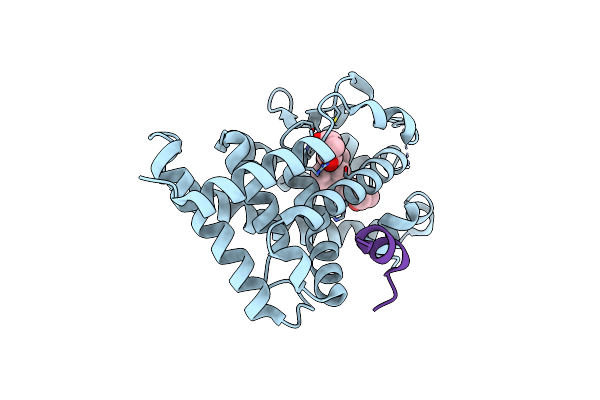
Crystal Structure Of The Rat Vitamin D Receptor Ligand Binding Domain Complexed With 2Md And A Synthetic Peptide Containing The Nr2 Box Of Drip 205
Organism: Rattus norvegicus
Method: X-RAY DIFFRACTION Resolution:1.99 Å Release Date: 2004-04-13 Classification: hormone/growth factor receptor Ligands: VDZ |
 |
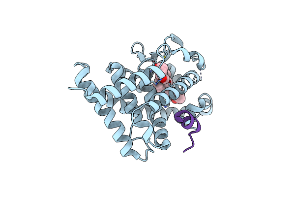
Crystal Structure Of The Rat Vitamin D Receptor Ligand Binding Domain Complexed With 2Mbisp And A Synthetic Peptide Containing The Nr2 Box Of Drip 205
Organism: Rattus norvegicus
Method: X-RAY DIFFRACTION Resolution:1.90 Å Release Date: 2004-04-13 Classification: hormone/growth factor receptor Ligands: VD1 |
 |
Crystal Structure Of The Rat Vitamin D Receptor Ligand Binding Domain Complexed With 2Am20R And A Synthetic Peptide Containing The Nr2 Box Of Drip 205
Organism: Rattus norvegicus
Method: X-RAY DIFFRACTION Resolution:2.28 Å Release Date: 2004-04-13 Classification: hormone/growth factor receptor Ligands: VD2 |
 |
Crystal Structure Of The Rat Vitamin D Receptor Ligand Binding Domain Complexed With 1,25-Dihydroxyvitamin D3 And A Synthetic Peptide Containing The Nr2 Box Of Drip 205
Organism: Rattus norvegicus
Method: X-RAY DIFFRACTION Resolution:2.20 Å Release Date: 2004-04-13 Classification: hormone/growth factor receptor Ligands: VDX |
 |
Insulin Receptor Ectodomain Construct Comprising Domains L1-Cr In Complex With High-Affinity Insulin Analogue [D-Pro-B26]-Dti-Nh2, Alphact Peptide(693-719) And Fab 83-7
Organism: Homo sapiens, Mus musculus, Homo sapiens
Method: X-RAY DIFFRACTION Resolution:4.30 Å Release Date: 2013-01-09 Classification: HORMONE RECEPTOR/HORMONE/IMMUNE SYSTEM Ligands: NAG |
 |
Insulin Receptor Ectodomain Construct Comprising Domains L1-Cr In Complex With Human Insulin, Alpha-Ct Peptide(704-719) And Fab 83-7
Organism: Homo sapiens, Mus musculus, Homo sapiens
Method: X-RAY DIFFRACTION Resolution:3.90 Å Release Date: 2013-01-09 Classification: HORMONE/HORMONE RECEPTOR/IMMUNE SYSTEM Ligands: NAG |
 |
Insulin Receptor Ectodomain Construct Comprising Domains L1-Cr In Complex With High-Affinity Insulin Analogue [D-Pro-B26]-Dti-Nh2, Alpha-Ct Peptide(704-719) And Fab 83-7
Organism: Homo sapiens, Mus musculus, Homo sapiens
Method: X-RAY DIFFRACTION Resolution:4.30 Å Release Date: 2013-01-09 Classification: HORMONE RECEPTOR/HORMONE/IMMUNE SYSTEM Ligands: NAG |
 |
Crystal Structure Of Rat Vitamin D Receptor Ligand Binding Domain Complexed With Vitiii 17-20E And The Nr2 Box Of Drip 205
Organism: Rattus norvegicus
Method: X-RAY DIFFRACTION Resolution:1.98 Å Release Date: 2007-01-30 Classification: HORMONE/GROWTH FACTOR RECEPTOR Ligands: VD5 |
 |
Crystal Structure Of Rat Vitamin D Receptor Ligand Binding Domain Complexed With Vitiii 17-20Z And The Nr2 Box Of Drip 205
Organism: Rattus norvegicus
Method: X-RAY DIFFRACTION Resolution:1.74 Å Release Date: 2007-01-30 Classification: HORMONE/GROWTH FACTOR RECEPTOR Ligands: VD4 |
 |
Fshr-Follicle Stimulating Hormone-Compound 716340-Gs Complex
Organism: Cricetulus griseus, Homo sapiens, Lama glama
Method: ELECTRON MICROSCOPY Release Date: 2023-03-29 Classification: HORMONE Ligands: CLR, PLM, MYR, NAG, O6F |
 |
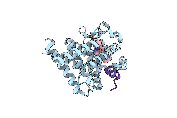
Cryo-Em Structure Of The Human Growth Hormone-Releasing Hormone Receptor-Gs Protein Complex
Organism: Homo sapiens, Rattus norvegicus, Bos taurus, Synthetic construct
Method: ELECTRON MICROSCOPY Release Date: 2020-11-18 Classification: MEMBRANE PROTEIN Ligands: PLM, CLR |