

Search Count: 14
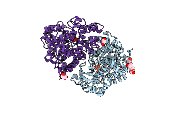 |
Structure Of Human Enolase 2 In Complex With Phosphoserine
Organism: Homo sapiens
Method: X-RAY DIFFRACTION Resolution:2.10 Å Release Date: 2022-11-09 Classification: LYASE Ligands: PO4, MG, ACT, EDO, PGE, SEP |
Organism: Homo sapiens
Method: X-RAY DIFFRACTION
Release Date: 2022-11-09
Ligands: PO4, MG, ACT, EDO, PGE, SEP
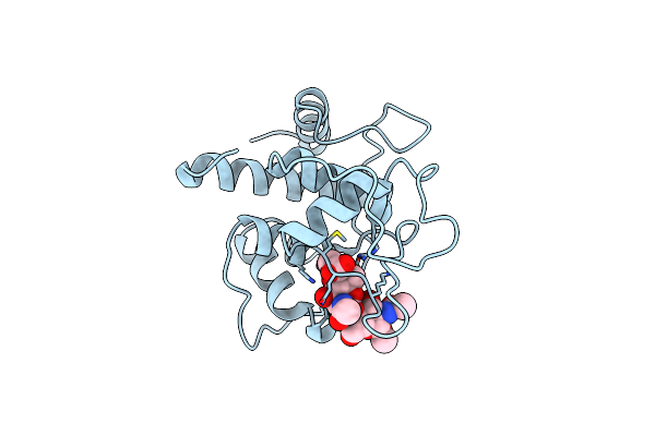 |
Crystal Structure Of Spaa-Slh In Complex With 4,6-Pyr-Beta-D-Mannac-(1->4)-Beta-D-Glcnacome
Organism: Paenibacillus alvei
Method: X-RAY DIFFRACTION Resolution:1.70 Å Release Date: 2022-03-09 Classification: SUGAR BINDING PROTEIN Ligands: D2Y |
Organism: Paenibacillus alvei
Method: X-RAY DIFFRACTION
Release Date: 2022-03-09
Ligands: D2Y
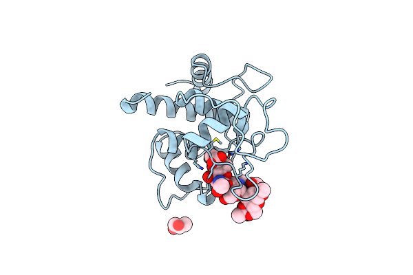 |
Crystal Structure Of Spaa-Slh In Complex With 4,6-Pyr-Beta-D-Mannac-(1->4)-Beta-D-Glcnac-(1->3)-4,6-Pyr-Beta-D-Mannacome
Organism: Paenibacillus alvei
Method: X-RAY DIFFRACTION Resolution:2.06 Å Release Date: 2022-03-09 Classification: SUGAR BINDING PROTEIN Ligands: GOL, D4I |
Organism: Paenibacillus alvei
Method: X-RAY DIFFRACTION
Release Date: 2022-03-09
Ligands: GOL, D4I
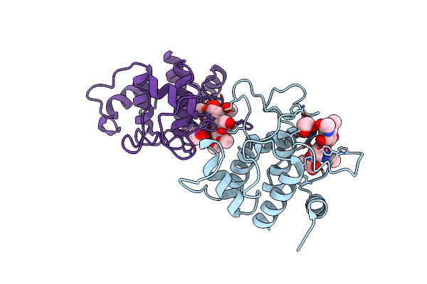 |
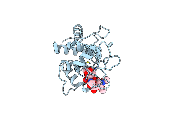
Crystal Structure Of Spaa-Slh/G109A In Complex With 4,6-Pyr-Beta-D-Mannac-(1->4)-Beta-D-Glcnacome
Organism: Paenibacillus alvei
Method: X-RAY DIFFRACTION Resolution:1.72 Å Release Date: 2022-03-09 Classification: SUGAR BINDING PROTEIN Ligands: GOL, D2Y |
Organism: Paenibacillus alvei
Method: X-RAY DIFFRACTION
Release Date: 2022-03-09
Ligands: GOL, D2Y
 |
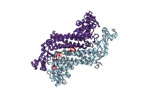
Crystal Structure Of Spaa-Slh/G46A/G109A In Complex With 4,6-Pyr-Beta-D-Mannac-(1->4)-Beta-D-Glcnacome
Organism: Paenibacillus alvei
Method: X-RAY DIFFRACTION Resolution:1.85 Å Release Date: 2022-03-09 Classification: SUGAR BINDING PROTEIN Ligands: D2Y |
Organism: Paenibacillus alvei
Method: X-RAY DIFFRACTION
Release Date: 2022-03-09
Ligands: D2Y
 |
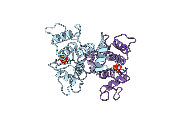
Crystal Structure Of Recombinant Human Fumarase In Complex With D-2-Amino-3-Phosphono-Propionic Acid
Organism: Homo sapiens
Method: X-RAY DIFFRACTION Resolution:2.15 Å Release Date: 2022-02-23 Classification: LYASE Ligands: APO, GOL |
Organism: Homo sapiens
Method: X-RAY DIFFRACTION
Release Date: 2022-02-23
Ligands: APO, GOL
 |
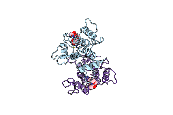
Crystal Structure Of Spaa-Slh
Organism: Paenibacillus alvei
Method: X-RAY DIFFRACTION Resolution:1.90 Å Release Date: 2018-08-15 Classification: SUGAR BINDING PROTEIN Ligands: SO4, CL |
Organism: Paenibacillus alvei
Method: X-RAY DIFFRACTION
Release Date: 2018-08-15
Ligands: SO4, CL
 |
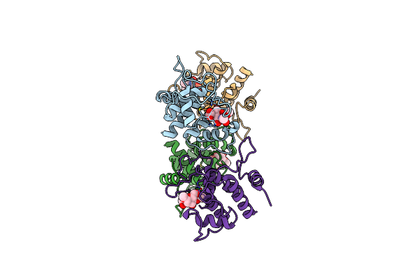
Crystal Structure Of Spaa-Slh In Complex With 4,6-Pyr-Beta-D-Mannacome
Organism: Paenibacillus alvei
Method: X-RAY DIFFRACTION Resolution:2.25 Å Release Date: 2018-08-15 Classification: SUGAR BINDING PROTEIN Ligands: 6LA |
Organism: Paenibacillus alvei
Method: X-RAY DIFFRACTION
Release Date: 2018-08-15
Ligands: 6LA
 |
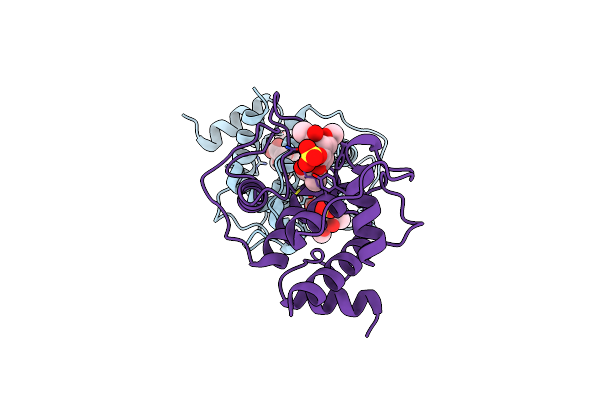
Crystal Structure Of Spaa-Slh In Complex With 4,6-Pyr-Beta-D-Mannacome (P1)
Organism: Paenibacillus alvei
Method: X-RAY DIFFRACTION Resolution:2.00 Å Release Date: 2018-08-15 Classification: SUGAR BINDING PROTEIN Ligands: 6LA |
Organism: Paenibacillus alvei
Method: X-RAY DIFFRACTION
Release Date: 2018-08-15
Ligands: 6LA
 |
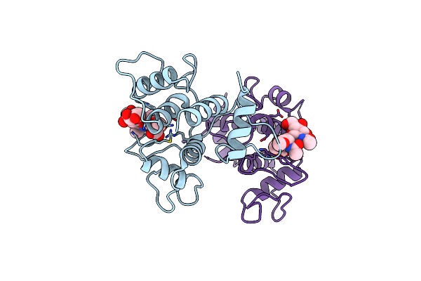
Crystal Structure Of Spaa-Slh In Complex With 4,6-Pyr-Beta-D-Mannacome (C2)
Organism: Paenibacillus alvei
Method: X-RAY DIFFRACTION Resolution:2.16 Å Release Date: 2018-08-15 Classification: SUGAR BINDING PROTEIN Ligands: 6LA, SO4 |
Organism: Paenibacillus alvei
Method: X-RAY DIFFRACTION
Release Date: 2018-08-15
Ligands: 6LA, SO4
 |
Crystal Structure Of Spaa-Slh In Complex With Beta-D-Glcnac-(1->3)-4,6-Pyr-Beta-D-Mannacome
Organism: Paenibacillus alvei
Method: X-RAY DIFFRACTION Resolution:2.15 Å Release Date: 2018-08-15 Classification: SUGAR BINDING PROTEIN Ligands: FHY |
Organism: Paenibacillus alvei
Method: X-RAY DIFFRACTION
Release Date: 2018-08-15
Ligands: FHY
 |
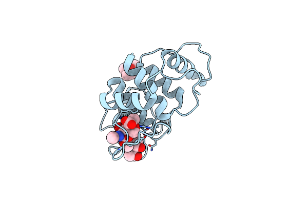
Crystal Structure Of Spaa-Slh/G109A
Organism: Paenibacillus alvei
Method: X-RAY DIFFRACTION Resolution:1.15 Å Release Date: 2018-08-15 Classification: SUGAR BINDING PROTEIN Ligands: CL |
Organism: Paenibacillus alvei
Method: X-RAY DIFFRACTION
Release Date: 2018-08-15
Ligands: CL
 |
Crystal Structure Of Spaa-Slh/G109A In Complex With 4,6-Pyr-Beta-D-Mannacome
Organism: Paenibacillus alvei
Method: X-RAY DIFFRACTION Resolution:1.53 Å Release Date: 2018-08-15 Classification: SUGAR BINDING PROTEIN Ligands: 6LA, MPD |
Organism: Paenibacillus alvei
Method: X-RAY DIFFRACTION
Release Date: 2018-08-15
Ligands: 6LA, MPD
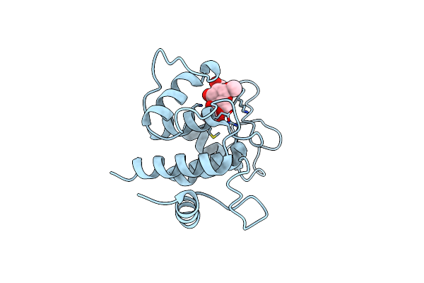 |
Crystal Structure Of Spaa-Slh/G46A/G109A In Complex With 4,6-Pyr-Beta-D-Mannacome
Organism: Paenibacillus alvei
Method: X-RAY DIFFRACTION Resolution:1.24 Å Release Date: 2018-08-15 Classification: SUGAR BINDING PROTEIN Ligands: 6LA |
Organism: Paenibacillus alvei
Method: X-RAY DIFFRACTION
Release Date: 2018-08-15
Ligands: 6LA