

Search Count: 25
 |
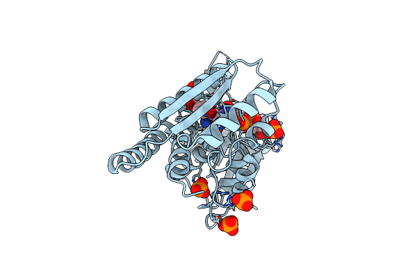
Structure Of Human Mthfd2 In Complex With Th9028
Organism: Homo sapiens
Method: X-RAY DIFFRACTION Resolution:1.95 Å Release Date: 2021-07-07 Classification: OXIDOREDUCTASE Ligands: KUK, PO4, NAD, MPD |
Organism: Homo sapiens
Method: X-RAY DIFFRACTION
Release Date: 2021-07-07
Ligands: KUK, PO4, NAD, MPD
 |
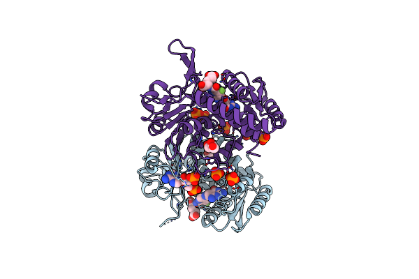
Structure Of Human Mthfd2 In Complex With Th7299
Organism: Homo sapiens
Method: X-RAY DIFFRACTION Resolution:1.90 Å Release Date: 2021-07-07 Classification: OXIDOREDUCTASE Ligands: 9L9, PO4, NAD |
Organism: Homo sapiens
Method: X-RAY DIFFRACTION
Release Date: 2021-07-07
Ligands: 9L9, PO4, NAD
 |
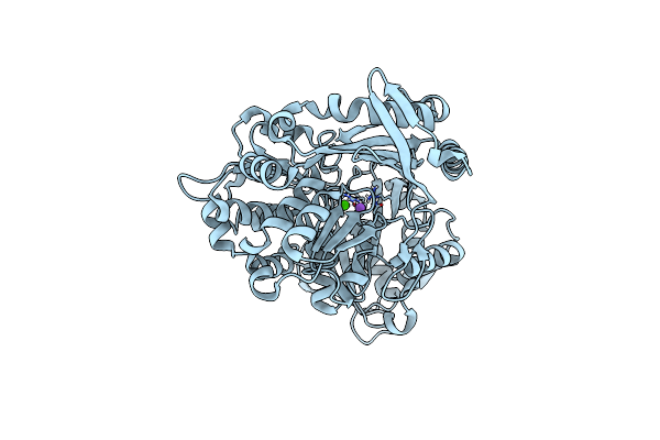
Structure Of Human Mthfd2 In Complex With Th9619
Organism: Homo sapiens
Method: X-RAY DIFFRACTION Resolution:2.20 Å Release Date: 2021-07-07 Classification: OXIDOREDUCTASE Ligands: KUN, PO4, ADP, GOL |
Organism: Homo sapiens
Method: X-RAY DIFFRACTION
Release Date: 2021-07-07
Ligands: KUN, PO4, ADP, GOL
 |
Structure Of Atlantic Herring (Clupea Harengus) Phosphoglucomutase 5 (Pgm5)
Organism: Clupea harengus
Method: X-RAY DIFFRACTION Resolution:2.25 Å Release Date: 2020-12-16 Classification: ISOMERASE Ligands: CA, NA |
Organism: Clupea harengus
Method: X-RAY DIFFRACTION
Release Date: 2020-12-16
Ligands: CA, NA
 |
Structure Of Baltic Herring (Clupea Harengus) Phosphoglucomutase 5 (Pgm5) With Bound Glucose-1-Phosphate
Organism: Clupea harengus
Method: X-RAY DIFFRACTION Resolution:1.95 Å Release Date: 2020-12-16 Classification: ISOMERASE Ligands: NI, CA, G1P, GOL, ACT, NA |
Organism: Clupea harengus
Method: X-RAY DIFFRACTION
Release Date: 2020-12-16
Ligands: NI, CA, G1P, GOL, ACT, NA
 |
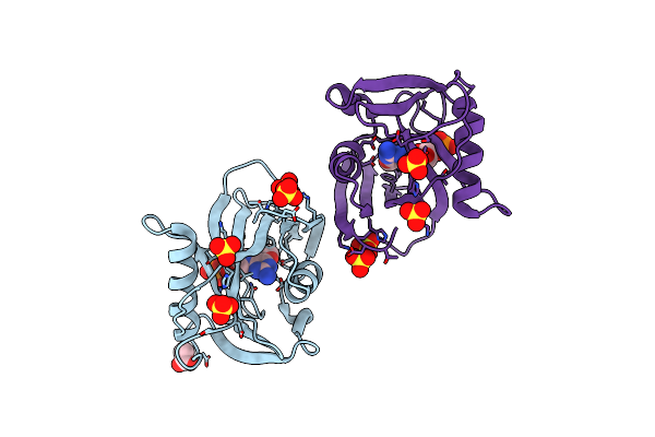
Structure Of Baltic Herring (Clupea Harengus) Phosphoglucomutase 5 (Pgm5)
Organism: Clupea harengus
Method: X-RAY DIFFRACTION Resolution:2.05 Å Release Date: 2020-12-16 Classification: ISOMERASE Ligands: NI, CA, NA, GOL, ACT |
Organism: Clupea harengus
Method: X-RAY DIFFRACTION
Release Date: 2020-12-16
Ligands: NI, CA, NA, GOL, ACT
 |
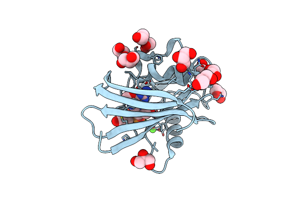
Crystal Structure Of Human Mth1 In Complex With O6-Methyl-Dgmp
Organism: Homo sapiens
Method: X-RAY DIFFRACTION Resolution:1.80 Å Release Date: 2018-09-05 Classification: HYDROLASE Ligands: SO4, ACT, 6OG |
Organism: Homo sapiens
Method: X-RAY DIFFRACTION
Release Date: 2018-09-05
Ligands: SO4, ACT, 6OG
 |
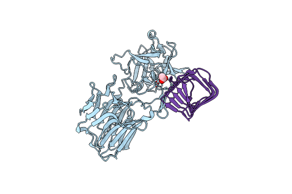
Crystal Structure Of Zebrafish Mth1 In Complex With O6-Methyl-Dgmp
Organism: Danio rerio
Method: X-RAY DIFFRACTION Resolution:0.99 Å Release Date: 2018-09-05 Classification: HYDROLASE Ligands: 6OG, GOL, CA, EDO, CL |
Organism: Danio rerio
Method: X-RAY DIFFRACTION
Release Date: 2018-09-05
Ligands: 6OG, GOL, CA, EDO, CL
 |
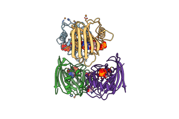
Crystal Structure Of The Binding Domain From Botulinum Neurotoxin A2 Bound To Extracellular Domain Of Human Receptor Sv2C
Organism: Clostridium botulinum, Homo sapiens
Method: X-RAY DIFFRACTION Resolution:2.00 Å Release Date: 2018-04-25 Classification: PROTEIN BINDING Ligands: ACT |
Organism: Clostridium botulinum, Homo sapiens
Method: X-RAY DIFFRACTION
Release Date: 2018-04-25
Ligands: ACT
 |
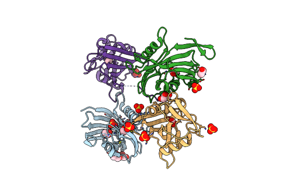
Crystal Structure Of Mouse Mth1 With 8-Oxo-Dgtp
Organism: Mus musculus
Method: X-RAY DIFFRACTION Resolution:2.10 Å Release Date: 2018-01-10 Classification: HYDROLASE Ligands: CU, PEG, 8DG, GOL |
Organism: Mus musculus
Method: X-RAY DIFFRACTION
Release Date: 2018-01-10
Ligands: CU, PEG, 8DG, GOL
 |
Crystal Structure Of Dog Mth1 Protein
Organism: Canis lupus familiaris
Method: X-RAY DIFFRACTION Resolution:2.00 Å Release Date: 2018-01-10 Classification: HYDROLASE Ligands: SO4, ACT, GOL, CL |
Organism: Canis lupus familiaris
Method: X-RAY DIFFRACTION
Release Date: 2018-01-10
Ligands: SO4, ACT, GOL, CL
 |
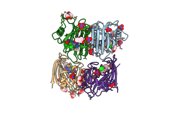
Crystal Structure Of Mouse Mth1 In Complex With Th588
Organism: Mus musculus
Method: X-RAY DIFFRACTION Resolution:1.85 Å Release Date: 2018-01-10 Classification: HYDROLASE Ligands: 2GE, SO4, SCN |
Organism: Mus musculus
Method: X-RAY DIFFRACTION
Release Date: 2018-01-10
Ligands: 2GE, SO4, SCN
 |
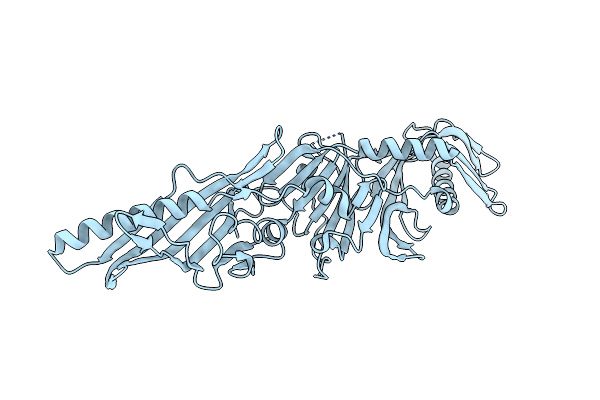
Crystal Structure Of Mouse Mth1 Mutant L116M With Inhibitor Th588
Organism: Mus musculus
Method: X-RAY DIFFRACTION Resolution:2.40 Å Release Date: 2018-01-10 Classification: HYDROLASE Ligands: 2GE, NO3, PEG, GOL, CU, MG |
Organism: Mus musculus
Method: X-RAY DIFFRACTION
Release Date: 2018-01-10
Ligands: 2GE, NO3, PEG, GOL, CU, MG
 |
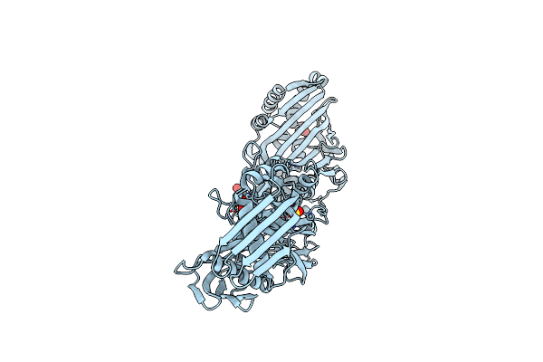
Structure Of P47 From Clostridium Botulinum A2
Organism: Clostridium botulinum (strain kyoto / type a2)
Method: X-RAY DIFFRACTION Resolution:1.75 Å Release Date: 2017-10-25 Classification: LIPID BINDING PROTEIN |
Organism: Clostridium botulinum (strain kyoto / type a2)
Method: X-RAY DIFFRACTION
Release Date: 2017-10-25
 |
Structure Of Orfx2 From Clostridium Botulinum A2
Organism: Clostridium botulinum (strain kyoto / type a2)
Method: X-RAY DIFFRACTION Resolution:2.10 Å Release Date: 2017-10-25 Classification: LIPID BINDING PROTEIN Ligands: SO4 |
Organism: Clostridium botulinum (strain kyoto / type a2)
Method: X-RAY DIFFRACTION
Release Date: 2017-10-25
Ligands: SO4
 |
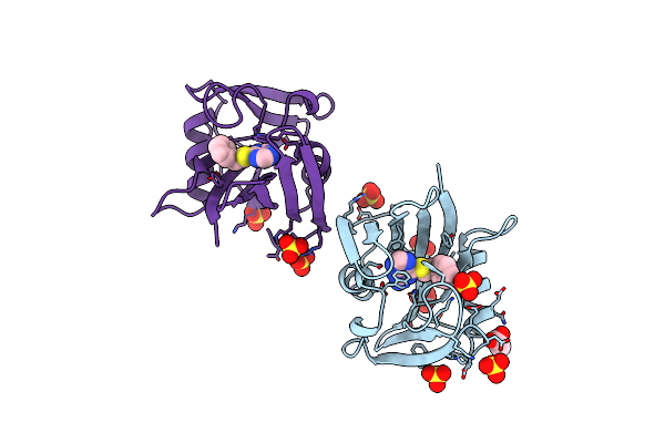
Crystal Structure Of Human Mth1 In Complex With Fragment Inhibitor 8-(Methylsulfanyl)-7H-Purin-6-Amine
Organism: Homo sapiens
Method: X-RAY DIFFRACTION Resolution:2.20 Å Release Date: 2017-10-04 Classification: HYDROLASE Ligands: 8WT, SO4 |
Organism: Homo sapiens
Method: X-RAY DIFFRACTION
Release Date: 2017-10-04
Ligands: 8WT, SO4
 |
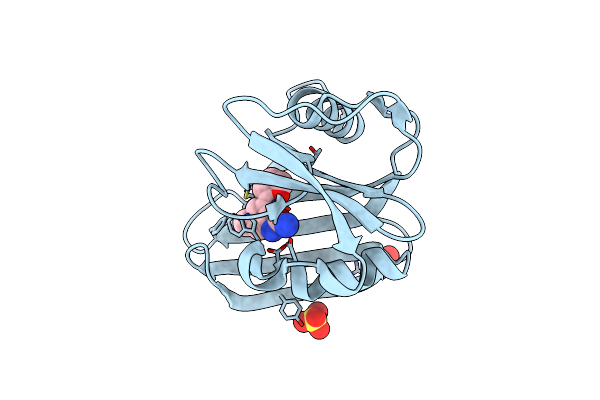
Crystal Structure Of Human Mth1 In Complex With Inhibitor 6-[(2-Phenylethyl)Sulfanyl]-7H-Purin-2-Amine
Organism: Homo sapiens
Method: X-RAY DIFFRACTION Resolution:1.85 Å Release Date: 2017-10-04 Classification: HYDROLASE Ligands: 8WW, SO4, ACT |
Organism: Homo sapiens
Method: X-RAY DIFFRACTION
Release Date: 2017-10-04
Ligands: 8WW, SO4, ACT
 |
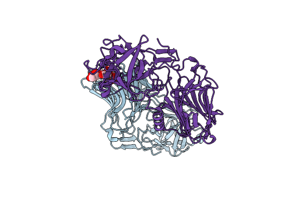
Crystal Structure Of Human Mth1 In Complex With Inhibitor 7-(Furan-2-Yl)-5-Methyl-1,3-Benzoxazol-2-Amine
Organism: Homo sapiens
Method: X-RAY DIFFRACTION Resolution:1.54 Å Release Date: 2017-10-04 Classification: HYDROLASE Ligands: 8WZ, SO4 |
Organism: Homo sapiens
Method: X-RAY DIFFRACTION
Release Date: 2017-10-04
Ligands: 8WZ, SO4
 |
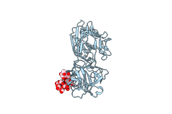
Binding Domain Of Bont/A Complexed With Ganglioside Variant
Organism: Clostridium botulinum
Method: X-RAY DIFFRACTION Resolution:2.60 Å Release Date: 2017-01-18 Classification: HYDROLASE Ligands: SIA |
Organism: Clostridium botulinum
Method: X-RAY DIFFRACTION
Release Date: 2017-01-18
Ligands: SIA
 |
Binding Domain Of Bont/A Complexed With Ganglioside
Organism: Clostridium botulinum
Method: X-RAY DIFFRACTION Resolution:2.00 Å Release Date: 2017-01-18 Classification: HYDROLASE |
Organism: Clostridium botulinum
Method: X-RAY DIFFRACTION
Release Date: 2017-01-18