
Search Count: 673
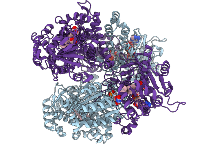 |
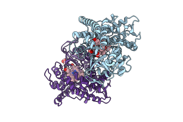
Organism: Shimazuella soli
Method: ELECTRON MICROSCOPY Resolution:2.72 Å Release Date: 2026-05-27 Classification: OXIDOREDUCTASE Ligands: HEM, FMN, FAD |
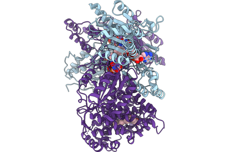 |
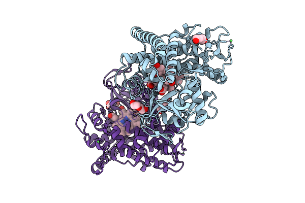
Organism: Shimazuella soli
Method: ELECTRON MICROSCOPY Resolution:3.08 Å Release Date: 2026-05-27 Classification: OXIDOREDUCTASE Ligands: FMN, FAD, HEM |
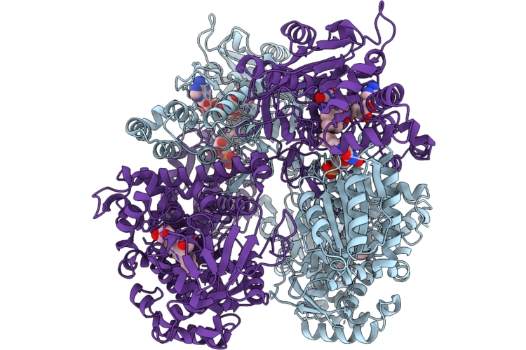 |
Organism: Shimazuella soli
Method: ELECTRON MICROSCOPY Resolution:2.34 Å Release Date: 2026-05-27 Classification: OXIDOREDUCTASE Ligands: HEM, FMN, FAD |
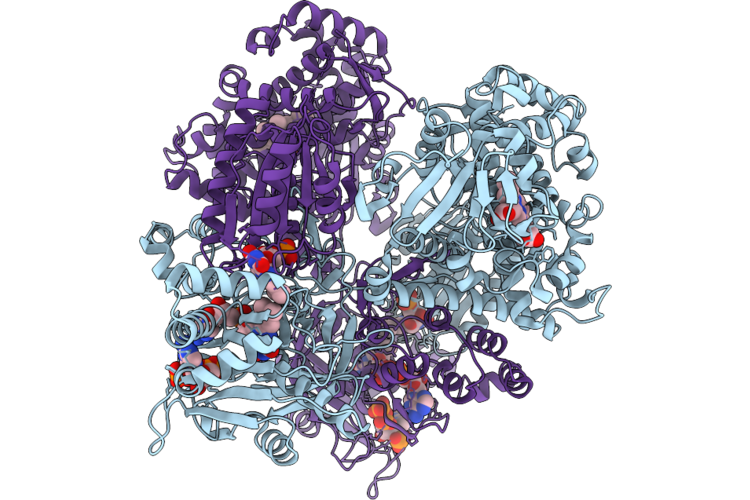 |
Organism: Shimazuella soli
Method: ELECTRON MICROSCOPY Resolution:2.68 Å Release Date: 2026-05-27 Classification: OXIDOREDUCTASE Ligands: HEM, FMN, FAD, NDP |
 |
Organism: Shimazuella soli
Method: X-RAY DIFFRACTION Resolution:3.38 Å Release Date: 2026-05-27 Classification: OXIDOREDUCTASE Ligands: HEM, FMN, FAD |
 |
Organism: Stenotrophomonas sp. cw117
Method: ELECTRON MICROSCOPY Release Date: 2026-05-13 Classification: HYDROLASE Ligands: ZN, 97U |
 |
Organism: Pseudoxanthomonas wuyuanensis
Method: ELECTRON MICROSCOPY Release Date: 2026-05-06 Classification: HYDROLASE Ligands: ZN, 97U |
 |
Organism: Novilysobacter luteus
Method: ELECTRON MICROSCOPY Resolution:2.69 Å Release Date: 2026-05-06 Classification: HYDROLASE Ligands: ZN, 97U |
 |
Organism: Novilysobacter luteus
Method: ELECTRON MICROSCOPY Resolution:2.90 Å Release Date: 2026-05-06 Classification: HYDROLASE Ligands: ZN |
 |
Organism: Mus musculus
Method: X-RAY DIFFRACTION Resolution:2.81 Å Release Date: 2026-04-22 Classification: ANTITUMOR PROTEIN |
 |
Organism: Serratia plymuthica 4rx13
Method: X-RAY DIFFRACTION Resolution:2.22 Å Release Date: 2026-03-18 Classification: LYASE Ligands: GOL |
 |
Organism: Serratia plymuthica 4rx13
Method: X-RAY DIFFRACTION Resolution:1.87 Å Release Date: 2026-03-18 Classification: LYASE Ligands: POP, MG, TRS |
 |
Organism: Serratia plymuthica 4rx13
Method: X-RAY DIFFRACTION Resolution:1.74 Å Release Date: 2026-03-18 Classification: LYASE Ligands: GPP, MG, TRS, POP |
 |
Organism: Priestia megaterium (strain nbrc 15308)
Method: X-RAY DIFFRACTION Resolution:1.57 Å Release Date: 2026-03-18 Classification: OXIDOREDUCTASE Ligands: HEM, IMD, PEG, TRS, CL |
 |
Organism: Priestia megaterium (strain nbrc 15308)
Method: X-RAY DIFFRACTION Resolution:1.80 Å Release Date: 2026-03-18 Classification: OXIDOREDUCTASE Ligands: HEM, ZER, TRS |
 |
Organism: Priestia megaterium (strain nbrc 15308)
Method: X-RAY DIFFRACTION Resolution:2.09 Å Release Date: 2026-03-18 Classification: OXIDOREDUCTASE Ligands: HEM, 36J, PEG, NI, TRS |
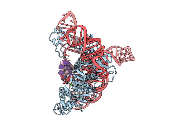 |
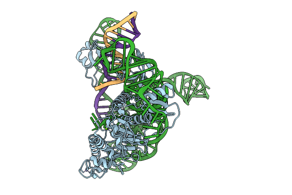
Organism: Synthetic construct
Method: ELECTRON MICROSCOPY Resolution:3.04 Å Release Date: 2026-01-21 Classification: RNA BINDING PROTEIN Ligands: ZN, MG |
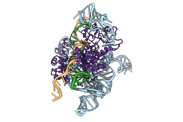 |
Organism: Synthetic construct
Method: ELECTRON MICROSCOPY Resolution:3.10 Å Release Date: 2026-01-21 Classification: DNA BINDING PROTEIN Ligands: MG, ZN |
 |
Organism: Synthetic construct
Method: ELECTRON MICROSCOPY Resolution:3.39 Å Release Date: 2026-01-21 Classification: RNA BINDING PROTEIN Ligands: ZN, MG |
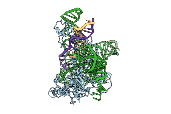 |
Organism: Synthetic construct
Method: ELECTRON MICROSCOPY Resolution:3.09 Å Release Date: 2026-01-21 Classification: DNA BINDING PROTEIN Ligands: ZN, MG |