

Search Count: 36
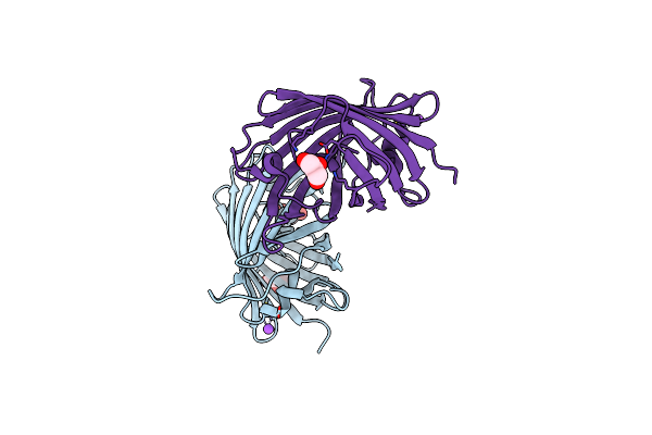 |
Crystal Structure Of A Bright Green Fluorescent Protein (Staygold) With Single Mutation (N137A) In Jellyfish Cytaeis Uchidae From Biortus
Organism: Cytaeis uchidae
Method: X-RAY DIFFRACTION Resolution:1.70 Å Release Date: 2023-12-13 Classification: FLUORESCENT PROTEIN Ligands: GOL, NA |
Organism: Cytaeis uchidae
Method: X-RAY DIFFRACTION
Release Date: 2023-12-13
Ligands: GOL, NA
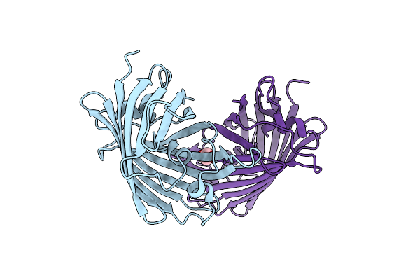 |
Crystal Structure Of A Bright Green Fluorescent Protein (Staygold) With Single Mutation (Q140S) In Jellyfish Cytaeis Uchidae From Biortus
Organism: Cytaeis uchidae
Method: X-RAY DIFFRACTION Resolution:1.75 Å Release Date: 2023-12-13 Classification: FLUORESCENT PROTEIN Ligands: EDO |
Organism: Cytaeis uchidae
Method: X-RAY DIFFRACTION
Release Date: 2023-12-13
Ligands: EDO
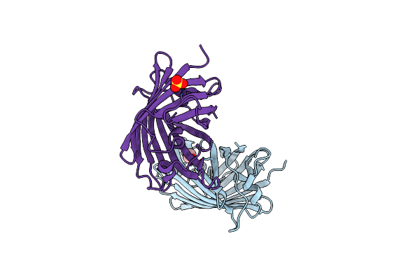 |
Crystal Structure Of A Bright Green Fluorescent Protein (Staygold) With Single Mutation (Y187F) In Jellyfish Cytaeis Uchidae From Biortus
Organism: Cytaeis uchidae
Method: X-RAY DIFFRACTION Resolution:1.90 Å Release Date: 2023-12-13 Classification: FLUORESCENT PROTEIN Ligands: EDO, SO4 |
Organism: Cytaeis uchidae
Method: X-RAY DIFFRACTION
Release Date: 2023-12-13
Ligands: EDO, SO4
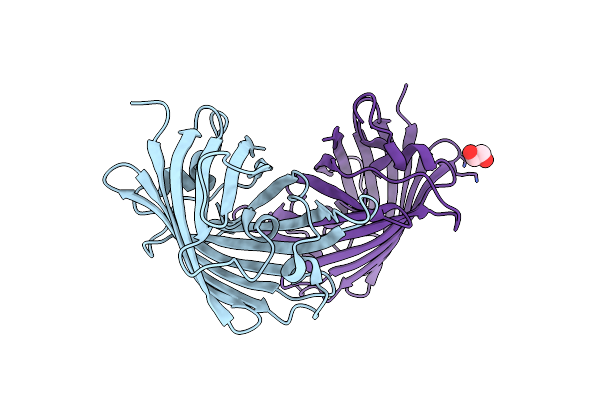 |
Crystal Structure Of A Bright Green Fluorescent Protein (Staygold) With Double Mutation (N137A, Q140S) In Jellyfish Cytaeis Uchidae From Biortus
Organism: Cytaeis uchidae
Method: X-RAY DIFFRACTION Resolution:1.70 Å Release Date: 2023-12-13 Classification: FLUORESCENT PROTEIN Ligands: EDO |
Organism: Cytaeis uchidae
Method: X-RAY DIFFRACTION
Release Date: 2023-12-13
Ligands: EDO
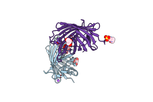 |
Crystal Structure Of A Bright Green Fluorescent Protein (Staygold) With Double Mutations (N137A, Y187F) In Jellyfish Cytaeis Uchidae From Biortus
Organism: Cytaeis uchidae
Method: X-RAY DIFFRACTION Resolution:1.70 Å Release Date: 2023-12-13 Classification: FLUORESCENT PROTEIN Ligands: GOL, NA, EPE |
Organism: Cytaeis uchidae
Method: X-RAY DIFFRACTION
Release Date: 2023-12-13
Ligands: GOL, NA, EPE
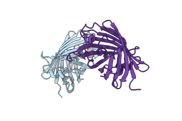 |
Crystal Structure Of A Bright Green Fluorescent Protein (Staygold) With Double Mutations (Q140S, Y187F) In Jellyfish Cytaeis Uchidae From Biortus
Organism: Cytaeis uchidae
Method: X-RAY DIFFRACTION Resolution:1.70 Å Release Date: 2023-12-13 Classification: FLUORESCENT PROTEIN Ligands: EDO |
Organism: Cytaeis uchidae
Method: X-RAY DIFFRACTION
Release Date: 2023-12-13
Ligands: EDO
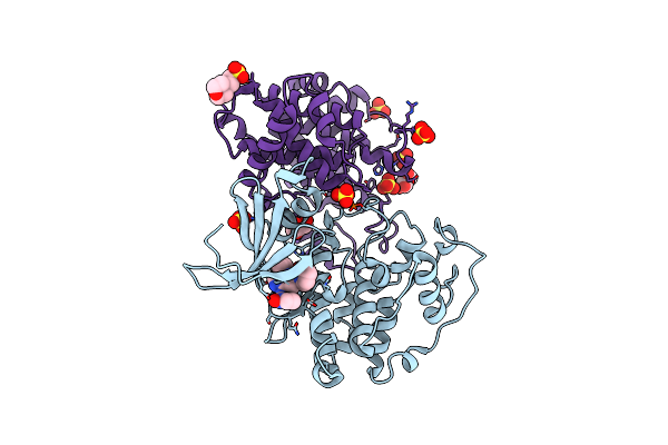 |
The Crystal Structure Of Cdk3 And Cycline1 Complex With Dinaciclib From Biortus
Organism: Schistosoma japonicum, Homo sapiens
Method: X-RAY DIFFRACTION Resolution:2.75 Å Release Date: 2023-10-11 Classification: CELL CYCLE Ligands: 1QK, SO4, GOL, MES |
Organism: Schistosoma japonicum, Homo sapiens
Method: X-RAY DIFFRACTION
Release Date: 2023-10-11
Ligands: 1QK, SO4, GOL, MES
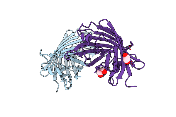 |
Crystal Structure Of A Bright Green Fluorescent Protein (Staygold) With Single Mutation (K192Y) In Jellyfish Cytaeis Uchidae From Biortus
Organism: Cytaeis uchidae
Method: X-RAY DIFFRACTION Resolution:2.00 Å Release Date: 2023-10-04 Classification: FLUORESCENT PROTEIN Ligands: EDO |
Organism: Cytaeis uchidae
Method: X-RAY DIFFRACTION
Release Date: 2023-10-04
Ligands: EDO
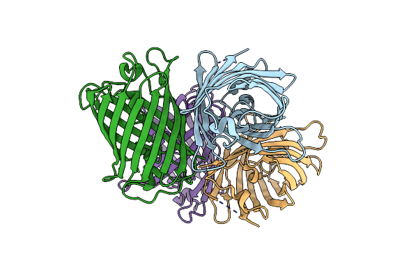 |
Crystal Structure Of A Bright Green Fluorescent Protein (Staygold) With Triple Mutations (N137A, Q140S, Y187F) In Jellyfish Cytaeis Uchidae From Biortus
Organism: Cytaeis uchidae
Method: X-RAY DIFFRACTION Resolution:2.30 Å Release Date: 2023-08-16 Classification: FLUORESCENT PROTEIN Ligands: EDO |
Organism: Cytaeis uchidae
Method: X-RAY DIFFRACTION
Release Date: 2023-08-16
Ligands: EDO
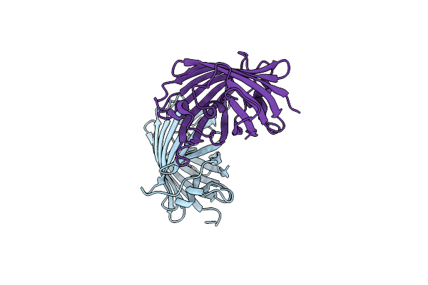 |
Crystal Structure Of A Bright Green Fluorescent Protein (Oxstaygold) In Jellyfish Cytaeis Uchidae From Biortus
Organism: Cytaeis uchidae
Method: X-RAY DIFFRACTION Resolution:1.50 Å Release Date: 2023-07-19 Classification: FLUORESCENT PROTEIN |
Organism: Cytaeis uchidae
Method: X-RAY DIFFRACTION
Release Date: 2023-07-19
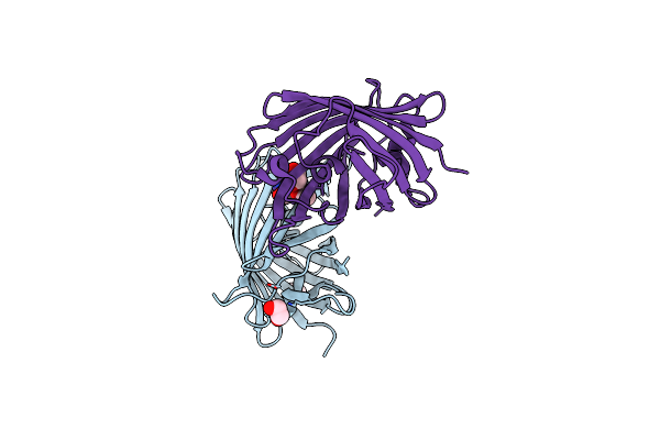 |
Crystal Structure Of A Bright Green Fluorescent Protein (Staygold) In Jellyfish Cytaeis Uchidae From Biortus
Organism: Cytaeis uchidae
Method: X-RAY DIFFRACTION Resolution:1.70 Å Release Date: 2023-07-05 Classification: FLUORESCENT PROTEIN Ligands: EDO |
Organism: Cytaeis uchidae
Method: X-RAY DIFFRACTION
Release Date: 2023-07-05
Ligands: EDO
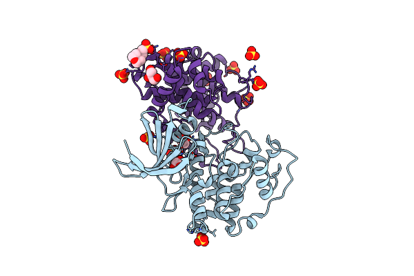 |
The Crystal Structure Of Cdk3 And Cycline1 Complex From Biortus.
Organism: Schistosoma japonicum, Homo sapiens
Method: X-RAY DIFFRACTION Resolution:2.25 Å Release Date: 2023-05-17 Classification: CELL CYCLE Ligands: SO4, GOL, MES |
Organism: Schistosoma japonicum, Homo sapiens
Method: X-RAY DIFFRACTION
Release Date: 2023-05-17
Ligands: SO4, GOL, MES
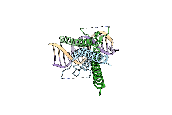 |
The Complex Of Dna With The C-Terminal Domain Of Tye7 From Saccharomyces Cerevisiae.
Organism: Saccharomyces cerevisiae (strain atcc 204508 / s288c), Synthetic construct
Method: X-RAY DIFFRACTION Resolution:2.55 Å Release Date: 2021-10-13 Classification: TRANSCRIPTION |
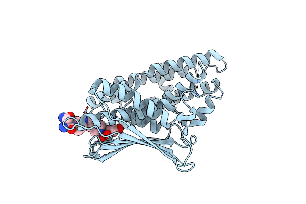 |
Crystal Structure Of Pyruvate Dehydrogenase Kinase Isoform 2 In Complex With Inhibitor Ps46
Organism: Homo sapiens
Method: X-RAY DIFFRACTION Resolution:2.05 Å Release Date: 2017-01-25 Classification: Transferase/Transferase Inhibitor Ligands: P46 |
Organism: Homo sapiens
Method: X-RAY DIFFRACTION
Release Date: 2017-01-25
Ligands: P46
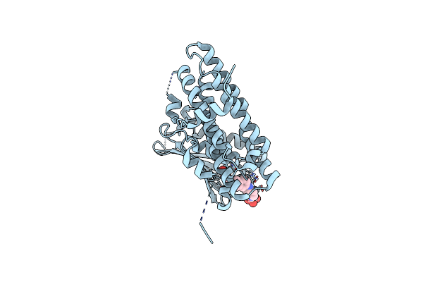 |
Crystal Structure Of Pyruvate Dehydrogenase Kinase Isoform 2 In Complex With Inhibitor Ps35
Organism: Homo sapiens
Method: X-RAY DIFFRACTION Resolution:1.65 Å Release Date: 2017-01-25 Classification: Transferase/Transferase Inhibitor Ligands: TLA, P35 |
Organism: Homo sapiens
Method: X-RAY DIFFRACTION
Release Date: 2017-01-25
Ligands: TLA, P35
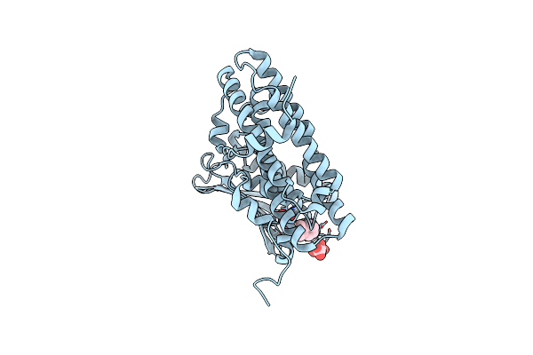 |
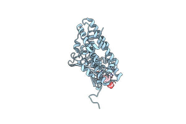
Crystal Structure Of Pyruvate Dehydrogenase Kinase Isoform 2 In Complex With Inhibitor Pa1
Organism: Homo sapiens
Method: X-RAY DIFFRACTION Resolution:1.75 Å Release Date: 2014-01-01 Classification: Transferase/Transferase Inhibitor Ligands: PV1, TLA |
Organism: Homo sapiens
Method: X-RAY DIFFRACTION
Release Date: 2014-01-01
Ligands: PV1, TLA
 |
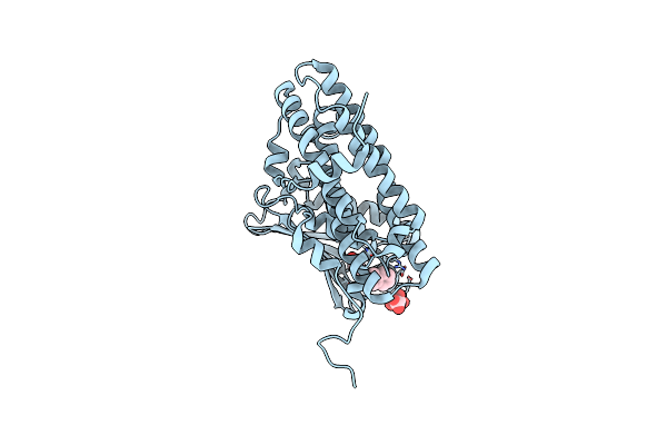
Crystal Structure Of Pyruvate Dehydrogenase Kinase Isoform 2 In Complex With Inhibitor Pa7
Organism: Homo sapiens
Method: X-RAY DIFFRACTION Resolution:1.80 Å Release Date: 2014-01-01 Classification: Transferase/Transferase Inhibitor Ligands: PFT, TLA |
Organism: Homo sapiens
Method: X-RAY DIFFRACTION
Release Date: 2014-01-01
Ligands: PFT, TLA
 |
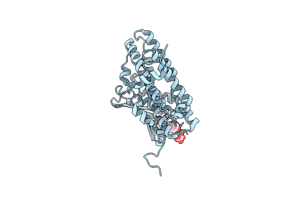
Crystal Structure Of Pyruvate Dehydrogenase Kinase Isoform 2 In Complex With Inhibitor Ps2
Organism: Homo sapiens
Method: X-RAY DIFFRACTION Resolution:1.70 Å Release Date: 2014-01-01 Classification: Transferase/Transferase Inhibitor Ligands: TLA, PV2 |
Organism: Homo sapiens
Method: X-RAY DIFFRACTION
Release Date: 2014-01-01
Ligands: TLA, PV2
 |
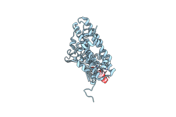
Crystal Structure Of Pyruvate Dehydrogenase Kinase Isoform 2 In Complex With Inhibitor Ps8
Organism: Homo sapiens
Method: X-RAY DIFFRACTION Resolution:1.95 Å Release Date: 2014-01-01 Classification: Transferase/Transferase Inhibitor Ligands: PV8, TLA |
Organism: Homo sapiens
Method: X-RAY DIFFRACTION
Release Date: 2014-01-01
Ligands: PV8, TLA
 |
Crystal Structure Of Pyruvate Dehydrogenase Kinase Isoform 2 In Complex With Inhibitor Ps10
Organism: Homo sapiens
Method: X-RAY DIFFRACTION Resolution:1.75 Å Release Date: 2014-01-01 Classification: TRANSFERASE Ligands: PV0, TLA |
Organism: Homo sapiens
Method: X-RAY DIFFRACTION
Release Date: 2014-01-01
Ligands: PV0, TLA