

Search Count: 30
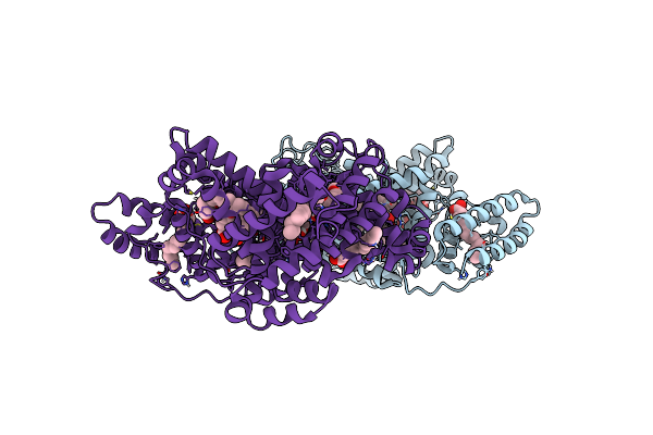 |
Structure Of Human Serum Albumin In Complex With Aristolochic Acid Ii At 1.9 A Resolution
Organism: Homo sapiens
Method: X-RAY DIFFRACTION Resolution:1.90 Å Release Date: 2024-06-26 Classification: LIPID BINDING PROTEIN Ligands: MYR, EDO, GOR |
Organism: Homo sapiens
Method: X-RAY DIFFRACTION
Release Date: 2024-06-26
Ligands: MYR, EDO, GOR
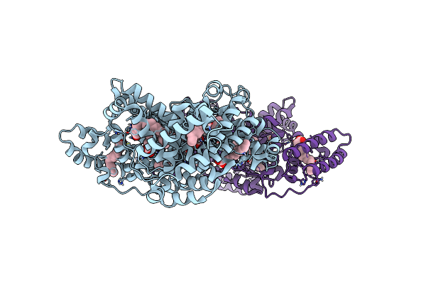 |
Structure Of Human Serum Albumin In Complex With Myristic Acid
Organism: Homo sapiens
Method: X-RAY DIFFRACTION Resolution:1.90 Å Release Date: 2024-06-26 Classification: LIPID BINDING PROTEIN Ligands: MYR, EDO |
Organism: Homo sapiens
Method: X-RAY DIFFRACTION
Release Date: 2024-06-26
Ligands: MYR, EDO
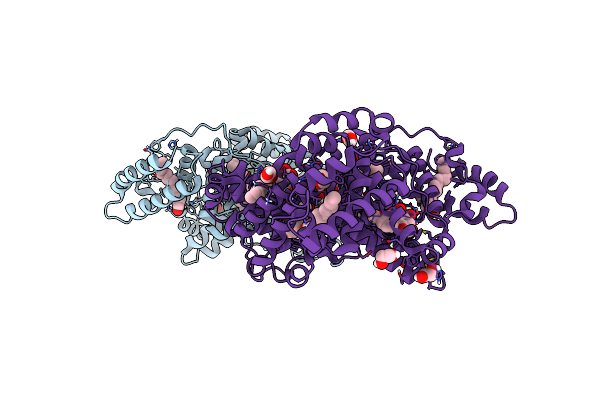 |
Structure Of Human Serum Albumin In Complex With Aristolochic Acid At 1.9 A Resolution
Organism: Homo sapiens
Method: X-RAY DIFFRACTION Resolution:1.90 Å Release Date: 2024-06-26 Classification: TRANSPORT PROTEIN Ligands: MYR, EDO, GOQ |
Organism: Homo sapiens
Method: X-RAY DIFFRACTION
Release Date: 2024-06-26
Ligands: MYR, EDO, GOQ
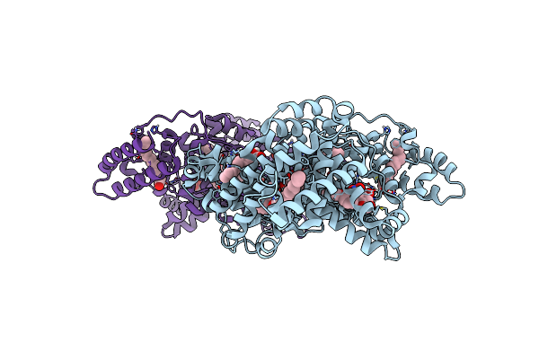 |
Structure Of Human Serum Albumin In Complex With Aristolochic Acid I At 1.9 A Resolution - Optimized
Organism: Homo sapiens
Method: X-RAY DIFFRACTION Resolution:1.90 Å Release Date: 2024-06-26 Classification: TRANSPORT PROTEIN Ligands: MYR, EDO, GOQ |
Organism: Homo sapiens
Method: X-RAY DIFFRACTION
Release Date: 2024-06-26
Ligands: MYR, EDO, GOQ
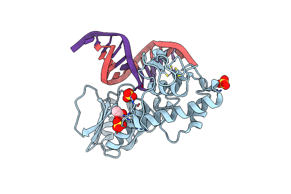 |
Crystal Structure Of The Dna Repair Enzyme Endonuclease-Viii (Nei) From E. Coli (E2Q) In Complex With Ap-Site Containing Dna Substrate
Organism: Escherichia coli, Synthetic construct
Method: X-RAY DIFFRACTION Resolution:2.00 Å Release Date: 2019-01-30 Classification: HYDROLASE Ligands: SO4, ZN, ACT |
Organism: Escherichia coli, Synthetic construct
Method: X-RAY DIFFRACTION
Release Date: 2019-01-30
Ligands: SO4, ZN, ACT
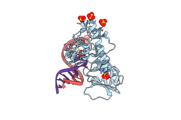 |
Crystal Structure Of The Dna Repair Enzyme Endonuclease-Viii (Nei) From E. Coli In Complex With Ap-Site Containing Dna Substrate
Organism: Escherichia coli
Method: X-RAY DIFFRACTION Resolution:1.40 Å Release Date: 2008-02-26 Classification: HYDROLASE/DNA Ligands: SO4, ZN, GOL |
Organism: Escherichia coli
Method: X-RAY DIFFRACTION
Release Date: 2008-02-26
Ligands: SO4, ZN, GOL
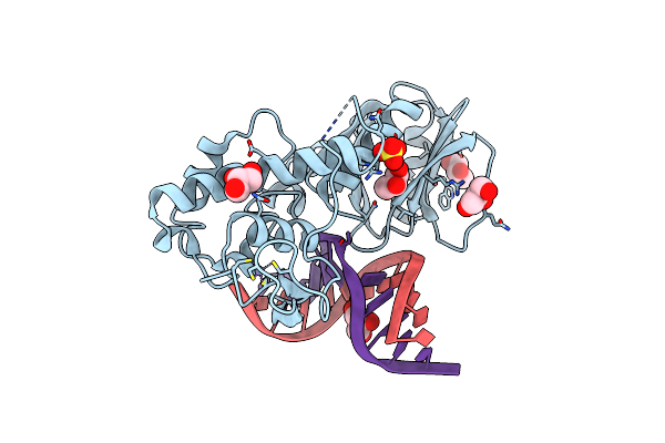 |
Crystal Structure Of The Dna Repair Enzyme Endonuclease-Viii (Nei) From E. Coli (R252A) In Complex With Ap-Site Containing Dna Substrate
Organism: Escherichia coli
Method: X-RAY DIFFRACTION Resolution:1.85 Å Release Date: 2008-02-26 Classification: HYDROLASE/DNA Ligands: GOL, ZN, SO4 |
Organism: Escherichia coli
Method: X-RAY DIFFRACTION
Release Date: 2008-02-26
Ligands: GOL, ZN, SO4
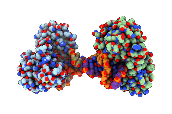 |
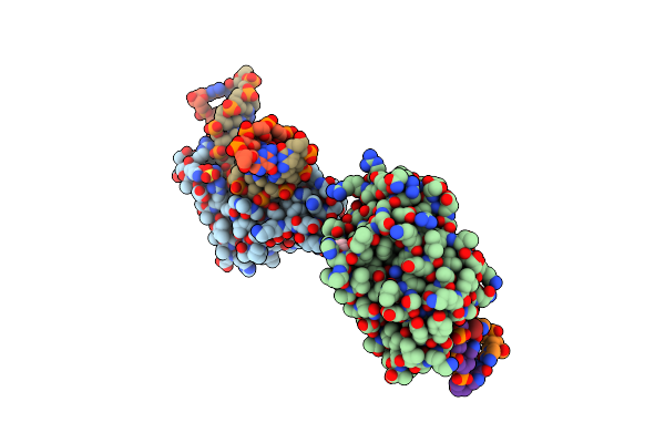
Crystal Structure Of The Dna Repair Enzyme Endonuclease-Viii (Nei) From E. Coli (E2Q) In Complex With Ap-Site Containing Dna Substrate
Organism: Escherichia coli
Method: X-RAY DIFFRACTION Resolution:2.60 Å Release Date: 2008-02-26 Classification: HYDROLASE/DNA |
Organism: Escherichia coli
Method: X-RAY DIFFRACTION
Release Date: 2008-02-26
 |
Crystal Structure Of T4 Pyrimidine Dimer Glycosylase (T4-Pdg) Covalently Complexed With A Dna Substrate Containing Abasic Site
Organism: Enterobacteria phage t4
Method: X-RAY DIFFRACTION Resolution:2.30 Å Release Date: 2006-10-03 Classification: HYDROLASE Ligands: SO4, GOL |
Organism: Enterobacteria phage t4
Method: X-RAY DIFFRACTION
Release Date: 2006-10-03
Ligands: SO4, GOL
 |
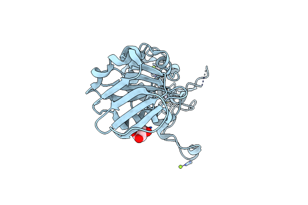
Crystal Structure Of The Dna Repair Enzyme Endonuclease-Viii (Nei) From E. Coli: The Wt Enzyme At 2.8 Resolution.
Organism: Escherichia coli
Method: X-RAY DIFFRACTION Resolution:2.80 Å Release Date: 2004-08-03 Classification: HYDROLASE Ligands: ZN, CA |
Organism: Escherichia coli
Method: X-RAY DIFFRACTION
Release Date: 2004-08-03
Ligands: ZN, CA
 |
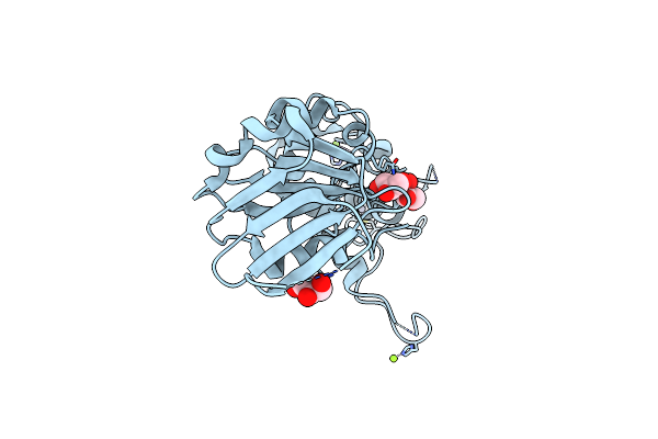
Crystal Structure Of The Dna Repair Enzyme Endonuclease-Viii (Nei) From E. Coli: The R252A Mutant At 2.05 Resolution.
Organism: Escherichia coli
Method: X-RAY DIFFRACTION Resolution:2.05 Å Release Date: 2004-08-03 Classification: HYDROLASE Ligands: ZN, MG, GOL |
Organism: Escherichia coli
Method: X-RAY DIFFRACTION
Release Date: 2004-08-03
Ligands: ZN, MG, GOL
 |
Crystal Structure Of The Dna Repair Enzyme Endonuclease-Viii (Nei) From E. Coli: The E2A Mutant At 2.3 Resolution.
Organism: Escherichia coli
Method: X-RAY DIFFRACTION Resolution:2.30 Å Release Date: 2004-08-03 Classification: HYDROLASE Ligands: ZN, MG, GOL |
Organism: Escherichia coli
Method: X-RAY DIFFRACTION
Release Date: 2004-08-03
Ligands: ZN, MG, GOL
 |
Crystal Structure Of Enterobacteria Phage Rb69 Gp43 Dna Polymerase Complexed With Tetrahydrofuran Containing Dna
Organism: Enterobacteria phage rb69
Method: X-RAY DIFFRACTION Resolution:2.69 Å Release Date: 2004-04-27 Classification: Transferase,replication/DNA Ligands: 3DR, DOC, DGP, CA |
Organism: Enterobacteria phage rb69
Method: X-RAY DIFFRACTION
Release Date: 2004-04-27
Ligands: 3DR, DOC, DGP, CA
 |
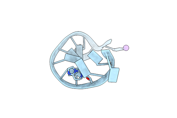
Crystal Structure Of Enterobacteria Phage Rb69 Gp43 Dna Polymerase Complexed With 8-Oxoguanosine Containing Dna
Organism: Enterobacteria phage rb69
Method: X-RAY DIFFRACTION Resolution:2.80 Å Release Date: 2004-04-27 Classification: TRANSFERASE, REPLICATION/DNA Ligands: DCP, CA |
Organism: Enterobacteria phage rb69
Method: X-RAY DIFFRACTION
Release Date: 2004-04-27
Ligands: DCP, CA
 |
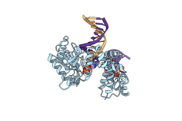
Crystal Structure Analysis Of The B-Dna Dodecamer Cgcgaatt(Ethenoc)Gcg
|
Method: X-RAY DIFFRACTION
Release Date: 2003-06-03
 |
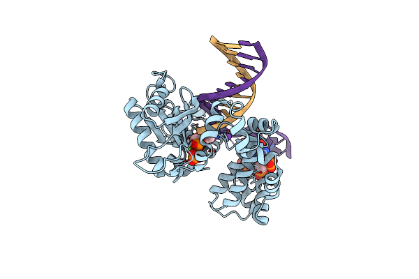
Human Dna Polymerase Beta Complexed With Gapped Dna Containing An 8-Oxo-7,8-Dihydro-Guanine And Damp
Organism: Homo sapiens
Method: X-RAY DIFFRACTION Resolution:3.10 Å Release Date: 2003-01-28 Classification: TRANSFERASE/DNA Ligands: D5M, NA |
Organism: Homo sapiens
Method: X-RAY DIFFRACTION
Release Date: 2003-01-28
Ligands: D5M, NA
 |
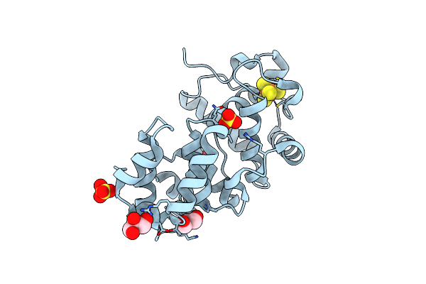
Human Dna Polymerase Beta Complexed With Gapped Dna Containing An 8-Oxo-7,8-Dihydro-Guanine Template Paired With Dctp
Organism: Homo sapiens
Method: X-RAY DIFFRACTION Resolution:2.80 Å Release Date: 2003-01-28 Classification: TRANSFERASE/DNA Ligands: MG, NA, DCP |
Organism: Homo sapiens
Method: X-RAY DIFFRACTION
Release Date: 2003-01-28
Ligands: MG, NA, DCP
 |
Crystal Structure Of The Core Fragment Of Muty From E.Coli At 1.2A Resolution
Organism: Escherichia coli
Method: X-RAY DIFFRACTION Resolution:1.20 Å Release Date: 2002-11-26 Classification: HYDROLASE Ligands: SO4, SF4, GOL |
Organism: Escherichia coli
Method: X-RAY DIFFRACTION
Release Date: 2002-11-26
Ligands: SO4, SF4, GOL
 |
Crystal Structure Of The Core Fragment Of Muty From E.Coli At 1.55A Resolution
Organism: Escherichia coli
Method: X-RAY DIFFRACTION Resolution:1.55 Å Release Date: 2002-11-26 Classification: HYDROLASE Ligands: SO4, SF4, GOL |
Organism: Escherichia coli
Method: X-RAY DIFFRACTION
Release Date: 2002-11-26
Ligands: SO4, SF4, GOL
 |
Crystal Structure Of The K142A Mutant Of E. Coli Muty (Core Fragment)
Organism: Escherichia coli
Method: X-RAY DIFFRACTION Resolution:1.60 Å Release Date: 2002-11-26 Classification: HYDROLASE Ligands: SO4, SF4, GOL |
Organism: Escherichia coli
Method: X-RAY DIFFRACTION
Release Date: 2002-11-26
Ligands: SO4, SF4, GOL