

Search Count: 17
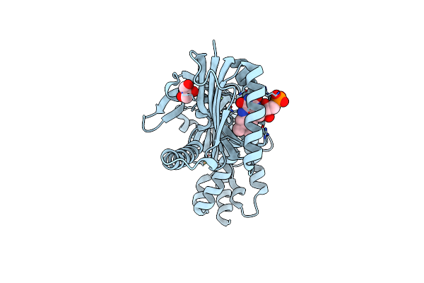 |
Crystal Structure Of Adenylyate Cyclase Variant Y6W
Organism: Oscillatoria acuminata
Method: X-RAY DIFFRACTION Resolution:1.95 Å Release Date: 2025-02-26 Classification: LYASE Ligands: FMN, GOL, CA |
Organism: Oscillatoria acuminata
Method: X-RAY DIFFRACTION
Release Date: 2025-02-26
Ligands: FMN, GOL, CA
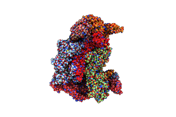 |
High-Resolution Structure Of The Anaphase-Promoting Complex/Cyclosome (Apc/C) Bound To Co-Activator Cdh1
Organism: Homo sapiens
Method: ELECTRON MICROSCOPY Release Date: 2024-08-14 Classification: CELL CYCLE Ligands: ZN |
Organism: Homo sapiens
Method: ELECTRON MICROSCOPY
Release Date: 2024-08-14
Ligands: ZN
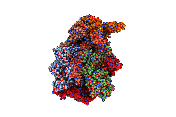 |
Cryo-Em Structure Of The Apo Anaphase-Promoting Complex/Cyclosome (Apc/C) At 3.2 Angstrom Resolution
Organism: Homo sapiens
Method: ELECTRON MICROSCOPY Release Date: 2023-07-19 Classification: CELL CYCLE Ligands: ZN |
Organism: Homo sapiens
Method: ELECTRON MICROSCOPY
Release Date: 2023-07-19
Ligands: ZN
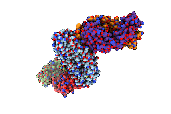 |
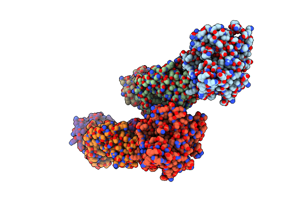
Pfrh5 Bound To Monoclonal Antibody R5.015 And R5.016 Fab Fragments
Organism: Plasmodium falciparum (isolate 3d7), Homo sapiens
Method: X-RAY DIFFRACTION Resolution:2.53 Å Release Date: 2022-02-09 Classification: IMMUNE SYSTEM |
Organism: Plasmodium falciparum (isolate 3d7), Homo sapiens
Method: X-RAY DIFFRACTION
Release Date: 2022-02-09
 |
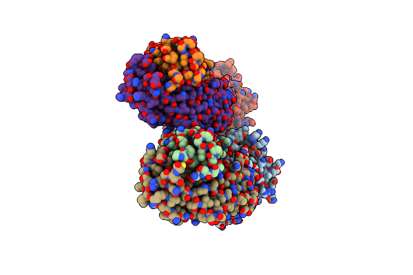
Pfcyrpa Bound To Monoclonal Antibody Cy.007 Fab Fragment
Organism: Plasmodium falciparum 3d7, Gallus gallus
Method: X-RAY DIFFRACTION Resolution:3.09 Å Release Date: 2022-02-09 Classification: IMMUNE SYSTEM Ligands: NH4 |
Organism: Plasmodium falciparum 3d7, Gallus gallus
Method: X-RAY DIFFRACTION
Release Date: 2022-02-09
Ligands: NH4
 |
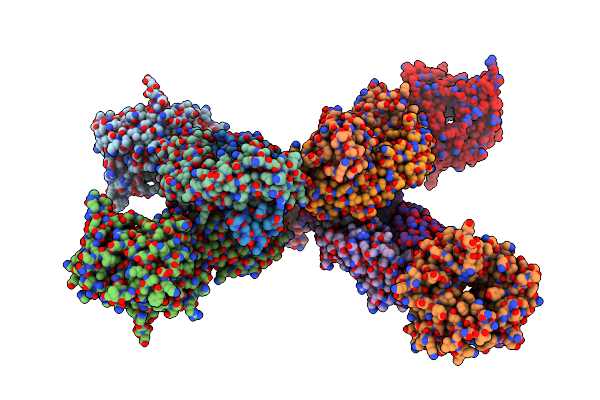
Pfcyrpa Bound To Monoclonal Antibody Cy.004 Fab Fragment
Organism: Plasmodium falciparum (isolate 3d7), Gallus gallus
Method: X-RAY DIFFRACTION Resolution:2.79 Å Release Date: 2022-02-09 Classification: IMMUNE SYSTEM Ligands: CA |
Organism: Plasmodium falciparum (isolate 3d7), Gallus gallus
Method: X-RAY DIFFRACTION
Release Date: 2022-02-09
Ligands: CA
 |
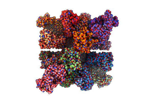
Pfcyrpa Bound To Monoclonal Antibody Cy.003 Fab Fragment
Organism: Plasmodium falciparum (isolate 3d7), Gallus gallus
Method: X-RAY DIFFRACTION Resolution:3.14 Å Release Date: 2022-02-09 Classification: IMMUNE SYSTEM |
Organism: Plasmodium falciparum (isolate 3d7), Gallus gallus
Method: X-RAY DIFFRACTION
Release Date: 2022-02-09
 |
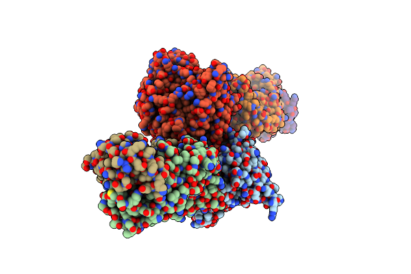
Pfcyrpa Bound To Fab Fragments From Monoclonal Antibodies Cy.003, Cy.004 And Cy.007
Organism: Plasmodium falciparum 3d7, Gallus gallus
Method: X-RAY DIFFRACTION Resolution:3.27 Å Release Date: 2022-02-09 Classification: IMMUNE SYSTEM Ligands: CA |
Organism: Plasmodium falciparum 3d7, Gallus gallus
Method: X-RAY DIFFRACTION
Release Date: 2022-02-09
Ligands: CA
 |
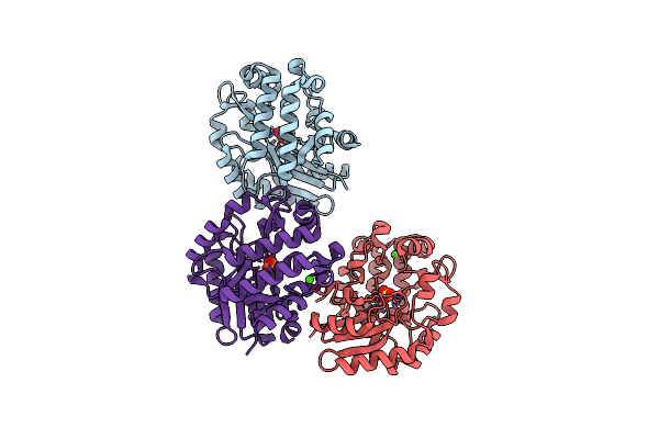
Pfcyrpa Bound To Monoclonal Antibody Cy.002 Fab Fragment
Organism: Plasmodium falciparum (isolate 3d7), Gallus gallus
Method: X-RAY DIFFRACTION Resolution:2.72 Å Release Date: 2022-02-09 Classification: IMMUNE SYSTEM |
Organism: Plasmodium falciparum (isolate 3d7), Gallus gallus
Method: X-RAY DIFFRACTION
Release Date: 2022-02-09
 |
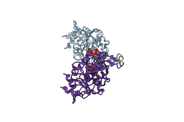
Crystal Structure Of The Putative Histidinol Phosphatase Hisk From Listeria Monocytogenes With Trinuclear Metals Determined By Pixe Revealing Sulphate Ion In Active Site. Based On Pixe Analysis And Original Date From 3Dcp
Organism: Listeria monocytogenes serotype 4b str. h7858
Method: X-RAY DIFFRACTION Resolution:2.10 Å Release Date: 2019-12-25 Classification: HYDROLASE Ligands: MN, CO, SO4, FE, CA |
Organism: Listeria monocytogenes serotype 4b str. h7858
Method: X-RAY DIFFRACTION
Release Date: 2019-12-25
Ligands: MN, CO, SO4, FE, CA
 |
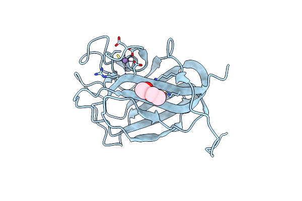
The Nucleotide-Binding Protein Af_226 In Complex With Adp From Archaeoglobus Fulgidus With Co Found By Pixe. Based On 3Kb1.
Organism: Archaeoglobus fulgidus
Method: X-RAY DIFFRACTION Resolution:2.87 Å Release Date: 2019-12-25 Classification: METAL TRANSPORT Ligands: ADP, CO |
Organism: Archaeoglobus fulgidus
Method: X-RAY DIFFRACTION
Release Date: 2019-12-25
Ligands: ADP, CO
 |
X-Ray Structure Of The C-Terminal Domain (277-440) Of Putative Chitobiase From Bacteroides Thetaiotaomicron. Northeast Structural Genomics Consortium Target Btr324A. Re-Refinement Of 3Ggl With Correct Metal Mn Replacing Zn. New Metal Confirmed With Pixe Analysis Of Original Sample.
Organism: Bacteroides thetaiotaomicron
Method: X-RAY DIFFRACTION Resolution:3.00 Å Release Date: 2019-12-25 Classification: HYDROLASE Ligands: PEG, MN |
Organism: Bacteroides thetaiotaomicron
Method: X-RAY DIFFRACTION
Release Date: 2019-12-25
Ligands: PEG, MN
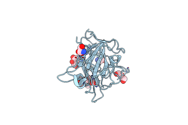 |
X-Ray Crystal Structure Of Lsaa9B
Organism: Lentinus similis
Method: X-RAY DIFFRACTION Resolution:1.60 Å Release Date: 2019-09-11 Classification: UNKNOWN FUNCTION Ligands: MAN, CL, PEG, GLY, SER |
Organism: Lentinus similis
Method: X-RAY DIFFRACTION
Release Date: 2019-09-11
Ligands: MAN, CL, PEG, GLY, SER
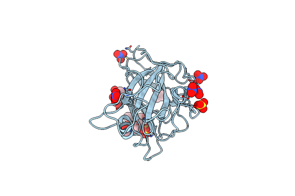 |
X-Ray Crystal Structure Of Lsaa9B (Deglycosylated Form)
Organism: Lentinus similis
Method: X-RAY DIFFRACTION Resolution:1.60 Å Release Date: 2019-09-11 Classification: UNKNOWN FUNCTION Ligands: MAN, MPD, SO4, NO3, PGE |
Organism: Lentinus similis
Method: X-RAY DIFFRACTION
Release Date: 2019-09-11
Ligands: MAN, MPD, SO4, NO3, PGE
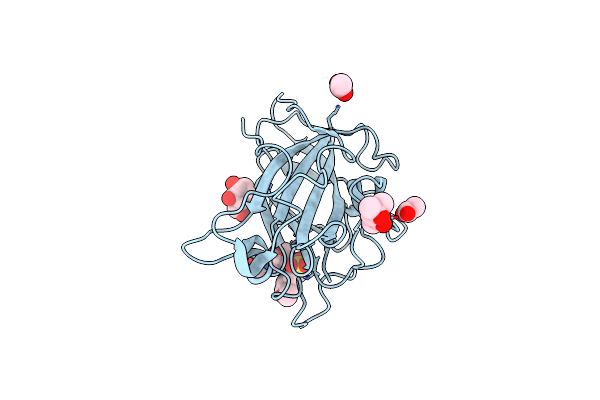 |
X-Ray Crystal Structure Of Lsaa9B (Transition Metals Soak)
Organism: Lentinus similis
Method: X-RAY DIFFRACTION Resolution:1.58 Å Release Date: 2019-09-11 Classification: UNKNOWN FUNCTION Ligands: MAN, MES, MPD, ACT |
Organism: Lentinus similis
Method: X-RAY DIFFRACTION
Release Date: 2019-09-11
Ligands: MAN, MES, MPD, ACT
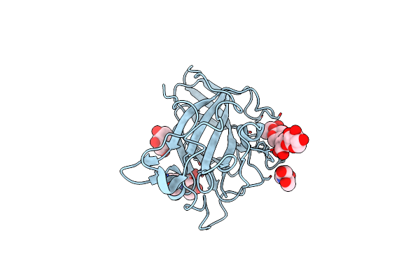 |
X-Ray Crystal Structure Of Lsaa9B (Xylotetraose Soak)
Organism: Lentinus similis
Method: X-RAY DIFFRACTION Resolution:1.40 Å Release Date: 2019-09-11 Classification: UNKNOWN FUNCTION Ligands: MAN, BCN |
Organism: Lentinus similis
Method: X-RAY DIFFRACTION
Release Date: 2019-09-11
Ligands: MAN, BCN
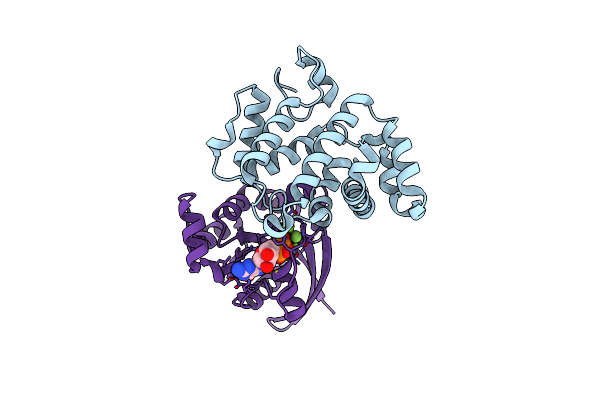 |
Crystal Structure Of Rhoa.Gdp.Mgf3-In Complex With Rhogap
Organism: Homo sapiens
Method: X-RAY DIFFRACTION Resolution:1.80 Å Release Date: 2003-05-06 Classification: GENE REGULATION/SIGNALING PROTEIN Ligands: MG, GDP, MGF |
Organism: Homo sapiens
Method: X-RAY DIFFRACTION
Release Date: 2003-05-06
Ligands: MG, GDP, MGF