

Search Count: 20
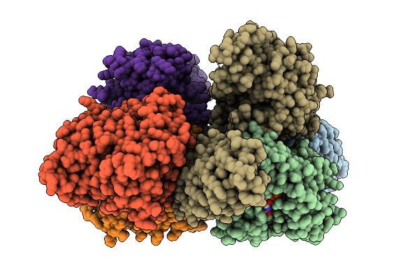 |
Human Kif18A-Darpin Fusion Protein Bound To Amp-Pnp And Tubulin
Organism: Homo sapiens, Synthetic construct, Sus scrofa
Method: X-RAY DIFFRACTION Resolution:2.41 Å Release Date: 2025-11-05 Classification: MOTOR PROTEIN Ligands: GTP, MG, EDO, GDP, ANP |
Organism: Homo sapiens, Synthetic construct, Sus scrofa
Method: X-RAY DIFFRACTION
Release Date: 2025-11-05
Ligands: GTP, MG, EDO, GDP, ANP
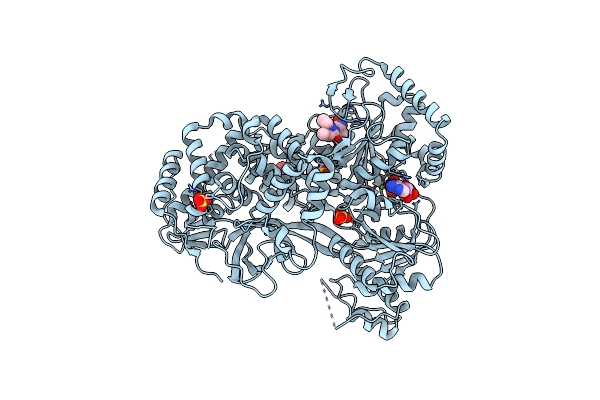 |
Human Dhx9 In Complex With Compound 1 And Adp
Organism: Homo sapiens
Method: X-RAY DIFFRACTION Resolution:3.26 Å Release Date: 2025-05-07 Classification: HYDROLASE Ligands: A1BK0, ADP, MG, SO4 |
Organism: Homo sapiens
Method: X-RAY DIFFRACTION
Release Date: 2025-05-07
Ligands: A1BK0, ADP, MG, SO4
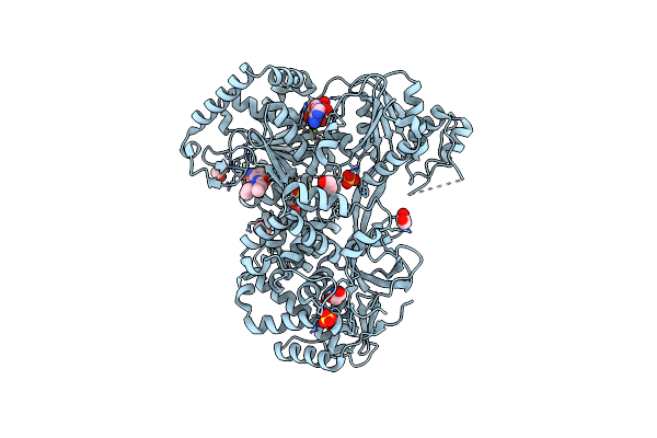 |
Cat Dhx9 In Complex With Compound 1 And Adp
Organism: Felis catus
Method: X-RAY DIFFRACTION Resolution:2.27 Å Release Date: 2025-05-07 Classification: HYDROLASE Ligands: A1BK0, ADP, MG, SO4, EDO |
Organism: Felis catus
Method: X-RAY DIFFRACTION
Release Date: 2025-05-07
Ligands: A1BK0, ADP, MG, SO4, EDO
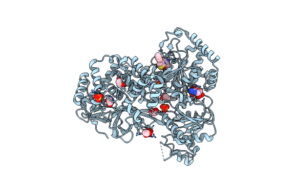 |
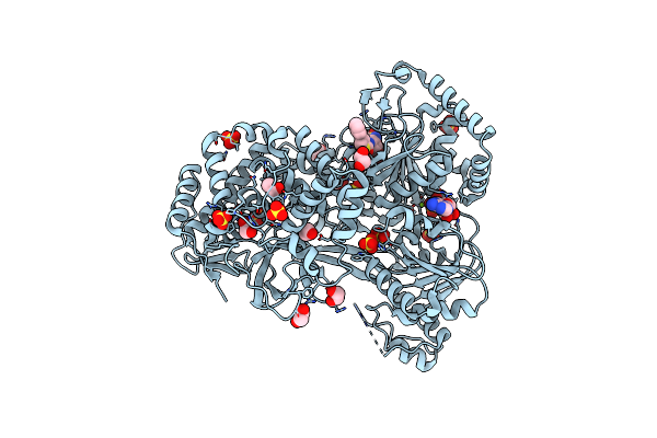
Cat Dhx9 In Complex With Compound 10 And Adp
Organism: Felis catus
Method: X-RAY DIFFRACTION Resolution:2.99 Å Release Date: 2025-05-07 Classification: HYDROLASE Ligands: A1BKZ, ADP, MG, SO4, EDO |
Organism: Felis catus
Method: X-RAY DIFFRACTION
Release Date: 2025-05-07
Ligands: A1BKZ, ADP, MG, SO4, EDO
 |
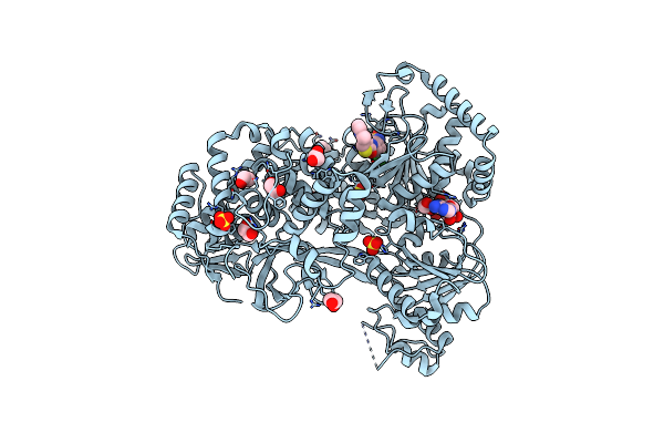
Cat Dhx9 In Complex With Compound 23 And Adp
Organism: Felis catus
Method: X-RAY DIFFRACTION Resolution:2.35 Å Release Date: 2025-05-07 Classification: HYDROLASE Ligands: A1BKY, ADP, MG, EDO, SO4, CL |
Organism: Felis catus
Method: X-RAY DIFFRACTION
Release Date: 2025-05-07
Ligands: A1BKY, ADP, MG, EDO, SO4, CL
 |
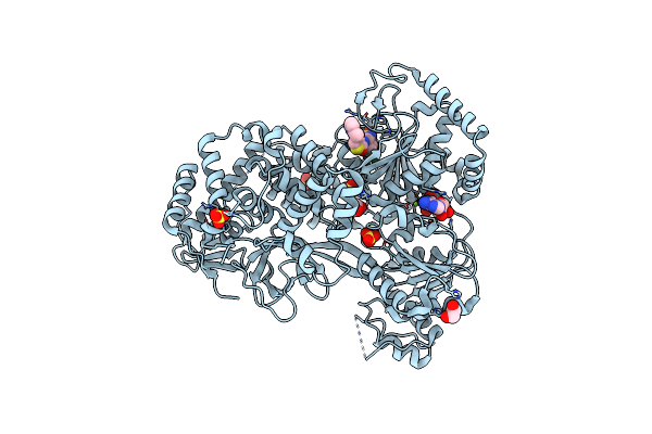
Cat Dhx9 In Complex With Atx968 And Adp
Organism: Felis catus
Method: X-RAY DIFFRACTION Resolution:2.43 Å Release Date: 2025-05-07 Classification: HYDROLASE Ligands: A1BKX, ADP, MG, EDO, SO4, CL |
Organism: Felis catus
Method: X-RAY DIFFRACTION
Release Date: 2025-05-07
Ligands: A1BKX, ADP, MG, EDO, SO4, CL
 |
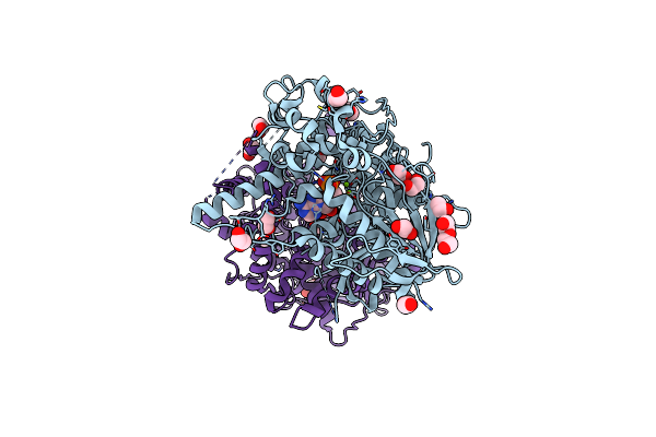
Human Dhx9 In Complex With Atx968 And Adp
Organism: Homo sapiens
Method: X-RAY DIFFRACTION Resolution:2.75 Å Release Date: 2025-05-07 Classification: HYDROLASE Ligands: A1BKX, ADP, MG, EDO, SO4 |
Organism: Homo sapiens
Method: X-RAY DIFFRACTION
Release Date: 2025-05-07
Ligands: A1BKX, ADP, MG, EDO, SO4
 |
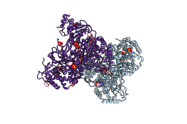
Human Xrn1 With Adenosine-3',5'-Bisphosphate (Pap) Bound
Organism: Homo sapiens
Method: X-RAY DIFFRACTION Resolution:2.10 Å Release Date: 2025-04-09 Classification: HYDROLASE Ligands: A3P, MG, EDO, CL, PEG |
Organism: Homo sapiens
Method: X-RAY DIFFRACTION
Release Date: 2025-04-09
Ligands: A3P, MG, EDO, CL, PEG
 |
Human Dhx9 Bound To Adp
Organism: Homo sapiens
Method: X-RAY DIFFRACTION Resolution:2.62 Å Release Date: 2023-08-02 Classification: HYDROLASE Ligands: ADP, MG, SO4, EDO |
Organism: Homo sapiens
Method: X-RAY DIFFRACTION
Release Date: 2023-08-02
Ligands: ADP, MG, SO4, EDO
 |
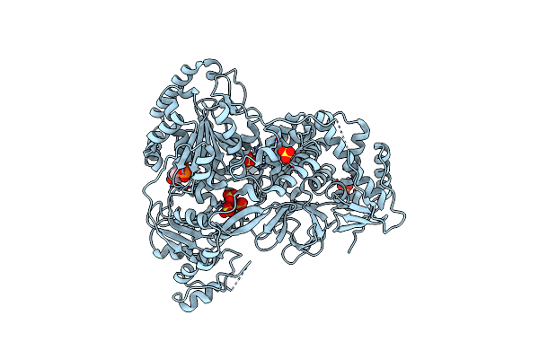
Cat Dhx9 Bound To Adp
Organism: Felis catus
Method: X-RAY DIFFRACTION Resolution:2.71 Å Release Date: 2023-08-02 Classification: HYDROLASE Ligands: ADP, MG, SO4, EDO |
Organism: Felis catus
Method: X-RAY DIFFRACTION
Release Date: 2023-08-02
Ligands: ADP, MG, SO4, EDO
 |
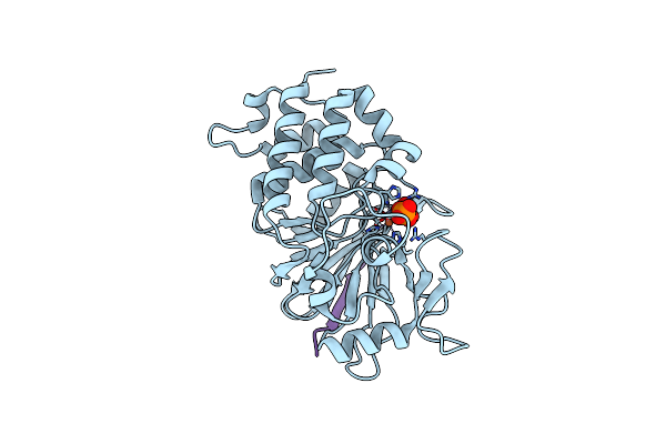
Dog Dhx9 Bound To Adp
Organism: Canis lupus familiaris
Method: X-RAY DIFFRACTION Resolution:2.97 Å Release Date: 2023-08-02 Classification: HYDROLASE Ligands: ADP, MG, SO4 |
Organism: Canis lupus familiaris
Method: X-RAY DIFFRACTION
Release Date: 2023-08-02
Ligands: ADP, MG, SO4
 |
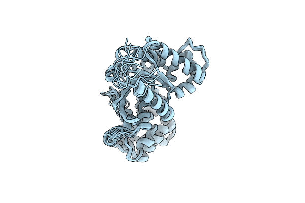
Structure Of Calcineurin Bound To Rcan1
Organism: Homo sapiens
Method: X-RAY DIFFRACTION Resolution:1.85 Å Release Date: 2020-09-09 Classification: HYDROLASE/Signaling Protein Ligands: PO4, ZN, FE |
Organism: Homo sapiens
Method: X-RAY DIFFRACTION
Release Date: 2020-09-09
Ligands: PO4, ZN, FE
 |
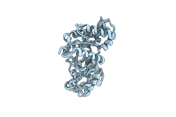
Structural Model Of A Apo G-Protein Alpha Subunit Determined With Nmr Residual Dipolar Couplings And Saxs
Organism: Homo sapiens
Method: SOLUTION NMR Release Date: 2016-06-29 Classification: SIGNALING PROTEIN |
Organism: Homo sapiens
Method: SOLUTION NMR
Release Date: 2016-06-29
 |
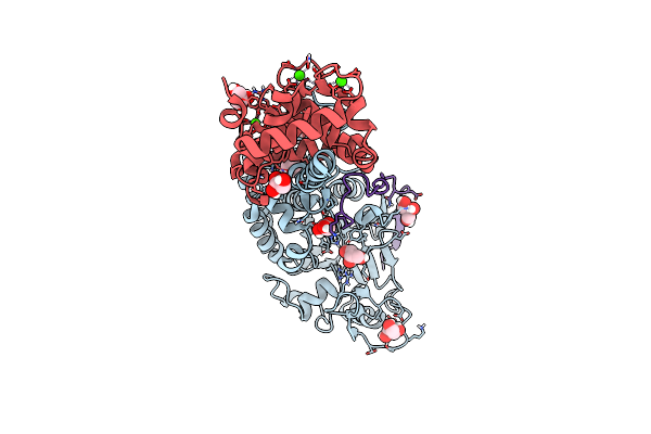
Structural Model Of A Protein Alpha Subunit In Complex With Gdp Obtained With Saxs And Nmr Residual Couplings
Organism: Homo sapiens
Method: SOLUTION NMR Release Date: 2016-06-29 Classification: SIGNALING PROTEIN |
Organism: Homo sapiens
Method: SOLUTION NMR
Release Date: 2016-06-29
 |
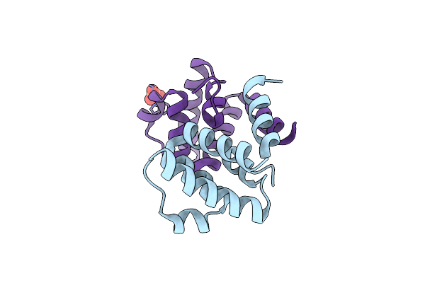
Crystal Structure Of Calcineurin In Complex With The Calcineurin-Inhibiting Domain Of The African Swine Fever Virus Protein A238L
Organism: Homo sapiens, African swine fever virus malawi lil 20/1
Method: X-RAY DIFFRACTION Resolution:1.70 Å Release Date: 2013-03-06 Classification: hydrolase/protein binding Ligands: GOL, CA |
Organism: Homo sapiens, African swine fever virus malawi lil 20/1
Method: X-RAY DIFFRACTION
Release Date: 2013-03-06
Ligands: GOL, CA
 |
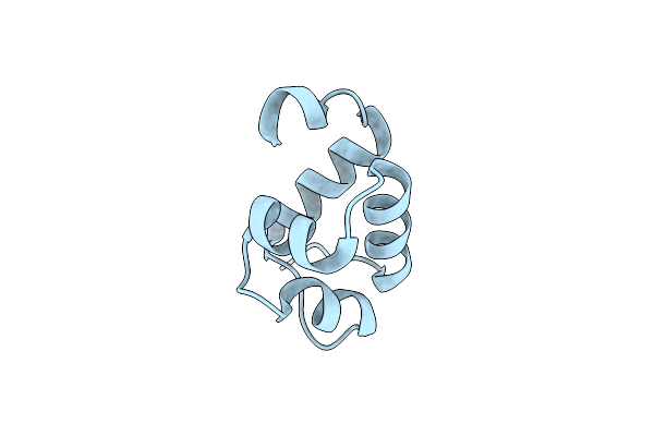
Crystal Structure Of The Nlrp4 Pyrin Domain
Organism: Homo sapiens
Method: X-RAY DIFFRACTION Resolution:2.28 Å Release Date: 2012-09-05 Classification: SIGNALING PROTEIN, PROTEIN BINDING Ligands: CL, SO4 |
Organism: Homo sapiens
Method: X-RAY DIFFRACTION
Release Date: 2012-09-05
Ligands: CL, SO4
 |
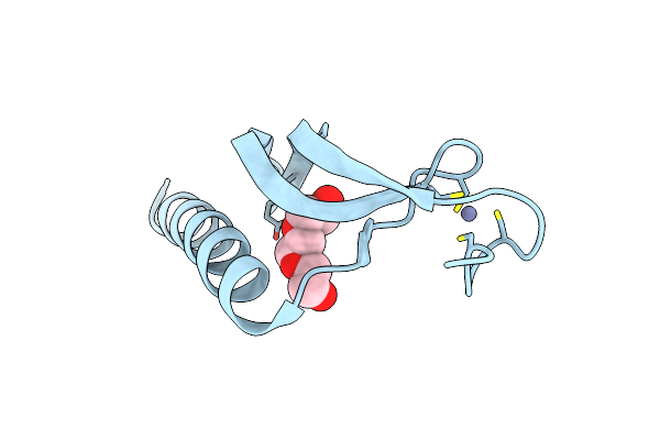
Structure Of The C-Terminal Domain Of The E. Coli Protein Mqsa (Ygit/B3021)
Organism: Escherichia coli k-12
Method: X-RAY DIFFRACTION Resolution:1.40 Å Release Date: 2010-01-12 Classification: DNA BINDING PROTEIN |
Organism: Escherichia coli k-12
Method: X-RAY DIFFRACTION
Release Date: 2010-01-12
 |
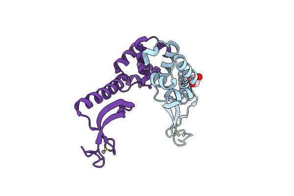
Structure Of The N-Terminal Domain Of The E. Coli Protein Mqsa (Ygit/B3021)
Organism: Escherichia coli k-12
Method: X-RAY DIFFRACTION Resolution:1.70 Å Release Date: 2010-01-12 Classification: DNA BINDING PROTEIN Ligands: ZN, PE4 |
Organism: Escherichia coli k-12
Method: X-RAY DIFFRACTION
Release Date: 2010-01-12
Ligands: ZN, PE4
 |
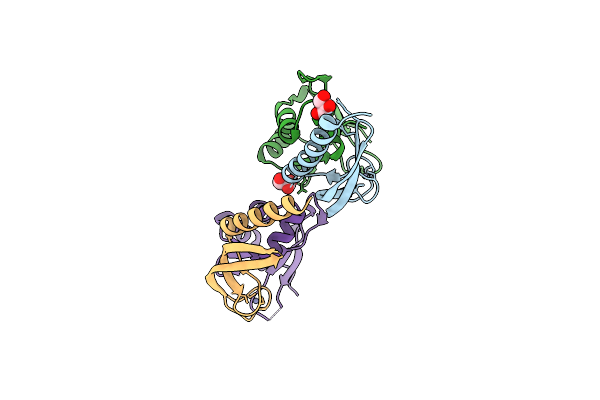
Structure Of The E. Coli Protein Mqsa (Ygit/B3021)
Organism: Escherichia coli k-12
Method: X-RAY DIFFRACTION Resolution:2.15 Å Release Date: 2010-01-12 Classification: DNA BINDING PROTEIN Ligands: ZN, GOL |
Organism: Escherichia coli k-12
Method: X-RAY DIFFRACTION
Release Date: 2010-01-12
Ligands: ZN, GOL
 |
Structure Of The N-Terminal Domain Of The E. Coli Antitoxin Mqsa (Ygit/B3021) In Complex With The E. Coli Toxin Mqsr (Ygiu/B3022)
Organism: Escherichia coli k-12
Method: X-RAY DIFFRACTION Resolution:2.00 Å Release Date: 2010-01-12 Classification: DNA BINDING PROTEIN/TOXIN Ligands: ZN, GOL |
Organism: Escherichia coli k-12
Method: X-RAY DIFFRACTION
Release Date: 2010-01-12
Ligands: ZN, GOL