

Search Count: 98
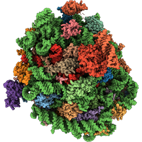 |
Rabbit 60S Ribosomal Subunit With Eef2 Domain Iv Closed
Organism: Oryctolagus cuniculus
Method: ELECTRON MICROSCOPY Release Date: 2026-09-02 Classification: RIBOSOME Ligands: ZN, GDP |
Organism: Oryctolagus cuniculus
Method: ELECTRON MICROSCOPY
Release Date: 2026-09-02
Ligands: ZN, GDP
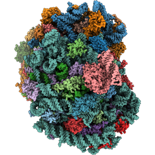 |
Rabbit 80S With Eef2,Ccdc124,And Larp1,40S-Head-Swiveled
Organism: Oryctolagus cuniculus
Method: ELECTRON MICROSCOPY Release Date: 2026-09-02 Classification: RIBOSOME Ligands: ZN, GDP |
Organism: Oryctolagus cuniculus
Method: ELECTRON MICROSCOPY
Release Date: 2026-09-02
Ligands: ZN, GDP
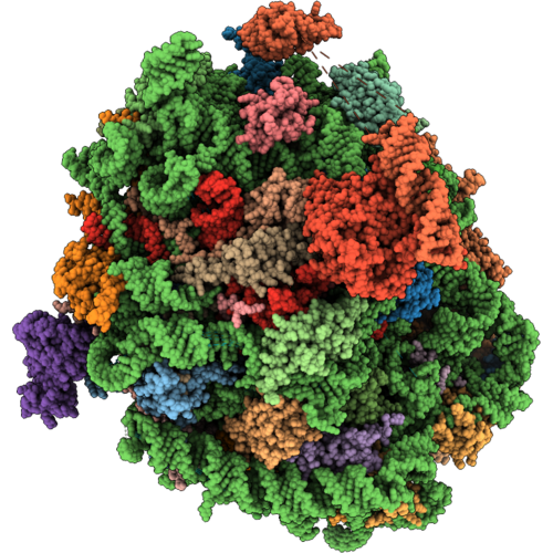 |
Rabbit 60S Ribosomal Subunit With Eef2 Domain Iv Open
Organism: Oryctolagus cuniculus
Method: ELECTRON MICROSCOPY Release Date: 2026-09-02 Classification: RIBOSOME Ligands: GDP, ZN |
Organism: Oryctolagus cuniculus
Method: ELECTRON MICROSCOPY
Release Date: 2026-09-02
Ligands: GDP, ZN
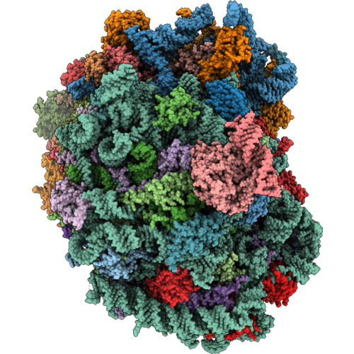 |
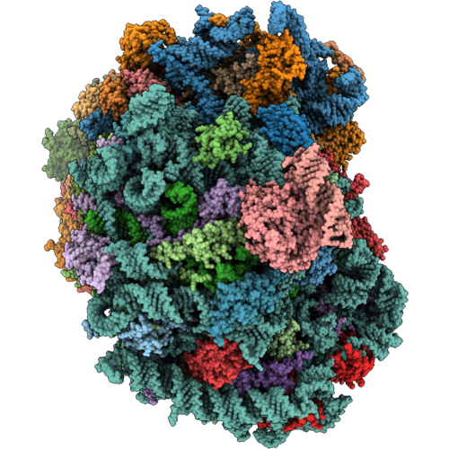
Rabbit 80S With Eef2,Eif5A And Serbp1
Organism: Oryctolagus cuniculus
Method: ELECTRON MICROSCOPY Release Date: 2026-09-02 Classification: RIBOSOME Ligands: ZN, GDP |
Organism: Oryctolagus cuniculus
Method: ELECTRON MICROSCOPY
Release Date: 2026-09-02
Ligands: ZN, GDP
 |
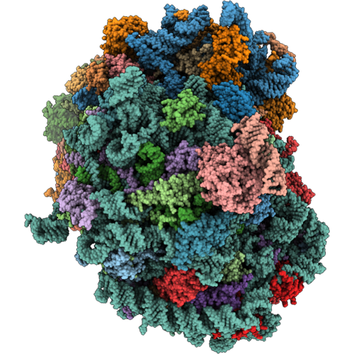
Rabbit 80S With Eef2,Ifrd2 And Larp1,40S-Head-Swiveled
Organism: Oryctolagus cuniculus
Method: ELECTRON MICROSCOPY Release Date: 2026-09-02 Classification: RIBOSOME Ligands: ZN, GDP |
Organism: Oryctolagus cuniculus
Method: ELECTRON MICROSCOPY
Release Date: 2026-09-02
Ligands: ZN, GDP
 |
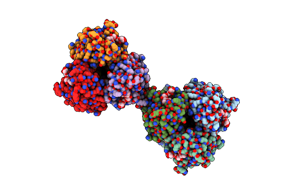
Rabbit 80S With Ifrd2 And Larp1, 40S Head-Swiveled
Organism: Oryctolagus cuniculus
Method: ELECTRON MICROSCOPY Release Date: 2026-09-02 Classification: RIBOSOME Ligands: ZN |
Organism: Oryctolagus cuniculus
Method: ELECTRON MICROSCOPY
Release Date: 2026-09-02
Ligands: ZN
 |
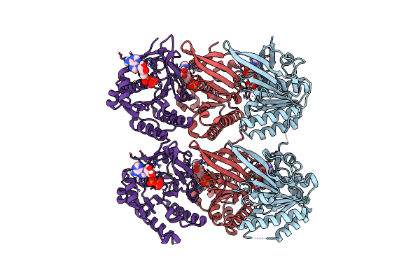
Subtomogram Average Of Dimers Of Influenza Ha Trimers
Organism: Influenza a virus (a/puerto rico/8/1934(h1n1))
Method: ELECTRON MICROSCOPY Release Date: 2025-04-16 Classification: VIRAL PROTEIN |
Organism: Influenza a virus (a/puerto rico/8/1934(h1n1))
Method: ELECTRON MICROSCOPY
Release Date: 2025-04-16
 |
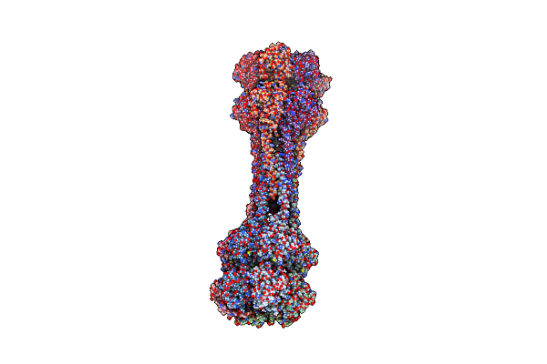
Crystal Structure Of Kaic With Truncated C-Terminal Coiled-Coil Domain
Organism: Cereibacter sphaeroides
Method: X-RAY DIFFRACTION Resolution:2.90 Å Release Date: 2023-03-22 Classification: CIRCADIAN CLOCK PROTEIN Ligands: ADP |
Organism: Cereibacter sphaeroides
Method: X-RAY DIFFRACTION
Release Date: 2023-03-22
Ligands: ADP
 |
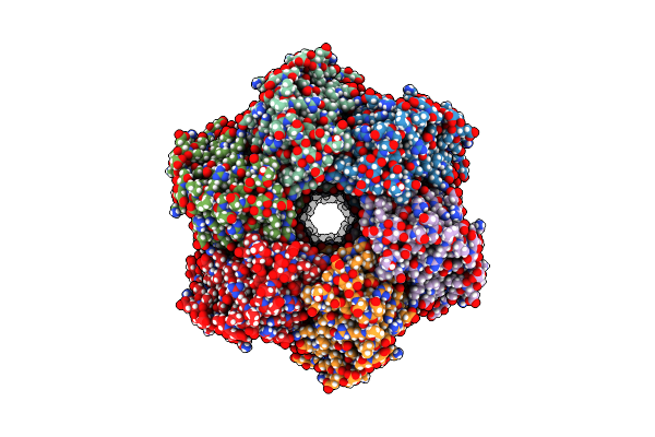
Crystal Structure Of Dodecameric Kaic
Organism: Cereibacter sphaeroides
Method: X-RAY DIFFRACTION Resolution:3.50 Å Release Date: 2023-03-22 Classification: CIRCADIAN CLOCK PROTEIN Ligands: ADP, MG |
Organism: Cereibacter sphaeroides
Method: X-RAY DIFFRACTION
Release Date: 2023-03-22
Ligands: ADP, MG
 |
Structure Of Dodecameric Kaic-Rs-S413E/S414E Solved By Cryo-Em
Organism: Cereibacter sphaeroides
Method: ELECTRON MICROSCOPY Release Date: 2023-03-22 Classification: CIRCADIAN CLOCK PROTEIN Ligands: ATP, ADP, MG |
Organism: Cereibacter sphaeroides
Method: ELECTRON MICROSCOPY
Release Date: 2023-03-22
Ligands: ATP, ADP, MG
 |
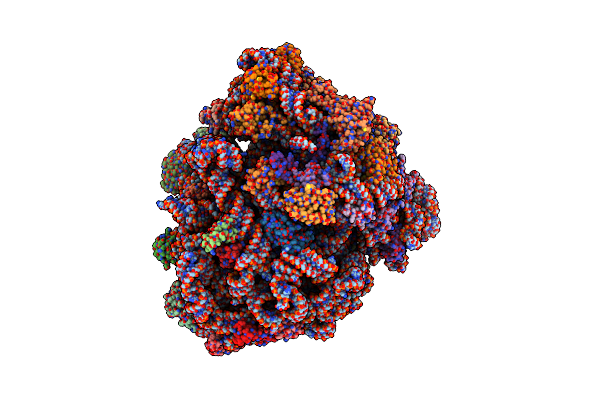
Structure Of Dodecameric Kaic-Rs-S413E/S414E Complexed With Kaib-Rs Solved By Cryo-Em
Organism: Cereibacter sphaeroides
Method: ELECTRON MICROSCOPY Release Date: 2023-03-22 Classification: CIRCADIAN CLOCK PROTEIN Ligands: ATP, ADP, MG |
Organism: Cereibacter sphaeroides
Method: ELECTRON MICROSCOPY
Release Date: 2023-03-22
Ligands: ATP, ADP, MG
 |
70S Ribosome Bound To Hiv Frameshifting Stem-Loop (Fss) And P/E Trna (Rotated Conformation)
Organism: Escherichia coli, Human immunodeficiency virus 1
Method: ELECTRON MICROSCOPY Release Date: 2020-06-03 Classification: RIBOSOME Ligands: MG |
Organism: Escherichia coli, Human immunodeficiency virus 1
Method: ELECTRON MICROSCOPY
Release Date: 2020-06-03
Ligands: MG
 |
70S Ribosome Bound To Hiv Frameshifting Stem-Loop (Fss) And P-Site Trna (Non-Rotated Conformation, Structure I)
Organism: Human immunodeficiency virus 1, Escherichia coli
Method: ELECTRON MICROSCOPY Release Date: 2020-06-03 Classification: RIBOSOME Ligands: MG |
Organism: Human immunodeficiency virus 1, Escherichia coli
Method: ELECTRON MICROSCOPY
Release Date: 2020-06-03
Ligands: MG
 |
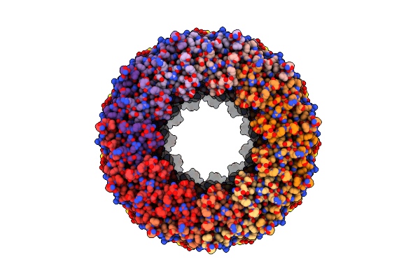
70S Ribosome Bound To Hiv Frameshifting Stem-Loop (Fss) And P-Site Trna (Non-Rotated Conformation, Structure Ii)
Organism: Human immunodeficiency virus 1, Escherichia coli
Method: ELECTRON MICROSCOPY Release Date: 2020-06-03 Classification: RIBOSOME Ligands: MG |
Organism: Human immunodeficiency virus 1, Escherichia coli
Method: ELECTRON MICROSCOPY
Release Date: 2020-06-03
Ligands: MG
 |
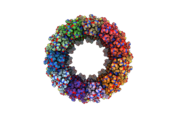
Cryo-Em Structure Of A Dimer Of Undecameric Human Calhm2
Organism: Homo sapiens
Method: ELECTRON MICROSCOPY Release Date: 2020-01-29 Classification: MEMBRANE PROTEIN |
Organism: Homo sapiens
Method: ELECTRON MICROSCOPY
Release Date: 2020-01-29
 |
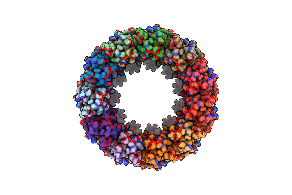
Cryo-Em Structure Of Human Calhm2
Organism: Homo sapiens
Method: ELECTRON MICROSCOPY Release Date: 2020-01-29 Classification: MEMBRANE PROTEIN |
Organism: Homo sapiens
Method: ELECTRON MICROSCOPY
Release Date: 2020-01-29
 |
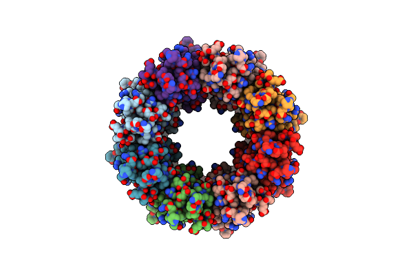
Cryo-Em Structure Of An Undecameric Chicken Calhm1 And Human Calhm2 Chimera
Organism: Aequorea victoria, Gallus gallus, Homo sapiens
Method: ELECTRON MICROSCOPY Release Date: 2020-01-29 Classification: MEMBRANE PROTEIN |
Organism: Aequorea victoria, Gallus gallus, Homo sapiens
Method: ELECTRON MICROSCOPY
Release Date: 2020-01-29
 |
Cryo-Em Structure Of Octameric Chicken Calhm1
Organism: Aequorea victoria, Gallus gallus
Method: ELECTRON MICROSCOPY Release Date: 2020-01-29 Classification: MEMBRANE PROTEIN |
Organism: Aequorea victoria, Gallus gallus
Method: ELECTRON MICROSCOPY
Release Date: 2020-01-29
 |
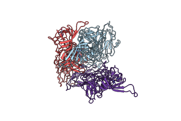
Structure Of The Vesicular Stomatitis Virus L Protein In Complex With Its Phosphoprotein Cofactor (3.0 A Resolution)
Organism: Vesicular stomatitis indiana virus (strain san juan)
Method: ELECTRON MICROSCOPY Release Date: 2020-01-22 Classification: VIRAL PROTEIN Ligands: ZN |
Organism: Vesicular stomatitis indiana virus (strain san juan)
Method: ELECTRON MICROSCOPY
Release Date: 2020-01-22
Ligands: ZN
 |
Symmetric Reconstruction Of Human Norovirus Gii.2 Snow Mountain Virus Strain Vlp In T=3 Symmetry
Organism: Snow mountain virus
Method: ELECTRON MICROSCOPY Release Date: 2019-06-26 Classification: VIRUS LIKE PARTICLE Ligands: ZN |
Organism: Snow mountain virus
Method: ELECTRON MICROSCOPY
Release Date: 2019-06-26
Ligands: ZN