

Search Count: 20
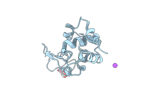 |
Multi-Hit Sfx Using Mhz Xfel Sources- First Hit
Organism: Gallus gallus
Method: X-RAY DIFFRACTION Resolution:3.20 Å Release Date: 2022-07-20 Classification: HYDROLASE Ligands: EDO, NA |
Organism: Gallus gallus
Method: X-RAY DIFFRACTION
Release Date: 2022-07-20
Ligands: EDO, NA
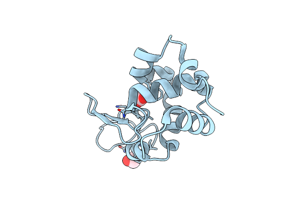 |
Multi-Hit Sfx Using Mhz Xfel Sources
Organism: Gallus gallus
Method: X-RAY DIFFRACTION Resolution:2.10 Å Release Date: 2021-10-13 Classification: HYDROLASE Ligands: ACT, EDO, CL |
Organism: Gallus gallus
Method: X-RAY DIFFRACTION
Release Date: 2021-10-13
Ligands: ACT, EDO, CL
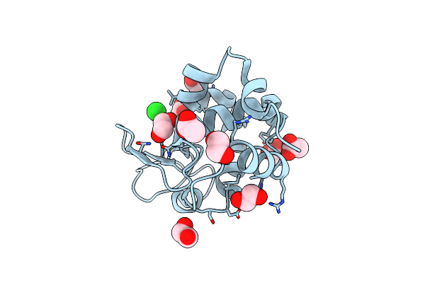 |
Multi-Hit Sfx Using Mhz Xfel Sources
Organism: Gallus gallus
Method: X-RAY DIFFRACTION Resolution:2.10 Å Release Date: 2021-10-13 Classification: HYDROLASE Ligands: CL, EDO, ACT |
Organism: Gallus gallus
Method: X-RAY DIFFRACTION
Release Date: 2021-10-13
Ligands: CL, EDO, ACT
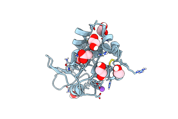 |
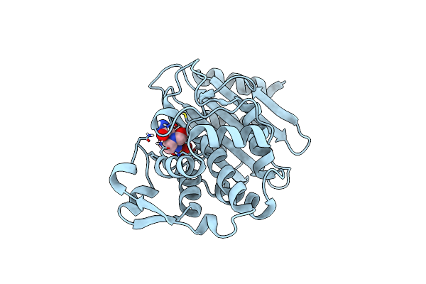
Serial Femtosecond Crystallography At Megahertz Pulse Rates
Organism: Gallus gallus
Method: X-RAY DIFFRACTION Resolution:1.76 Å Release Date: 2018-10-10 Classification: HYDROLASE Ligands: CL, NA, ACT, EDO |
Organism: Gallus gallus
Method: X-RAY DIFFRACTION
Release Date: 2018-10-10
Ligands: CL, NA, ACT, EDO
 |
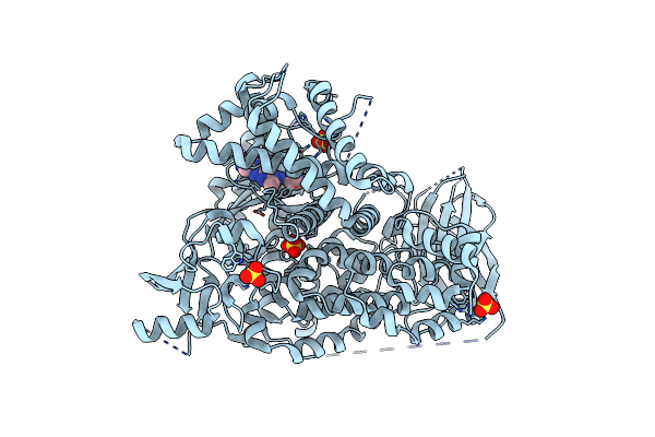
Serial Femtosecond Crystallography At Megahertz Pulse Rates
Organism: Klebsiella pneumoniae
Method: X-RAY DIFFRACTION Resolution:1.69 Å Release Date: 2018-10-10 Classification: ANTIBIOTIC Ligands: NXL |
Organism: Klebsiella pneumoniae
Method: X-RAY DIFFRACTION
Release Date: 2018-10-10
Ligands: NXL
 |
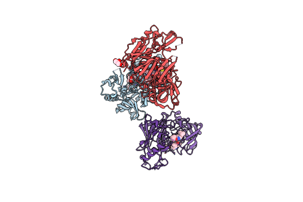
Crystal Structure Of Pi3K-Gamma In Complex With Imidazopyridazine 19E
Organism: Homo sapiens
Method: X-RAY DIFFRACTION Resolution:3.00 Å Release Date: 2013-04-10 Classification: Transferase/Transferase Inhibitor Ligands: SO4, 0U0 |
Organism: Homo sapiens
Method: X-RAY DIFFRACTION
Release Date: 2013-04-10
Ligands: SO4, 0U0
 |
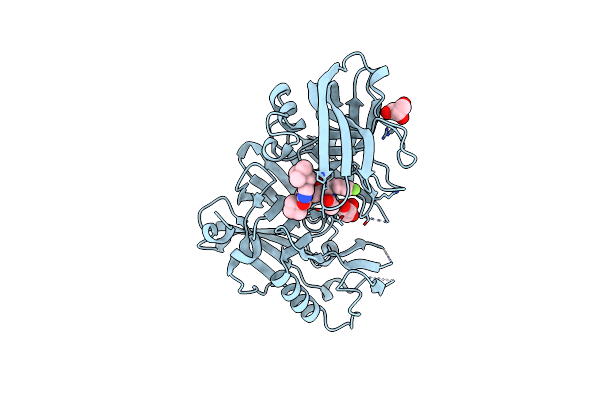
Crystal Structure Of Bace1 In Complex With Hydroxyethylamine Inhibitor 37
Organism: Homo sapiens
Method: X-RAY DIFFRACTION Resolution:2.00 Å Release Date: 2012-10-10 Classification: HYDROLASE/HYDROLASE INHIBITOR Ligands: 0K9, GOL |
Organism: Homo sapiens
Method: X-RAY DIFFRACTION
Release Date: 2012-10-10
Ligands: 0K9, GOL
 |
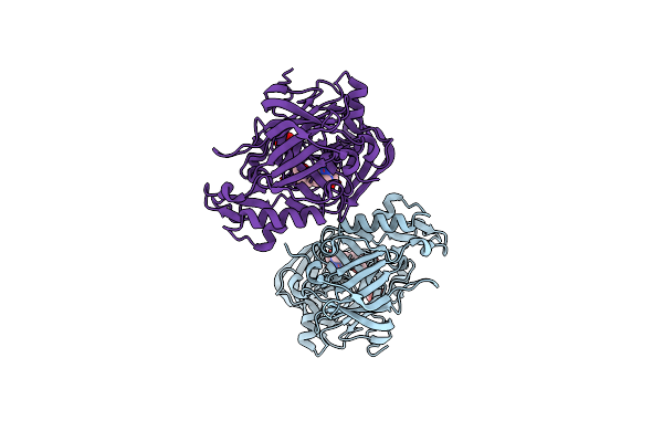
Structure Of Bace-1 (Beta-Secretase) In Complex With N-((2S,3R)-1-(4-Fluorophenyl)-3-Hydroxy-4-((6'-Neopentyl-3',4'-Dihydrospiro[Cyclobutane-1,2'-Pyrano[2,3-B]Pyridin]-4'-Yl)Amino)Butan-2-Yl)Acetamide
Organism: Homo sapiens
Method: X-RAY DIFFRACTION Resolution:2.50 Å Release Date: 2012-10-10 Classification: HYDROLASE/Hydrolase Inhibitor Ligands: IOD, GOL, 0MP |
Organism: Homo sapiens
Method: X-RAY DIFFRACTION
Release Date: 2012-10-10
Ligands: IOD, GOL, 0MP
 |
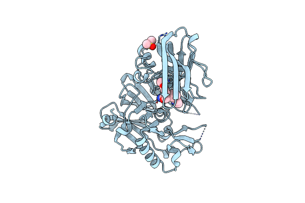
Structure Of Bace Bound To (S)-4-(3'-Methoxy-[1,1'-Biphenyl]-3-Yl)-1,4-Dimethyl-6-Oxotetrahydropyrimidin-2(1H)-Iminium
Organism: Homo sapiens
Method: X-RAY DIFFRACTION Resolution:1.74 Å Release Date: 2012-10-10 Classification: HYDROLASE/HYDROLASE INHIBITOR Ligands: H24, TLA |
Organism: Homo sapiens
Method: X-RAY DIFFRACTION
Release Date: 2012-10-10
Ligands: H24, TLA
 |
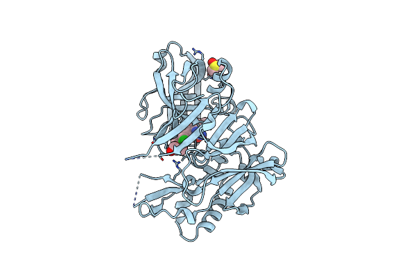
Crystal Structure Of Bace1 In Complex With Biarylspiro Aminooxazoline 6
Organism: Homo sapiens
Method: X-RAY DIFFRACTION Resolution:2.30 Å Release Date: 2012-09-12 Classification: HYDROLASE/INHIBITOR Ligands: IOD, DWA, DMS |
Organism: Homo sapiens
Method: X-RAY DIFFRACTION
Release Date: 2012-09-12
Ligands: IOD, DWA, DMS
 |
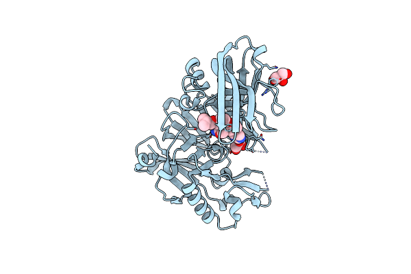
Crystal Structure Of Bace1 In Complex With Aminooxazoline Xanthene 9L
Organism: Homo sapiens
Method: X-RAY DIFFRACTION Resolution:1.95 Å Release Date: 2012-09-12 Classification: HYDROLASE/INHIBITOR Ligands: IOD, DWB, GOL, DMS |
Organism: Homo sapiens
Method: X-RAY DIFFRACTION
Release Date: 2012-09-12
Ligands: IOD, DWB, GOL, DMS
 |
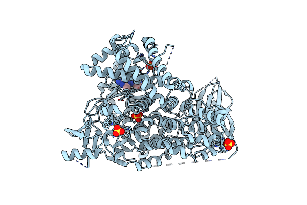
Crystal Structure Of Bace1 In Complex With Aminooxazoline Xanthene 11A
Organism: Homo sapiens
Method: X-RAY DIFFRACTION Resolution:2.10 Å Release Date: 2012-09-12 Classification: HYDROLASE/INHIBITOR Ligands: IOD, DWD, GOL |
Organism: Homo sapiens
Method: X-RAY DIFFRACTION
Release Date: 2012-09-12
Ligands: IOD, DWD, GOL
 |
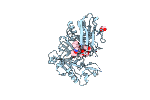
Crystal Structure Of Pi3K-Gamma In Complex With Imidazopyridine 2
Organism: Homo sapiens
Method: X-RAY DIFFRACTION Resolution:2.60 Å Release Date: 2012-07-18 Classification: Transferase/Inhibitor Ligands: SO4, 0TZ |
Organism: Homo sapiens
Method: X-RAY DIFFRACTION
Release Date: 2012-07-18
Ligands: SO4, 0TZ
 |
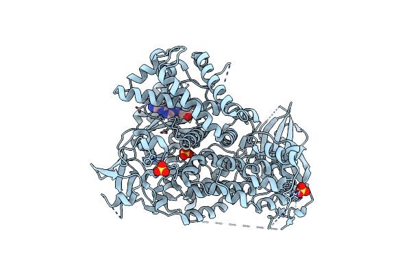
Structure Of Bace-1 (Beta-Secretase) In Complex With (2R)-N-((2S,3R)-1-(Benzo[D][1,3]Dioxol-5-Yl)-3-Hydroxy-4-((S)-6'-Neopentyl-3',4'-Dihydrospiro[Cyclobutane-1,2'-Pyrano[2,3-B]Pyridine]-4'-Ylamino)Butan-2-Yl)-2-Methoxypropanamide
Organism: Homo sapiens
Method: X-RAY DIFFRACTION Resolution:2.50 Å Release Date: 2012-04-18 Classification: HYDROLASE/HYDROLASE INHIBITOR Ligands: IOD, GOL, 0KN |
Organism: Homo sapiens
Method: X-RAY DIFFRACTION
Release Date: 2012-04-18
Ligands: IOD, GOL, 0KN
 |
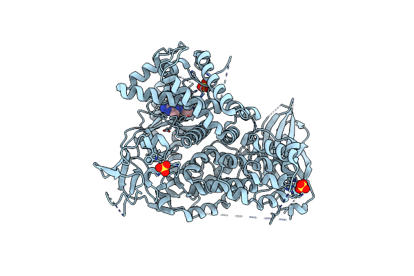
Crystal Structure Of Pi3K-Gamma In Complex With Triazine-Benzimidazole 1
Organism: Homo sapiens
Method: X-RAY DIFFRACTION Resolution:2.90 Å Release Date: 2011-03-30 Classification: TRANSFERASE/TRANSFERASE INHIBITOR Ligands: SO4, QAQ |
Organism: Homo sapiens
Method: X-RAY DIFFRACTION
Release Date: 2011-03-30
Ligands: SO4, QAQ
 |
Crystal Structure Of Pi3K-Gamma In Complex With Triazine-Benzimidazole 32
Organism: Homo sapiens
Method: X-RAY DIFFRACTION Resolution:2.65 Å Release Date: 2011-03-30 Classification: TRANSFERASE/TRANSFERASE INHIBITOR Ligands: SO4, QAR |
Organism: Homo sapiens
Method: X-RAY DIFFRACTION
Release Date: 2011-03-30
Ligands: SO4, QAR
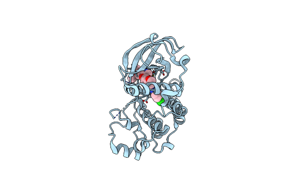 |
Crystal Structure Of The Vegfr2 Kinase Domain In Complex With A 2,3-Dihydro-1,4-Benzoxazine Inhibitor
Organism: Homo sapiens
Method: X-RAY DIFFRACTION Resolution:2.65 Å Release Date: 2008-04-08 Classification: TRANSFERASE Ligands: 2RL |
Organism: Homo sapiens
Method: X-RAY DIFFRACTION
Release Date: 2008-04-08
Ligands: 2RL
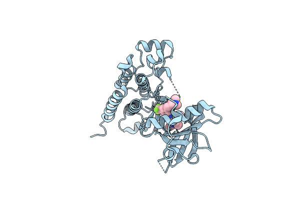 |
Crystal Structure Of The Vegfr2 Kinase Domain In Complex With A Benzamide Inhibitor
Organism: Homo sapiens
Method: X-RAY DIFFRACTION Resolution:1.75 Å Release Date: 2008-04-08 Classification: TRANSFERASE Ligands: RAJ |
Organism: Homo sapiens
Method: X-RAY DIFFRACTION
Release Date: 2008-04-08
Ligands: RAJ
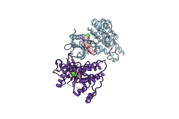 |
Crystal Structure Of The Vegfr2 Kinase Domain In Complex With A Naphthamide Inhibitor
Organism: Homo sapiens
Method: X-RAY DIFFRACTION Resolution:2.75 Å Release Date: 2008-04-01 Classification: TRANSFERASE Ligands: 900 |
Organism: Homo sapiens
Method: X-RAY DIFFRACTION
Release Date: 2008-04-01
Ligands: 900
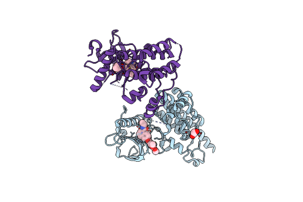 |
Crystal Structure Of The Vegfr2 Kinase Domain In Complex With A Naphthamide Inhibitor
Organism: Homo sapiens
Method: X-RAY DIFFRACTION Resolution:2.70 Å Release Date: 2008-04-01 Classification: TRANSFERASE Ligands: EDO, 887 |
Organism: Homo sapiens
Method: X-RAY DIFFRACTION
Release Date: 2008-04-01
Ligands: EDO, 887