

Search Count: 14
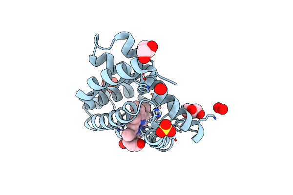 |
Crystal Structure Of Peroxymyoglobin Generated By Cryoradiolytic Reduction Of Myoglobin Compound Iii
Organism: Equus caballus
Method: X-RAY DIFFRACTION Resolution:1.30 Å Release Date: 2009-02-10 Classification: OXYGEN TRANSPORT Ligands: HEM, PER, SO4, GOL, PEO |
Organism: Equus caballus
Method: X-RAY DIFFRACTION
Release Date: 2009-02-10
Ligands: HEM, PER, SO4, GOL, PEO
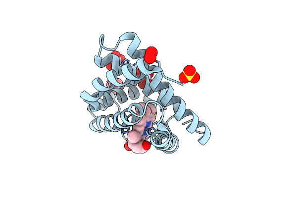 |
Crystal Structure Of Myoglobin Compound Iii (Radiation-Induced)
Organism: Equus caballus
Method: X-RAY DIFFRACTION Resolution:1.60 Å Release Date: 2008-01-29 Classification: OXYGEN TRANSPORT Ligands: HEM, OXY, SO4, GOL, PEO |
Organism: Equus caballus
Method: X-RAY DIFFRACTION
Release Date: 2008-01-29
Ligands: HEM, OXY, SO4, GOL, PEO
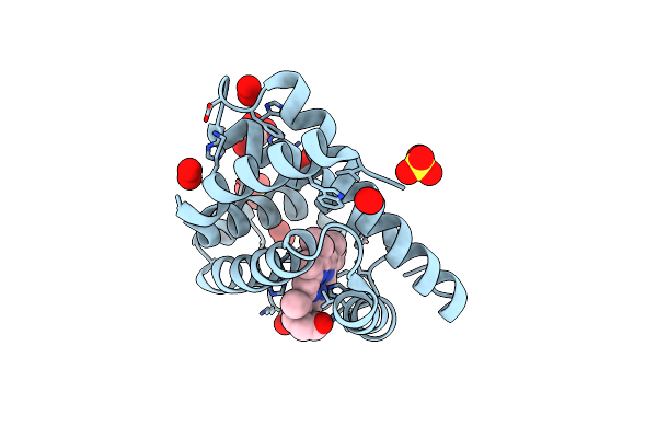 |
Crystal Structure Of Peroxymyoglobin Generated By Cryoradiolytic Reduction Of Myoglobin Compound Iii
Organism: Equus caballus
Method: X-RAY DIFFRACTION Resolution:1.50 Å Release Date: 2008-01-29 Classification: OXYGEN TRANSPORT Ligands: HEM, PER, SO4, GOL, PEO |
Organism: Equus caballus
Method: X-RAY DIFFRACTION
Release Date: 2008-01-29
Ligands: HEM, PER, SO4, GOL, PEO
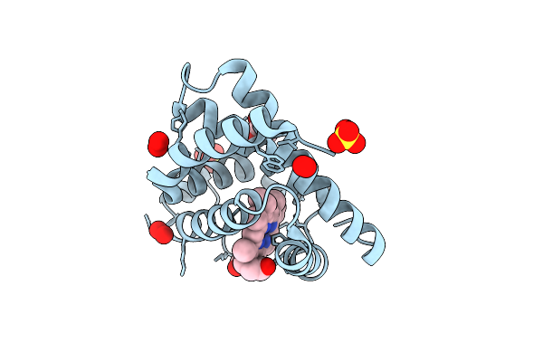 |
Crystal Structure Of Radiation-Induced Myoglobin Compound Ii Generated After Annealing Of Peroxymyoglobin
Organism: Equus caballus
Method: X-RAY DIFFRACTION Resolution:1.60 Å Release Date: 2008-01-29 Classification: OXYGEN TRANSPORT Ligands: HEM, OH, SO4, GOL, PEO |
Organism: Equus caballus
Method: X-RAY DIFFRACTION
Release Date: 2008-01-29
Ligands: HEM, OH, SO4, GOL, PEO
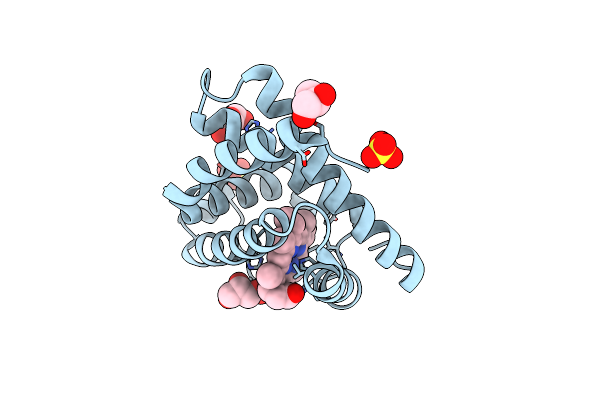 |
Crystal Structure Of Radiation-Induced Myoglobin Compound Ii - Intermediate H At Ph 6.8
Organism: Equus caballus
Method: X-RAY DIFFRACTION Resolution:1.30 Å Release Date: 2007-06-12 Classification: OXYGEN TRANSPORT Ligands: HEM, OH, SO4, GOL |
Organism: Equus caballus
Method: X-RAY DIFFRACTION
Release Date: 2007-06-12
Ligands: HEM, OH, SO4, GOL
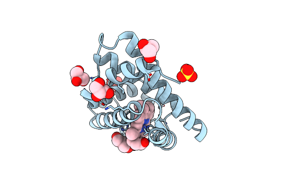 |
Crystal Structure Of Radiation-Induced Myoglobin Compound Ii - Intermediate H At Ph 8.7
Organism: Equus caballus
Method: X-RAY DIFFRACTION Resolution:1.20 Å Release Date: 2007-06-12 Classification: OXYGEN TRANSPORT Ligands: HEM, OH, SO4, GOL |
Organism: Equus caballus
Method: X-RAY DIFFRACTION
Release Date: 2007-06-12
Ligands: HEM, OH, SO4, GOL
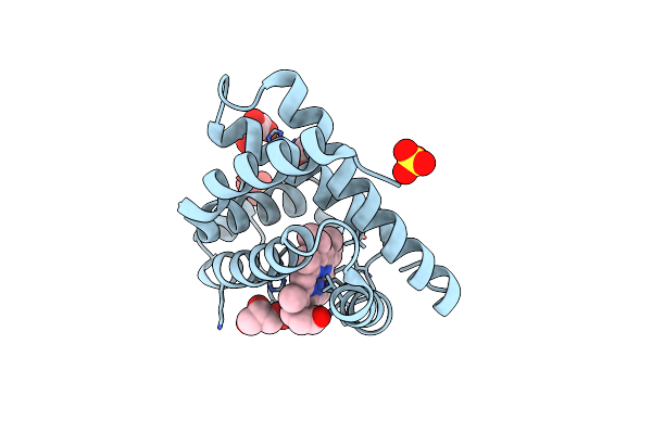 |
Crystal Structure Of Radiation-Induced Myoglobin Compound Ii - Intermediate H At Ph 5.2
Organism: Equus caballus
Method: X-RAY DIFFRACTION Resolution:1.35 Å Release Date: 2007-06-12 Classification: OXYGEN TRANSPORT Ligands: HEM, OH, SO4, GOL |
Organism: Equus caballus
Method: X-RAY DIFFRACTION
Release Date: 2007-06-12
Ligands: HEM, OH, SO4, GOL
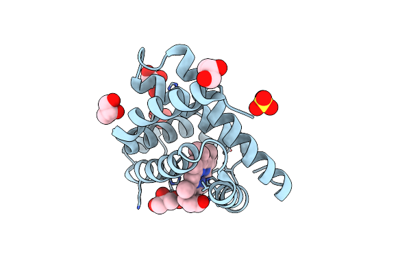 |
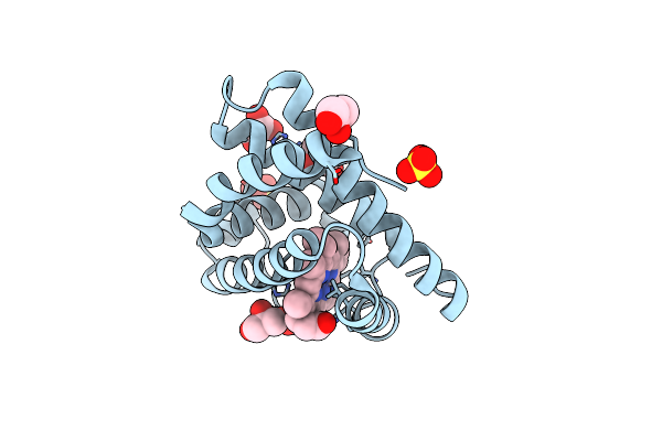
Crystal Structure Of Radiation-Induced Metmyoglobin - Aqua Ferrous Myoglobin At Ph 5.2
Organism: Equus caballus
Method: X-RAY DIFFRACTION Resolution:1.30 Å Release Date: 2007-06-12 Classification: OXYGEN TRANSPORT Ligands: HEM, SO4, GOL |
Organism: Equus caballus
Method: X-RAY DIFFRACTION
Release Date: 2007-06-12
Ligands: HEM, SO4, GOL
 |
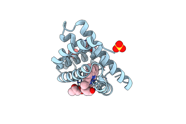
Crystal Structure Of Radiation-Induced Metmyoglobin - Aqua Ferrous Myoglobin At Ph 6.8
Organism: Equus caballus
Method: X-RAY DIFFRACTION Resolution:1.20 Å Release Date: 2007-06-12 Classification: OXYGEN TRANSPORT Ligands: HEM, SO4, GOL |
Organism: Equus caballus
Method: X-RAY DIFFRACTION
Release Date: 2007-06-12
Ligands: HEM, SO4, GOL
 |
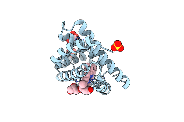
Crystal Structure Of Radiation-Induced Metmyoglobin - Aqua Ferrous Myoglobin At Ph 8.7
Organism: Equus caballus
Method: X-RAY DIFFRACTION Resolution:1.40 Å Release Date: 2007-06-12 Classification: OXYGEN TRANSPORT Ligands: HEM, SO4, GOL |
Organism: Equus caballus
Method: X-RAY DIFFRACTION
Release Date: 2007-06-12
Ligands: HEM, SO4, GOL
 |
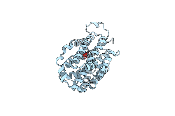
Crystal Structure Of Ferrous Deoxymyoglobin At Ph 6.8
Organism: Equus caballus
Method: X-RAY DIFFRACTION Resolution:1.25 Å Release Date: 2007-06-12 Classification: OXYGEN TRANSPORT Ligands: HEM, SO4, GOL |
Organism: Equus caballus
Method: X-RAY DIFFRACTION
Release Date: 2007-06-12
Ligands: HEM, SO4, GOL
 |
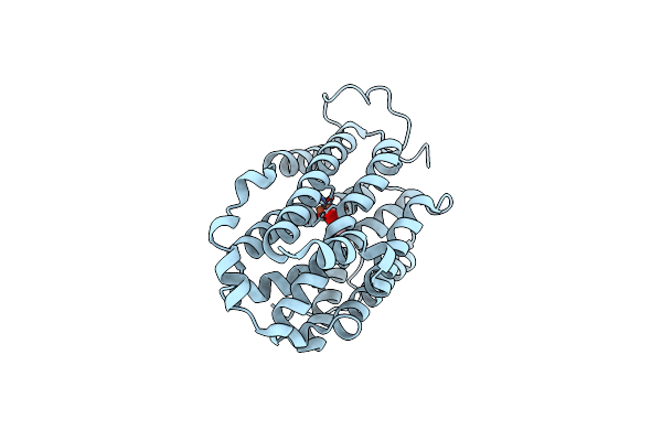
Crystal Structure Of Mouse Ribonucleotide Reductase Subunit R2 Under Oxidizing Conditions. A Fully Occupied Dinuclear Iron Cluster.
Organism: Mus musculus
Method: X-RAY DIFFRACTION Resolution:2.20 Å Release Date: 2004-08-26 Classification: REDUCTASE Ligands: FEO |
Organism: Mus musculus
Method: X-RAY DIFFRACTION
Release Date: 2004-08-26
Ligands: FEO
 |
Crystal Structure Of Mouse Ribonucleotide Reductase Subunit R2 Under Reducing Conditions. A Fully Occupied Dinuclear Iron Cluster And Bound Acetate.
Organism: Mus musculus
Method: X-RAY DIFFRACTION Resolution:2.20 Å Release Date: 2004-08-26 Classification: REDUCTASE Ligands: FE2, ACY |
Organism: Mus musculus
Method: X-RAY DIFFRACTION
Release Date: 2004-08-26
Ligands: FE2, ACY
 |
Hydrogen Peroxide Derived Myoglobin Compound Ii At Ph 5.2
Organism: Equus caballus
Method: X-RAY DIFFRACTION Resolution:1.35 Å Release Date: 2002-03-01 Classification: OXYGEN TRANSPORT Ligands: HEM, OH, SO4 |
Organism: Equus caballus
Method: X-RAY DIFFRACTION
Release Date: 2002-03-01
Ligands: HEM, OH, SO4