

Search Count: 34
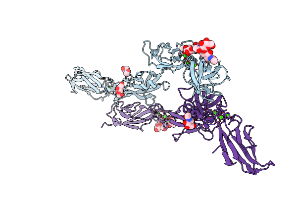 |
Crystal Structure Of Li-Cadherin Ec1-4
Organism: Homo sapiens
Method: X-RAY DIFFRACTION Resolution:2.70 Å Release Date: 2021-08-18 Classification: CELL ADHESION Ligands: NAG, CA |
Organism: Homo sapiens
Method: X-RAY DIFFRACTION
Release Date: 2021-08-18
Ligands: NAG, CA
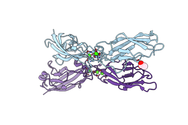 |
Crystal Structure Of Li-Cadherin Ec1-2
Organism: Homo sapiens
Method: X-RAY DIFFRACTION Resolution:1.38 Å Release Date: 2021-06-16 Classification: CELL ADHESION Ligands: CA, ACT, NA |
Organism: Homo sapiens
Method: X-RAY DIFFRACTION
Release Date: 2021-06-16
Ligands: CA, ACT, NA
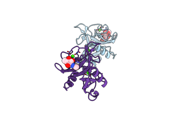 |
Spl-1 - Glcnac Complex
Organism: Saxidomus purpuratus
Method: X-RAY DIFFRACTION Resolution:1.70 Å Release Date: 2020-06-10 Classification: SUGAR BINDING PROTEIN Ligands: CA, NAG |
Organism: Saxidomus purpuratus
Method: X-RAY DIFFRACTION
Release Date: 2020-06-10
Ligands: CA, NAG
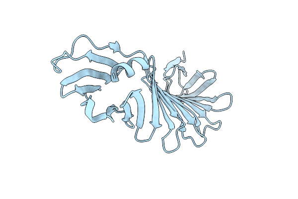 |
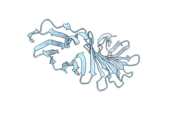
Loop Deletion Mutant (Deleting Two Residues)
Organism: Borrelia burgdorferi
Method: X-RAY DIFFRACTION Resolution:1.30 Å Release Date: 2019-07-03 Classification: DE NOVO PROTEIN |
Organism: Borrelia burgdorferi
Method: X-RAY DIFFRACTION
Release Date: 2019-07-03
 |
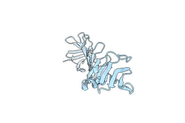
Loop Deletion Mutant (Deleting Four Residues)
Organism: Borrelia burgdorferi
Method: X-RAY DIFFRACTION Resolution:1.40 Å Release Date: 2019-07-03 Classification: DE NOVO PROTEIN |
Organism: Borrelia burgdorferi
Method: X-RAY DIFFRACTION
Release Date: 2019-07-03
 |
Loop Deletion And Proline Insertion Mutant (Deleting Six Residues And Inserted Six Proline Residues)
Organism: Borrelia burgdorferi
Method: X-RAY DIFFRACTION Resolution:2.01 Å Release Date: 2019-07-03 Classification: DE NOVO PROTEIN |
Organism: Borrelia burgdorferi
Method: X-RAY DIFFRACTION
Release Date: 2019-07-03
 |
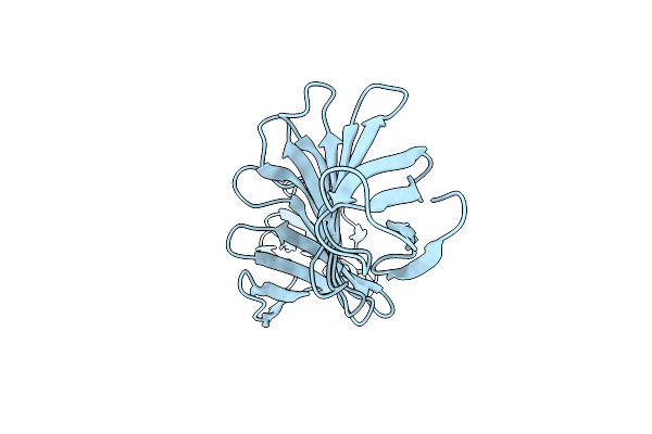
Loop Deletion And Proline Insertion Mutant (Deleting Six Residues And Inserted Five Proline Residues)
Organism: Borrelia burgdorferi b31
Method: X-RAY DIFFRACTION Resolution:2.40 Å Release Date: 2019-07-03 Classification: DE NOVO PROTEIN |
Organism: Borrelia burgdorferi b31
Method: X-RAY DIFFRACTION
Release Date: 2019-07-03
 |
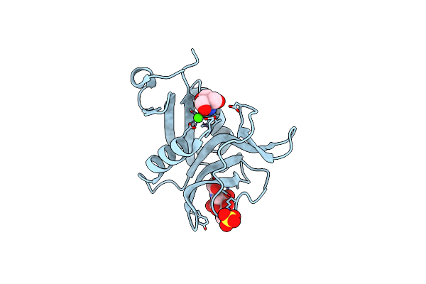
Loop Deletion And Proline Insertion Mutant (Deleting Six Residues And Inserted Three Proline Residues)
Organism: Borrelia burgdorferi (strain atcc 35210 / b31 / cip 102532 / dsm 4680)
Method: X-RAY DIFFRACTION Resolution:3.00 Å Release Date: 2019-07-03 Classification: LIPID BINDING PROTEIN |
 |
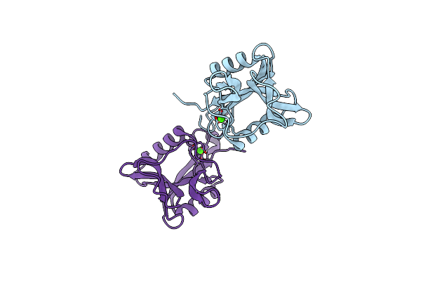
Ca2+-Independent C-Type Lectin Spl-2 From Saxidomus Purpuratus
Organism: Saxidomus purpuratus
Method: X-RAY DIFFRACTION Resolution:2.00 Å Release Date: 2019-03-20 Classification: SUGAR BINDING PROTEIN Ligands: NGA, TRS, CA, SO4 |
Organism: Saxidomus purpuratus
Method: X-RAY DIFFRACTION
Release Date: 2019-03-20
Ligands: NGA, TRS, CA, SO4
 |
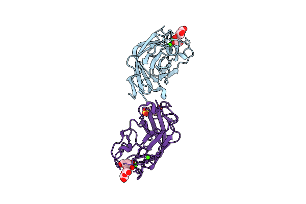
Ca2+-Independent C-Type Lectin Spl-1 From Saxidomus Purpuratus
Organism: Saxidomus purpuratus
Method: X-RAY DIFFRACTION Resolution:1.60 Å Release Date: 2019-03-20 Classification: SUGAR BINDING PROTEIN Ligands: CA |
Organism: Saxidomus purpuratus
Method: X-RAY DIFFRACTION
Release Date: 2019-03-20
Ligands: CA
 |
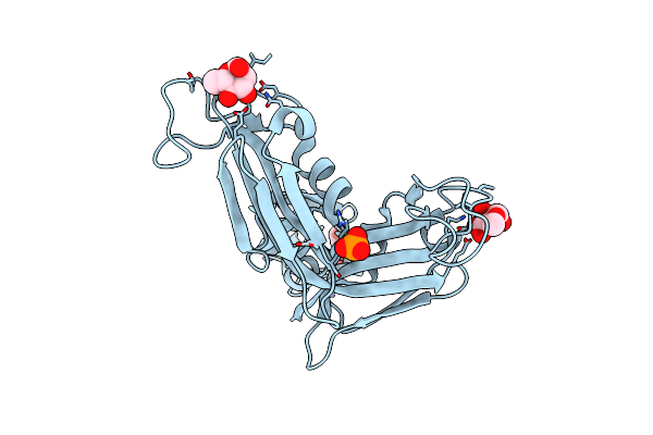
Ajlec From The Sea Anemone Anthopleura Japonica
Organism: Anthopleura japonica
Method: X-RAY DIFFRACTION Resolution:1.20 Å Release Date: 2018-07-25 Classification: SUGAR BINDING PROTEIN Ligands: CA, EPE |
Organism: Anthopleura japonica
Method: X-RAY DIFFRACTION
Release Date: 2018-07-25
Ligands: CA, EPE
 |
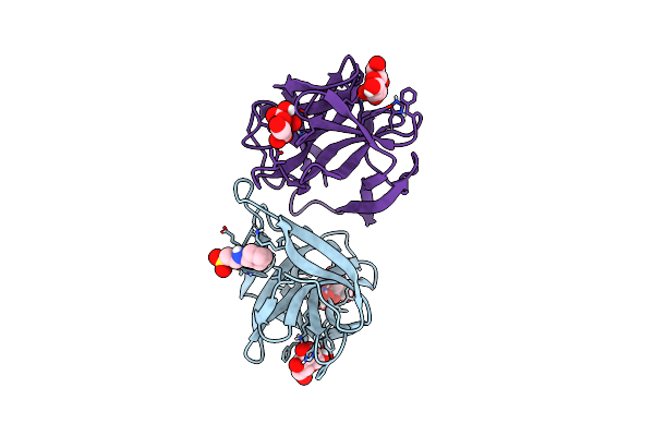
Crystal Structure Of A Rhamnose-Binding Lectin Sul-I From The Toxopneustid Sea Urchin Toxopneustes Pileolus
Organism: Toxopneustes pileolus
Method: X-RAY DIFFRACTION Resolution:1.80 Å Release Date: 2017-05-17 Classification: SUGAR BINDING PROTEIN Ligands: RAM, PO4 |
Organism: Toxopneustes pileolus
Method: X-RAY DIFFRACTION
Release Date: 2017-05-17
Ligands: RAM, PO4
 |
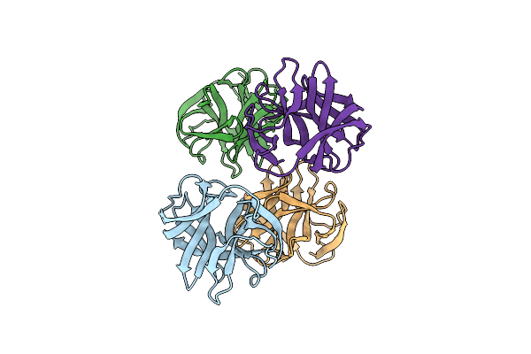
Crystal Structure Of Cgl1 From Crassostrea Gigas, Mannose-Bound Form (Cgl1/Man2)
Organism: Crassostrea gigas
Method: X-RAY DIFFRACTION Resolution:1.00 Å Release Date: 2016-07-06 Classification: SUGAR BINDING PROTEIN Ligands: BMA, MAN, NHE |
Organism: Crassostrea gigas
Method: X-RAY DIFFRACTION
Release Date: 2016-07-06
Ligands: BMA, MAN, NHE
 |
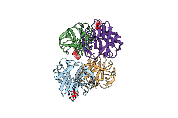
Crystal Structure Of Cgl1 From Crassostrea Gigas, Ligand Free Form (Cgl1/Free)
Organism: Crassostrea gigas
Method: X-RAY DIFFRACTION Resolution:1.10 Å Release Date: 2016-06-29 Classification: SUGAR BINDING PROTEIN |
Organism: Crassostrea gigas
Method: X-RAY DIFFRACTION
Release Date: 2016-06-29
 |
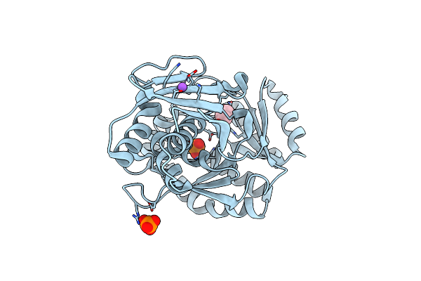
Crystal Structure Of Cgl1 From Crassostrea Gigas, Mannose-Bound Form (Cgl1/Man)
Organism: Crassostrea gigas
Method: X-RAY DIFFRACTION Resolution:1.10 Å Release Date: 2016-06-29 Classification: SUGAR BINDING PROTEIN Ligands: BMA |
Organism: Crassostrea gigas
Method: X-RAY DIFFRACTION
Release Date: 2016-06-29
Ligands: BMA
 |
Diphosphomevalonate Decarboxylase From The Sulfolobus Solfataricus, Space Group H32
Organism: Sulfolobus solfataricus (strain atcc 35092 / dsm 1617 / jcm 11322 / p2)
Method: X-RAY DIFFRACTION Resolution:2.20 Å Release Date: 2015-09-16 Classification: LYASE Ligands: PO4, NA, GOL |
Organism: Sulfolobus solfataricus (strain atcc 35092 / dsm 1617 / jcm 11322 / p2)
Method: X-RAY DIFFRACTION
Release Date: 2015-09-16
Ligands: PO4, NA, GOL
 |
Diphosphomevalonate Decarboxylase From The Sulfolobus Solfataricus, Space Group P21
Organism: Sulfolobus solfataricus (strain atcc 35092 / dsm 1617 / jcm 11322 / p2)
Method: X-RAY DIFFRACTION Resolution:2.70 Å Release Date: 2015-09-16 Classification: LYASE Ligands: SO4 |
Organism: Sulfolobus solfataricus (strain atcc 35092 / dsm 1617 / jcm 11322 / p2)
Method: X-RAY DIFFRACTION
Release Date: 2015-09-16
Ligands: SO4
 |
Structure Of Epnh Mutant Of Cel-I
Organism: Cucumaria echinata
Method: X-RAY DIFFRACTION Resolution:1.70 Å Release Date: 2015-04-29 Classification: SUGAR BINDING PROTEIN Ligands: CA, MAN |
Organism: Cucumaria echinata
Method: X-RAY DIFFRACTION
Release Date: 2015-04-29
Ligands: CA, MAN
 |
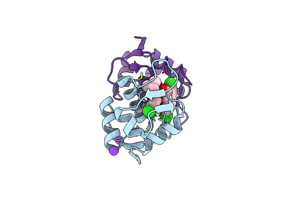
Pore-Forming Cel-Iii
Organism: Cucumaria echinata
Method: X-RAY DIFFRACTION Resolution:2.90 Å Release Date: 2014-03-19 Classification: TOXIN Ligands: CA, MG |
Organism: Cucumaria echinata
Method: X-RAY DIFFRACTION
Release Date: 2014-03-19
Ligands: CA, MG
 |
Structure Of A Novel Submicromolar Mdm2 Inhibitor
Organism: Homo sapiens
Method: X-RAY DIFFRACTION Resolution:2.10 Å Release Date: 2012-09-12 Classification: PROTEIN BINDING/INHIBITOR Ligands: TJ2, K |
Organism: Homo sapiens
Method: X-RAY DIFFRACTION
Release Date: 2012-09-12
Ligands: TJ2, K