

Search Count: 37
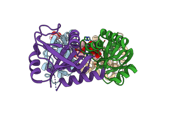 |
Crystal Structure Of The Mmi1 Yth Domain
Organism: Schizosaccharomyces pombe
Method: X-RAY DIFFRACTION Resolution:1.45 Å Release Date: 2018-04-25 Classification: RNA BINDING PROTEIN Ligands: GOL |
Organism: Schizosaccharomyces pombe
Method: X-RAY DIFFRACTION
Release Date: 2018-04-25
Ligands: GOL
 |
Nmr Structure Of The C-Terminal Domain Of The Bacteriophage T5 Decoration Protein Pb10.
Organism: Escherichia phage t5
Method: SOLUTION NMR Release Date: 2017-08-02 Classification: VIRAL PROTEIN |
Organism: Escherichia phage t5
Method: SOLUTION NMR
Release Date: 2017-08-02
 |
Nmr Structure Of The N-Terminal Domain Of The Bacteriophage T5 Decoration Protein Pb10
Organism: Escherichia phage t5
Method: SOLUTION NMR Release Date: 2017-04-19 Classification: VIRAL PROTEIN |
Organism: Escherichia phage t5
Method: SOLUTION NMR
Release Date: 2017-04-19
 |
T5 Bacteriophage Major Capsid Protein - One Pb8 Hexon
Organism: Escherichia phage t5
Method: ELECTRON MICROSCOPY Release Date: 2017-02-22 Classification: VIRAL PROTEIN |
Organism: Escherichia phage t5
Method: ELECTRON MICROSCOPY
Release Date: 2017-02-22
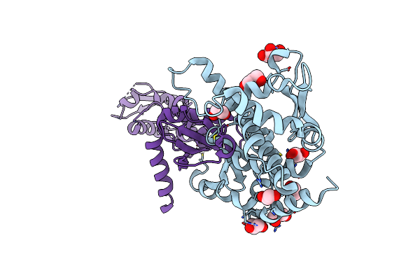 |
Structure Of The C-Terminal Domain Of The Saccharomyces Cerevisiae Mutl Alpha (Mlh1/Pms1) Heterodimer
Organism: Saccharomyces cerevisiae s288c
Method: X-RAY DIFFRACTION Resolution:2.50 Å Release Date: 2013-02-20 Classification: HYDROLASE Ligands: GOL, EDO, ZN |
Organism: Saccharomyces cerevisiae s288c
Method: X-RAY DIFFRACTION
Release Date: 2013-02-20
Ligands: GOL, EDO, ZN
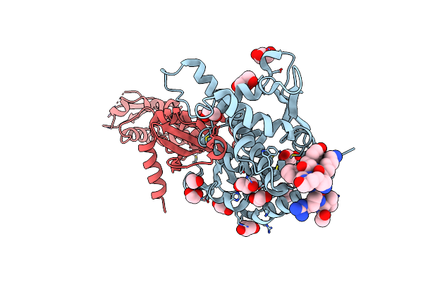 |
Structure Of The C-Terminal Domain Of The Saccharomyces Cerevisiae Mutl Alpha (Mlh1/Pms1) Heterodimer Bound To A Fragment Of Ntg2
Organism: Saccharomyces cerevisiae
Method: X-RAY DIFFRACTION Resolution:2.69 Å Release Date: 2013-02-20 Classification: HYDROLASE Ligands: GOL, EDO, ZN |
Organism: Saccharomyces cerevisiae
Method: X-RAY DIFFRACTION
Release Date: 2013-02-20
Ligands: GOL, EDO, ZN
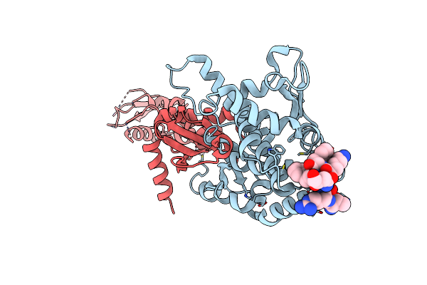 |
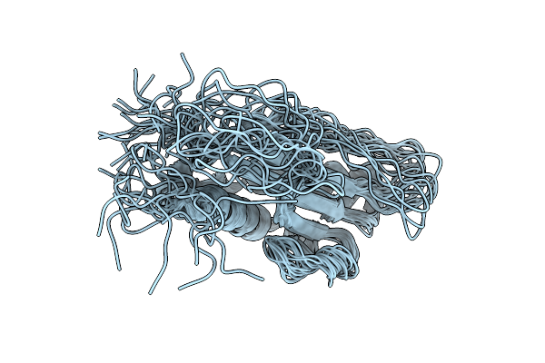
Structure Of The C-Terminal Domain Of The Saccharomyces Cerevisiae Mutl Alpha (Mlh1/Pms1) Heterodimer Bound To A Fragment Of Exo1
Organism: Saccharomyces cerevisiae
Method: X-RAY DIFFRACTION Resolution:3.04 Å Release Date: 2013-02-20 Classification: HYDROLASE Ligands: MG, ZN |
Organism: Saccharomyces cerevisiae
Method: X-RAY DIFFRACTION
Release Date: 2013-02-20
Ligands: MG, ZN
 |
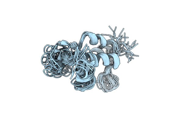
Structure Of Bacteriophage Spp1 Gp17 Protein
Organism: Bacillus phage spp1
Method: SOLUTION NMR Release Date: 2011-09-28 Classification: VIRAL PROTEIN |
Organism: Bacillus phage spp1
Method: SOLUTION NMR
Release Date: 2011-09-28
 |
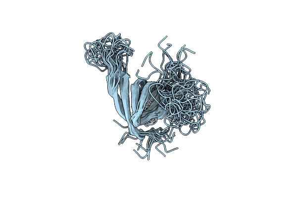
Nmr Structure Of Protein Gp15 Of Bacteriophage Spp1
Organism: Bacillus phage spp1
Method: SOLUTION NMR Release Date: 2009-06-09 Classification: VIRAL PROTEIN |
Organism: Bacillus phage spp1
Method: SOLUTION NMR
Release Date: 2009-06-09
 |
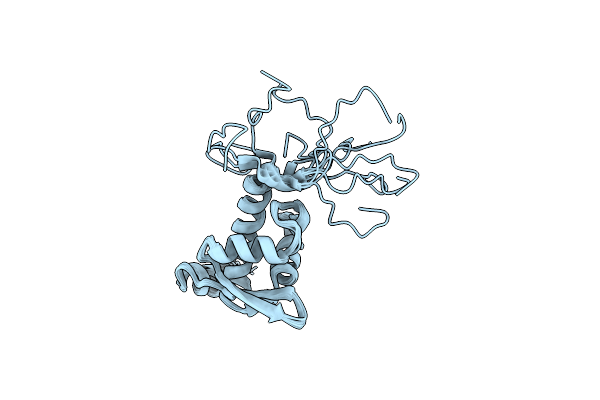
Gp16
Organism: Bacillus phage spp1
Method: SOLUTION NMR Release Date: 2009-06-02 Classification: VIRAL PROTEIN |
Organism: Bacillus phage spp1
Method: SOLUTION NMR
Release Date: 2009-06-02
 |
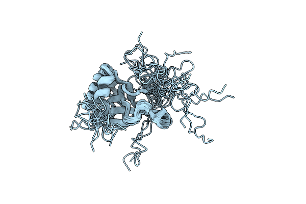
Solution Structure Of The Region 51-160 Of Human Kin17 Reveals A Winged Helix Fold
Organism: Homo sapiens
Method: SOLUTION NMR Release Date: 2007-11-27 Classification: NUCLEAR PROTEIN |
Organism: Homo sapiens
Method: SOLUTION NMR
Release Date: 2007-11-27
 |
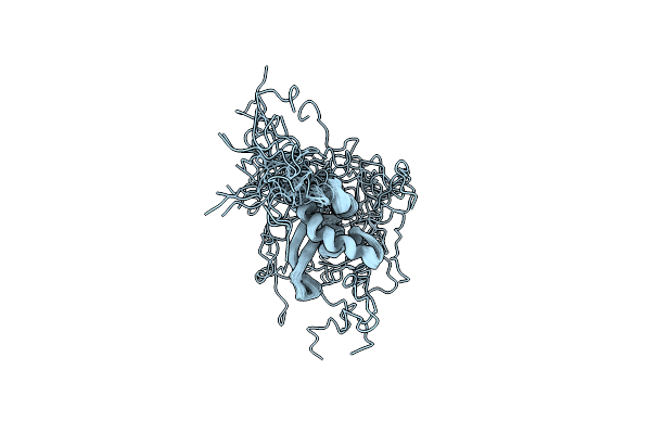
Nmr Solution Structure Of The Pwwp Domain Of Mismatch Repair Protein Hmsh6
Organism: Homo sapiens
Method: SOLUTION NMR Release Date: 2007-04-10 Classification: DNA BINDING PROTEIN |
Organism: Homo sapiens
Method: SOLUTION NMR
Release Date: 2007-04-10
 |
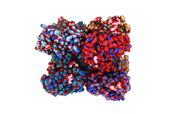
Solution Structure Of The Human Man1 C-Terminal Domain (Residues 655- 775)
Organism: Homo sapiens
Method: SOLUTION NMR Release Date: 2006-05-16 Classification: NUCLEAR PROTEIN |
Organism: Homo sapiens
Method: SOLUTION NMR
Release Date: 2006-05-16
 |
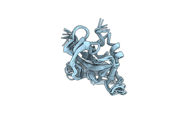
Crystal Structure Of Urokinase Plasminogen Activator Receptor
Organism: Homo sapiens
Method: X-RAY DIFFRACTION Resolution:2.70 Å Release Date: 2005-05-10 Classification: HYDROLASE RECEPTOR Ligands: NAG, SO4 |
Organism: Homo sapiens
Method: X-RAY DIFFRACTION
Release Date: 2005-05-10
Ligands: NAG, SO4
 |
Solution Structure Of The Mouse 53Bp1 Fragment (Residues 1463-1617)
|
Organism: Mus musculus
Method: SOLUTION NMR
Release Date: 2004-09-14
 |
Crystal Structure At 1.8 A Of Toxin A From Naja Nigricollis Venom
Organism: Naja nigricollis
Method: X-RAY DIFFRACTION Resolution:1.80 Å Release Date: 2003-11-25 Classification: TOXIN Ligands: SO4 |
Organism: Naja nigricollis
Method: X-RAY DIFFRACTION
Release Date: 2003-11-25
Ligands: SO4
 |
Psae Subunit Of The Photosystem I Of The Cyanobacterium Synechocystis Sp. Pcc 6803
Organism: Synechocystis sp.
Method: SOLUTION NMR Release Date: 2003-04-04 Classification: PHOTOSYNTHESIS |
Organism: Synechocystis sp.
Method: SOLUTION NMR
Release Date: 2003-04-04
 |
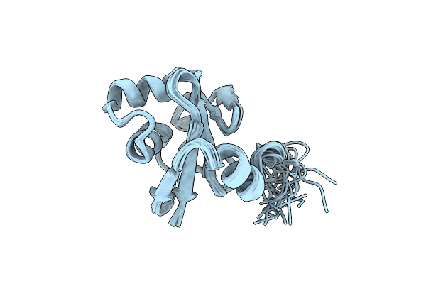
Nmr Structures Of The C-Terminal Globular Domain Of Human Lamin A/C
Organism: Homo sapiens
Method: SOLUTION NMR Release Date: 2002-08-21 Classification: STRUCTURAL PROTEIN |
Organism: Homo sapiens
Method: SOLUTION NMR
Release Date: 2002-08-21
 |
Solution Structure Of Tyrosyl-Trna Synthetase C-Terminal Domain.
Organism: Geobacillus stearothermophilus
Method: SOLUTION NMR Release Date: 2002-03-20 Classification: LIGASE |
Organism: Geobacillus stearothermophilus
Method: SOLUTION NMR
Release Date: 2002-03-20
 |
Crystal Structure Of Fab-Estradiol Complexes
Organism: Mus musculus
Method: X-RAY DIFFRACTION Resolution:2.70 Å Release Date: 2002-02-06 Classification: IMMUNE SYSTEM |
Organism: Mus musculus
Method: X-RAY DIFFRACTION
Release Date: 2002-02-06